Background
Perforating disorders are characterized by transepidermal elimination of altered keratin or dermal connective tissue material and are divided into primary and secondary forms. The primary subtypes include perforating folliculitis (as shown below), Kyrle disease, elastosis perforans serpiginosa, reactive perforating collagenosis. A secondary form is acquired perforating dermatosis (APD) and usually occurs in association with diabetes mellitus or chronic renal failure, but has also been reported in patients with malignant, hepatic and endocrinological disorders, HIV infection, tuberculosis, pulmonary aspergillosis, neurodermatitis, atopic dermatitis, and scabies; it has also been diagnosed in patients without any known medical comorbidity. [1, 2] Cases of perforating folliculitis have also been reported after hematopoietic stem cell transplantation and solid organ transplantation. [3, 4]
Cases of overlap between the subtypes are described, and diagnostic criteria are not well-defined for all the entities. [5] Clinically, the lesions are hyperkeratotic to verrucous papules and nodules. Histologic findings are often required to differentiate among the four conditions and confirm diagnosis. [2]
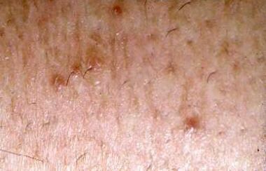 Typical appearance of lesions of perforating folliculitis consisting of keratotic follicular papules.
In perforating folliculitis, keratotic follicular papules develop, particularly over extensor surfaces. Microscopically, the disorder is characterized by disruption of the infundibular portion of the follicular wall, with transepidermal (transfollicular) elimination of connective-tissue elements and cellular debris.
Perforating folliculitis may present as an isolated finding, apparently unrelated to other disease states, but also can be associated with chronic renal failure and diabetes mellitus.

-
Typical appearance of lesions of perforating folliculitis consisting of keratotic follicular papules.
Tables

What would you like to print?
- FDA Warns Of Potential Risk From Hologic's Devices Implanted in Soft Tissue
- Posttransplant Skin Disease: Consider Skin Cancer, Infection Risks
- Presurgery Skin Antisepsis Affects Patient Infection Risk
-
 In Lupus, How to Spot Hidden Heart Risk
-
Is Microwave Ablation Better for Treating Thyroid Nodules?
-
Is Scarless Thyroidectomy an Option for Your Patient?


