Practice Essentials
Viral conjunctivitis, or pinkeye (see the image below), is a common, self-limiting condition that typically is caused by adenovirus. [1, 2, 3] Other viruses that can be responsible for conjunctival infection include herpes simplex virus (HSV), varicella-zoster virus (VZV), picornavirus (enterovirus 70, Coxsackie A24), poxvirus (molluscum contagiosum, vaccinia), and human immunodeficiency virus (HIV).
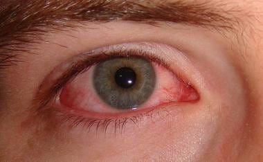 Viral conjunctivitis. Image courtesy of Wikimedia Commons.
Viral conjunctivitis is highly contagious, usually for 10-12 days from onset as long as the eyes are red. [1, 2, 3] Patients should avoid touching their eyes, shaking hands, and sharing towels, napkins, pillow cases, and other fomites, among other activities. Transmission may occur through accidental inoculation of viral particles from the patient's hands or by direct eye contact with infected upper respiratory droplets, fomites, or contaminated swimming pools. The infection usually resolves spontaneously within 2-4 weeks.
Signs and symptoms
Signs and symptoms of viral conjunctivitis may include the following:
-
Itchy eyes
-
Tearing
-
Redness
-
Discharge
-
Light sensitivity (when corneal involvement is present)
See Clinical Presentation for more details.
Diagnosis
Generally, a diagnosis of viral conjunctivitis is made on the clinical features alone. Laboratory tests typically are unnecessary, but they may be extremely helpful in some cases, particularly when an epidemic of adenoviral keratoconjunctivitis threatens a community or clinic. Specimens can be obtained by culture and conjunctival cytology smear if inflammation is severe, in chronic or recurrent infections, with atypical conjunctival reactions, and in patients who fail to respond to treatment. Giemsa staining of conjunctival scrapings may aid in characterizing the inflammatory response. A rapid point-of-service immunoassay is readily available to guide the clinician’s recommendations upon initial presentation (Adenoplus, RPS, Sarasota, FL). Approximately 1 in 4 patients with acute conjunctivitis have confirmed adenoviral conjunctivitis. Adenoplus detects all known serotypes of adenoviral conjunctivitis. [4, 5]
See Workup for more details.
Management
Treatment of adenoviral conjunctivitis is supportive. [1, 2, 3] Patients should be instructed to use cold compresses and lubricants, such as chilled artificial tears, for comfort. Topical vasoconstrictors and antihistamines may be used for severe itching but generally are not indicated. For patients who may be susceptible, a topical astringent or antibiotic may be used to prevent bacterial superinfection. There is clinical evidence that topical ganciclovir is effective against at least Adenovirus serotype 8, thus compelling many clinicians to prescribe this agent off-label for compelling cases of epidemic keratoconjunctivitis (EKC), particularly when corneal lesions are noted.
Virus-specific treatments
Patients with conjunctivitis caused by HSV usually are treated with topical antiviral agents, including ganciclovir (Zirgan, Bausch & Lomb, Bridgewater, NJ), idoxuridine solution and ointment, vidarabine ointment, and trifluridine solution (Viroptic, Alcon, Fort Worth, TX).
Treatment of VZV eye disease includes high-dose oral acyclovir to terminate viral replication.
For conjunctivitis associated with molluscum contagiosum, disease will persist until the skin lesion is treated. Removal of the central core of the lesion or inducement of bleeding within the lesion usually is enough to cure the infection.
Prevention
Preventing transmission of viral conjunctivitis is important. Both patient and provider should wash hands thoroughly and often, keep hands away from the infected eye and contralateral eye, and avoid sharing towels, linens, and cosmetics. Infected patients should be advised to stay home from school and work. Those who wear contact lenses should be instructed to discontinue lens wear until signs and symptoms have resolved.
See Treatment and Medication for more details.
Background
Viruses are a common cause of conjunctivitis in patients of all ages. A variety of viruses can be responsible for conjunctival infection; however, adenovirus is by far the most common cause, and herpes simplex virus (HSV) is the most problematic. [1, 2, 3] Less common causes include varicella-zoster virus (VZV), picornavirus (enterovirus 70, Coxsackie A24), poxvirus (molluscum contagiosum, vaccinia), and human immunodeficiency virus (HIV). Rarely, conjunctivitis is seen during systemic infection with influenza virus, Epstein-Barr virus, paramyxovirus (measles, mumps, Newcastle), or rubella. (See Etiology.) [6]
Viral conjunctivitis, although usually benign and self-limited, tends to follow a longer course than acute bacterial conjunctivitis, lasting for approximately 2-4 weeks. Viral infection commonly is characterized by an acute follicular conjunctival reaction and preauricular adenopathy. (See History and Physical Examination.)
See the following for more information:
Etiology
Adenoviral conjunctivitis is the most common cause of viral conjunctivitis. [1, 2, 3] Particular subtypes of adenoviral conjunctivitis include epidemic keratoconjunctivitis (EKC; pink eye) and pharyngoconjunctival fever (PCF).
Viral conjunctivitis is highly contagious, usually for 10-12 days from onset as long as the eyes are red, in addition to a prodromal period of 3-7 days. Patients should avoid touching their eyes, shaking hands, and sharing towels, among other activities. Transmission may occur through accidental inoculation of viral particles from the patient's hands or by contact with infected upper respiratory droplets, fomites, or contaminated swimming pools.
Primary ocular herpes simplex infection is common in children and usually is associated with a follicular conjunctivitis. Infection usually is caused by HSV type I, although HSV type II may be a cause, especially in neonates. Recurrent infection, typically seen in adults, often is associated with superficial epithelial or deep stromal corneal involvement.
VZV can affect the conjunctiva during primary infection (chickenpox) or secondary infection (zoster). Infection can be caused by direct contact with VZV or zoster skin lesions or by inhalation of infectious respiratory secretions.
Picornaviruses cause an acute hemorrhagic conjunctivitis (AHC) that is clinically similar to adenoviral conjunctivitis but is more severe and hemorrhagic. Infection is highly contagious and occurs in epidemics.
Molluscum contagiosum may produce a chronic follicular conjunctivitis that occurs secondary to shedding of viral particles into the conjunctival sac from an irritative eyelid lesion.
Vaccinia virus has become a rare cause of conjunctivitis because, with the elimination of smallpox, the vaccination rarely is administered. Infection occurs through accidental inoculation of viral particles from the patient's hands.
HIV is the etiologic agent of acquired immunodeficiency syndrome (AIDS). Ocular abnormalities in patients with AIDS primarily affect the posterior segment, but anterior segment findings have been reported. When conjunctivitis occurs in a patient with AIDS, it tends to follow a more severe and prolonged course than in patients without AIDS. In general, patients with AIDS may develop a transient, nonspecific conjunctivitis, characterized by irritation, hyperemia, and tearing, that requires no specific treatment. Microsporidia has been isolated from the cornea and conjunctiva of several patients with AIDS and keratoconjunctivitis. In these patients, symptoms included foreign body sensation, blurred vision, and photophobia; most cases resolved without antimicrobial therapy.
Epidemiology
United States and international occurrence
Viral conjunctivitis is a common ocular disease in the United States and worldwide. Because it is so common, and because many cases are not brought to medical attention, accurate statistics on the frequency of the disease are unavailable. An estimated 6 million new cases of viral conjunctivitis occur annually in the United States. [7] Viral infection frequently occurs in epidemics within families, schools, offices, shipyards, athletic teams, residential communities, and military organizations.
Sex predilection
Viral conjunctivitis can occur equally in men and women.
Age predilection
Viral conjunctivitis can affect all age groups, depending on the specific viral etiology. Usually, adenovirus affects patients aged 20-40 years. HSV and primary VZV infection usually affect young children and infants. Herpes zoster ophthalmicus results from reactivation of latent VZV infection and may present in any age group. Typically, the picornaviruses affect children and young adults in the lower socioeconomic classes. [8]
Prognosis
Most cases of viral conjunctivitis are acute, benign, and self-limited, although chronic infections have been reported. Long-term ocular sequelae are uncommon but may be severe and even debilitating in rare highly susceptible individuals. The infection usually resolves spontaneously within 2-4 weeks. Subepithelial infiltrates may last for several months, and, if in the visual axis, they may cause decreased vision or glare.
Morbidity
Complications include the following: punctate keratitis with subepithelial infiltrates, bacterial superinfection, conjunctival scarring and symblepharon, severe dry eye, irregular astigmatism, corneal ulceration with persistent keratoconjunctivitis, corneal scarring, and chronic infection.
Epithelial keratitis may accompany viral conjunctivitis. Punctate epithelial erosions that stain with fluorescein are commonly associated with viral keratitis. Rarely, these changes are sufficiently distinctive morphologically to allow identification of a specific type of virus as the etiologic agent. If the conjunctivitis persists or is severe, disturbances in the anterior stroma beneath the epithelial abnormalities may occur. In general, the stromal or subepithelial abnormalities are transient and resolve despite persistence of epithelial keratitis. However, in cases of specific adenoviral serotype infection, the stromal abnormalities may persist for months to years, long after the epithelial changes have resolved. In such cases, these subepithelial infiltrates are considered to be immunologic in origin, the result of antigen-antibody reaction. If they are in the pupillary axis, they may cause decreased vision and/or glare. Rarely, these corneal changes or the accompanying severe dry eye caused by partial obliteration of the conjunctival lacrimal ductules can lead to career-ending photophobia, eye pain, or visual disturbances.
Patient Education
To allay patient anxiety, it is helpful to inform patients that their symptoms may worsen during the first 4-7 days after onset before they begin to improve and may not resolve for 2-4 weeks. The contagiousness of the infection also should be emphasized. Proper isolation from the workplace or school is advisable and essential to prevent epidemics.
Patients with conjunctivitis who wear contact lenses should be instructed to discontinue lens wear until signs and symptoms have resolved.
For patient education information, see the Eye and Vision Center and the Skin, Hair, and Nails Center, as well as Pinkeye, How to Instill Your Eyedrops, and Molluscum Contagiosum.
-
Viral conjunctivitis. Image courtesy of Wikimedia Commons.
Tables

What would you like to print?
- Optimal Management of Perianal Disease in Crohn's Disease: The Importance of a Multidisciplinary Approach
- Elevated Estradiol Linked to Bulging Cornea in Premenopausal Women
- Psoriatic Disease and MASLD: What Is the Connection?
- Cornea and External Disease
- Artificial Intelligence and Corneal Diseases
- Perioperative Care of the Patient With eye Pathologies Undergoing Nonocular Surgery


