Practice Essentials
Hand injuries are very common in all sports, especially in ball-playing athletes. Most athletic hand injuries are closed hand injuries and include ligamentous injuries, fractures and fracture-dislocations, tendon injuries, and neurovascular problems. There is increasing recognition that fractures and dislocations of the hand can result in long-term pain and disability if they are not recognized and treated early. [1, 2, 3, 4, 5]
Extra-articular fractures of the distal phalanx are common and are associated with significant soft-tissue injury. Most distal phalangeal fractures are crush injuries from a perpendicular force. They can be associated with significant debility, usually in the form of soft-tissue loss, nail bed injury, or posttraumatic neuromas. Intra-articular fractures of the distal phalanx can result from avulsion of either the extensor tendon, also known as mallet fractures, or of the flexor digitorum profundus, also known as jersey fractures. These can be associated with either small dorsal fragments or larger articular fragments with volar subluxation of the volar fragment. Conservative management is usually the standard of treatment.
Fractures of the proximal phalanx are more common than fractures of the middle phalanges. Dorsal or palmar angulation may occur with these fractures, depending on their location. Nondisplaced fractures are usually stable and are treated with closed reduction and fixation. [2, 6] If significant comminution or segmental bone loss is present, these unstable fractures may require either internal or external fixation.
The proximal interphalangeal (PIP) joint is particularly vulnerable to injury as either an ligamentous or intra-articular fracture, with or without subluxation or dislocation. Middle phalangeal articular fractures at the PIP joint include dorsal lip fractures, palmar lip fractures, and central articular disruptions or pilon fractures. Avulsion and impaction sheer are 2 fracture mechanisms.
Middle phalanx palmar lip fractures are the most common form of osseous injury associated with PIP joint fracture-dislocations. Dorsal fracture-dislocation of the PIP joint is reported to occur in 9 of every 100,000 people each year. Many of these injuries are frequently ignored or treated inappropriately. As a result, there can be permanent swelling, pain, and variable degrees of stiffness, angulation, and degenerative changes.
If a serious phalangeal injury is suspected, radiographs should be performed before more forceful testing. Hand fractures in the athlete are treated with adequate alignment, immobilization, and then motion. In general, intra-articular fractures must be reduced anatomically. Reduction requires early recognition of the exact location of the fracture and having a complete understanding of the muscle pull on the fragments, then minimizing the deforming force.
See the image below.
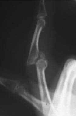 Acute dorsal proximal interphalangeal joint fracture-dislocation.
Functional Anatomy
The phalanges do not contain muscle bellies, and motor function is accomplished only by the flexor and extensor tendons. An overview of the muscles and tendons of the hand is necessary. The thenar muscles consist of 3 intrinsic muscles including the abductor pollicis brevis (which abducts the thumb), the flexor pollicis brevis (which flexes the proximal phalanx of the thumb), and the opponens pollicis (which produces opposition of the thumb).
All 3 intrinsic thenar muscles are supplied by the recurrent branch of the median nerve. The adductor pollicis adducts the thumb and is supplied by the deep branch of the ulnar nerve. The hypothenar muscles are also supplied by the deep branch of the ulnar nerve.
The abductor digiti minimi abducts the fifth digit and flexes its proximal phalanx. The flexor digiti minimi is deeper and also flexes the proximal phalanx of the fifth digit. The opponens digiti minimi, as its name implies, opposes the fifth digit.
The lumbricals are 4 muscles that arise from the tendons of flexor digitorum profundus. Their tendons insert into the radial side of each of the proximal phalanges of the fingers and into the dorsal hood. They flex the metacarpophalangeal joints and extend the interphalangeal joints. The first and second lumbricals are supplied by the median nerve, and the third and fourth lumbricals are supplied by the ulnar nerve.
The palmar and dorsal interossei arise from the metacarpals. The palmar interossei insert into the proximal phalanx and the expansion of the extensor digitorum communis. The palmar interossei are adductor muscles. Dorsal interossei are abductors and insert into the proximal phalanges and the dorsal digital hood. The interosseous muscles are all supplied by the deep branch of the ulnar nerve.
As the tendons of the long flexor and extensor muscles reach the hand, the flexor tendons must first pass deep to the flexor retinaculum and the extensor tendons must pass under the extensor retinaculum. Flexor tendons on the palmar side are anchored to the phalanges by fibrous flexor sheaths to prevent "bow-stringing." Synovial sheaths prevent friction from occurring between fibrous flexor sheaths and the tendons. Synovial sheaths are present on the dorsum of the hand deep to the extensor retinaculum. They extend from a point proximal to the retinaculum to a point in the proximal one third of the dorsum of the hand.
Anatomy of the distal interphalangeal (DIP) joint includes the insertion of the extensor tendon on the distal phalanx.
Sport-Specific Biomechanics
The PIP joint is the most commonly injured area in the hand. There is both anatomic and functional complexity to this joint, which consists of the articulation of the proximal end of the middle phalanx and the distal end of the proximal phalanx. It is a hinge joint with range of motion from 0 º to 120 º in the extension-flexion plane, with the bulk of static and dynamic stability provided by the surrounding ligaments and tendons.
The capsule surrounding the articular surface is composed of the volar plate, thick collateral ligaments, and the extensor tendon dorsally, which divides into 3 slips as it passes over the proximal phalanx. The central slip of the extensor tendon passes directly over the joint and inserts on the dorsal base of the middle phalanx. The lateral bands of the extensor tendon combine distally with the tendons of the intrinsic hand muscles (the retinacular ligaments) to form the extensor tendon that attach to the distal phalanx.
The thick ulnar and radial collateral ligaments of the PIP joint combine with the volar plate to provide lateral stability. The volar plate, a thick fibrocartilaginous structure, forms a sturdy attachment to the middle phalanx where it becomes continuous with the articular cartilage. This limits extension of the PIP joint beyond 0 º.
Proximally, the volar plate forms a thin continuous attachment with the synovial reflection. The lateral margins remain thick strong ligaments. This results in a cul-de-sac between the proximal half of the volar plate and the head of the proximal phalanx, which allows the base of the middle phalanx to glide along the articular surface of the proximal phalanx as the finger flexes. Thus, the volar plate becomes both a static stabilizer limiting hyperextension beyond 0 º and a dynamic stabilizer that influences the position of the flexor tendons at initiation of PIP joint flexion.
Epidemiology
United States statistics
In a retrospective study that included 1747 adults with phalangeal fractures, Moura et al reported that these injuries most often occurred in the small finger (26%), followed by the ring finger (24%), middle finger (19%), and thumb and index finger (both 16%). The most common fracture site was the phalangeal shaft (36%), followed by the base (32%), tuft (19%), head (6%), neck (4%), and complex multilevel fractures (4%). The majority of phalangeal fractures (65%) occurred in men. [7]
International statistics
A retrospective study (N = 245) by Dizin et al of pediatric hand injuries requiring emergency surgery found that 69% of the injuries occurred at home, 11% at school, and 4% at a sports center. Most of the injuries involved the dorsal aspect, and the fingers were affected more frequently than the hand. The most common lesion (36% of cases) was a crush injury of a distal phalanx. Fractures and dislocations accounted for 12% of cases. [8]
Prognosis
Most phalangeal fractures heal without significant complications. Fractures that involve a joint are more prone to prolonged stiffness and decreased range of motion.
Onishi et al conducted a study consisting of 70 patients with 75 unstable proximal phalangeal fractures to determine the risk factors for postoperative finger stiffness after open reduction and internal fixation of unstable proximal phalangeal fractures using a low-profile plate and/or screw system. The study found that plate fixation and dorsal placement were independent risk factors for finger stiffness. The study recommended the use of screw fixation as much as possible for unstable proximal phalangeal fractures using a midlateral approach. [9]
Complications
Phalangeal fractures, as with all fractures, are subject to the risks of delayed union, malunion, and nonunion. These can be the result of inadequate immobilization and patient noncompliance with immobilization.
-
Acute dorsal proximal interphalangeal joint fracture-dislocation.
Tables

What would you like to print?
- Christmas: A Time for Love and... Penile Fractures
- Vertebral Fractures and Myeloma: Link Is Questionable
- Hip Fractures in Patients With Dementia: To Operate or Not?
-
 American Geriatrics Society (AGS) 2025 Annual Scientific Meeting
- Transcatheter Aortic Valve Replacement Beyond Severe Aortic Stenosis
-
Apr 25, 2025 This Week in Cardiology Podcast