Background
Cornelia de Lange syndrome is a syndrome of multiple congenital anomalies characterized by a distinctive facial appearance, prenatal and postnatal growth deficiency, feeding difficulties, psychomotor delay, behavioral problems, and associated malformations that mainly involve the upper extremities.
The syndrome has been attributed to mutations in the genes NIPBL, SMC3, RAD21, SMC1A, HDAC8, BRD4, and ANKRD11. Early intervention in patients with Cornelia de Lange syndrome is necessary for feeding problems, hearing and visual impairment, congenital heart disease, and urinary system abnormalities.
Cornelia de Lange first described the condition as a distinct syndrome in 1933, [1] although Brachmann had described a child with similar features in 1916. [2] Diagnosing classic cases of Cornelia de Lange syndrome is usually straightforward; however, diagnosing mild cases may be challenging, even for an experienced clinician. See the images below.
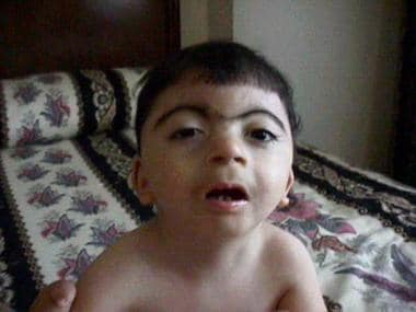 Facial appearance of a patient with Cornelia de Lange syndrome. Courtesy of Ian Krantz, MD, Children's Hospital of Philadelphia.
 Facial profile of a patient with Cornelia de Lange syndrome. Courtesy of Ian Krantz, MD, Children's Hospital of Philadelphia.
 Severe upper-extremity malformations in a patient with Cornelia de Lange syndrome. Courtesy of Ian Krantz, MD, Children's Hospital of Philadelphia.
Pathophysiology
More than 99% of cases are sporadic. Cornelia de Lange syndrome is occasionally transmitted in an autosomal dominant pattern, according to several instances in which a usually mildly affected parent had one or more affected offspring. Twins with concordance and discordance have been reported. Although possible autosomal recessive inheritance has been reported in some families, these instances were likely to be due to germline mosaicism. [3] The recurrence risk is 0.5-1.5% if parents are unaffected, and 50% if a parent is affected.
Heterozygous mutations in the gene NIPBL, the human homolog of the Drosophila melanogaster Nipped-B gene, [4] have been identified in approximately 50% of individuals with Cornelia de Lange syndrome. [5, 6, 7] Although the exact function of the protein product of NIPBL in humans (delangin) remains unknown, its homologs in other species are known to play roles in developmental regulation and in cohesion of sister chromatids.
Mutations in genes coding for two other proteins involved in cohesion of sister chromatids, SMC1A and SMC3, have been reported in 5% and 1% of patients with Cornelia de Lange syndrome, respectively. [8] Thus, Cornelia de Lange syndrome is considered to be a cohesinopathy, along with Roberts syndrome/SC phocomelia.
Inheritance is autosomal dominant in families with NIPBL and SMC3 mutations and is X-linked dominant in families with SMC1A mutations.
All types of NIPBL mutations, including missense, splice-site, nonsense, and frameshift mutations, have been reported to result in the Cornelia de Lange syndrome phenotype. The most likely effect of these mutations is haploinsufficiency. The mutation-detection rate is approximately 50%. Genomic deletions and duplications of the NIPBL locus are rare. [9, 10] Reported mutations of SMC1A include missense mutations and in-frame deletions. One reported SMC3 mutation is an in-frame deletion.
The correlation between genotype and phenotype suggests that individuals with an identifiable mutation in NIPBL have a phenotype more severe than that of patients without mutations.
SMC1A and SMC3 mutations are responsible for a milder craniofacial phenotype of the syndrome, with moderate psychomotor delay and less visible major structural defects, including absence of severe limb defects, when compared with typical Cornelia de Lange syndrome. More fullness of the eyebrows and a more prominent nose have been noted in SMC1A patients. SMC1A mutation can be associated with epilepsy and epileptic encephalopathy. [11] [12]
A study by Gil-Rodríguez et al of SMC3-associated phenotypes in 16 patients found a tendency toward the following [13] :
-
Less distinctive syndrome-related craniofacial features (although, as in other cases of Cornelia de Lange syndrome, microcephaly is characteristic)
-
Fewer associated congenital heart defects
-
Normal limbs (as mentioned above)
-
Milder prenatal growth retardation (although it becomes more severe in childhood)
The mutations in the X-linked gene HDAC8 ( Xq13.1) were identified as the cause of some cases of Cornelia de Lange syndrome. More HDAC8-positive cases are heterozygous females, each with marked skewing of X inactivation in peripheral blood DNA. Hemizygous males are more severely affected than heterozygous females. [14] [15]
Patients with HDAC8 mutations demonstrate the classic phenotype, but without significant limb malformations. Some distinct clinical features include delayed anterior fontanelle closure, hypertelorism, and hooding of the eyelids. [14] [15, 16]
Persons who are positive for RAD21, an autosomal gene, demonstrate only minor skeletal anomalies and less distinctive facial dysmorphism. [11]
Autosomal genes behind Cornelia de Lange syndrome also include BRD4 and ANKRD11. Because so few patients with the BRD4 form have been found, the most common phenotype has yet to be discerned. ANKRD11 gives rise to a non-classic phenotype for the syndrome. [17, 18]
Some autopsy data have indicated cerebral dysgenesis, with a decreased number of neurons, neuronal heterotopias, and focal gyral folding abnormalities, as causes of psychomotor delay.
Kaur et al showed that expression levels of NIPBL, as well as other genes, correlate with the phenotypic severity in Cornelia de Lange syndrome. Nonsense mutations in NIPBL led to about 35% reduced expression and gave rise to severe phenotypes. Missense and in-frame deletions in NIPBL and other genes were associated with 20% reduced expression and produced milder phenotypes. [19]
A study by Di Nardo et al of in vitro cell lines indicated that spontaneous genome instability, increased oxidative stress, and premature senescence occur in cells containing NIPBL, SMC1A, and HDAC8 gene variants and can serve as biomarkers for Cornelia de Lange syndrome. According to the investigators, these results also demonstrate the importance of cohesin in proper cellular function. [20]
Etiology
Mutations in NIPBL, SMC3, RAD21, SMC1A, HDAC8, BRD4, and ANKRD11 have been shown to cause Cornelia de Lange syndrome. NIPBL, SMC3, RAD21, BRD4, and ANKRD11 are autosomal dominant. SMC1A and HDAC8 are X-linked dominant. There is emerging evidence that mutation in EP300 may also cause the syndrome.
Most cases of Cornelia de Lange syndrome are sporadic, due to de novo mutations, but familial occurrence and parental consanguinity have been recorded. Autosomal dominant transmission, both maternal and paternal, has been documented in a number of families. [21]
Recurrence when parents were clinically unaffected has also been noted, and this has been explained by the possibility of germline mosaicism. [5] [22] [23] For example, paternal germline mosaicism of the NIPBL mutation was documented in analyzed sperm. [24]
A study by Aoi et al suggested that abnormalities in the genes ZMYND11, MED13L, and PHIP may also cause Cornelia de Lange syndrome or a similar condition. [25]
Parenti et al found heterozygous loss-of-function mutations in the ANKRD11 gene (16q24,3) in two patients with the clinical diagnosis of Cornelia de Lange syndrome. [26] As previously stated, ANKRD11 gives rise to a non-classic phenotype for the syndrome.
The etiology of a significant proportion of cases of Cornelia de Lange syndrome remains undetermined.
Epidemiology
Frequency
Estimation of the overall prevalence of Cornelia de Lange syndrome is difficult because of the unknown proportion of milder cases. Birth prevalence was calculated from 1 in 100,000 live births [27] to as high as 1 in 10,000 live births, when patients with either the severe or mild form were considered. [28] A population-based epidemiologic study of the classic form of Cornelia de Lange syndrome using the European Surveillance of Congenital Anomalies (EUROCAT) database established a prevalence for the classic form of 1.24 cases per 100,000 births and the overall prevalence of Cornelia de Lange syndrome to be 1.6-2.2 cases per 100,000 births. [29]
Mortality/Morbidity
Gastrointestinal (GI) disease complications are one of the most common causes of death in this syndrome. They include diaphragmatic hernia in infancy and aspiration pneumonia and volvulus at an older age.
A retrospective review of 295 propositi with Cornelia de Lange syndrome found that respiratory issues (eg, aspiration, reflux, pneumonia) were the most common causes of death (31%), followed by GI disease, including obstruction/volvulus (19%), congenital anomalies (15%), neurologic causes, (8%), accidents (8%), sepsis (4%), acquired cardiac disease (3%), cancer (2%), renal disease (1.7%), and other causes (9%). [30]
Sex and race
No differences based on sex or race have been described. In a study of 246 individuals with Cornelia de Lange syndrome from 15 nations, Dowsett et al found that 49% were female. [31]
Special Concerns
A prenatal diagnosis is made after Cornelia de Lange syndrome–related abnormalities are carefully evaluated using prenatal ultrasonography. These abnormalities include growth retardation, limb defects, diaphragmatic hernia, hypoplastic forearms, underdeveloped hands, and typical facial defects.
The availability of molecular diagnosis should substantially improve prenatal diagnosis. Prenatal diagnosis with molecular genetic techniques is currently available if a mutation is known in the family.
Failure to detect a mildly affected parent may result in incorrect risk estimation for future pregnancies.
Anatomic abnormalities of the face and neck may cause difficulties during intubation. GI obstruction or feeding difficulties may occur. Early feeding management is important.
Severe speech delay and poor communication are concerns. The patient may have congenital heart disease.
Prognosis
Very few cases of adults with Cornelia de Lange syndrome have been reported in the literature. In research into two cohorts of adults, totaling 122 patients, individuals ranged in age from 15-50 years, with an average age of 17 and 24. Premature aging was seen among these patients. The most common medical issues included the following [32] :
-
Gastroesophageal reflux diseases (GERD) - 71-82%
-
Overweight/obesity - 16-31%
-
Limb length discrepancy - 26-46%
-
Epilepsy - 26%
-
Hearing loss - 42-65%
-
Vision problems - 38-50%
-
Behavioral issues - 50%
All individuals reported having not required continuing assistance with nasogastric tubes and were able to feed orally in adulthood.
Epilepsy in Cornelia de Lange syndrome has proven to be amenable to medications, while cardiac issues typically do not require surgery in adulthood (18%). Intellectual disabilities are variable, as follows:
-
Borderline - 11%
-
Mild - 29%
-
Moderate - 18%
-
Severe - 16%
-
Profound - 19%
A prognostic index created by Cereda et al for Cornelia de Lange syndrome uses a point system to help predict the severity of a child's intellectual disabilities early in development. [33]
Patient Education
Information can be obtained from the Cornelia de Lange Syndrome Foundation, Inc., at https://www.cdlsusa.org/.
-
Facial appearance of a patient with Cornelia de Lange syndrome. Courtesy of Ian Krantz, MD, Children's Hospital of Philadelphia.
-
Facial profile of a patient with Cornelia de Lange syndrome. Courtesy of Ian Krantz, MD, Children's Hospital of Philadelphia.
-
Severe upper-extremity malformations in a patient with Cornelia de Lange syndrome. Courtesy of Ian Krantz, MD, Children's Hospital of Philadelphia.
Tables
Parameter |
1 point |
2 point |
3 point |
Birth weight |
Above 2500 g |
2000-2500 g |
Below 2,000 g |
Sitting alone |
< 9 mo |
9-20 mo |
>20 mo |
Walking alone |
< 18 mo |
18-42 months |
>42 mo |
Saying first word |
< 24 mo |
24-48 mo |
>48 mo |
Upper limb malformation |
No defect |
Partial defect (>2 digits) |
Severe defect (<2 digits) |
Number of other major malformations |
0-1 |
2-3 |
>3 |
Hearing loss |
Absent |
... |
... |
A score of less than 15 points indicates mild involvement, a score of 15-22 points indicates moderate involvement, and a score of more than 22 points indicates severe involvement. |
|||
