Background
Erythema multiforme (EM) is an acute, self-limited, and sometimes recurring skin condition that is considered to be a type IV hypersensitivity reaction associated with certain infections, medications, and various other triggers. [1] It has a wide spectrum of severity and occurs in both minor and major forms.
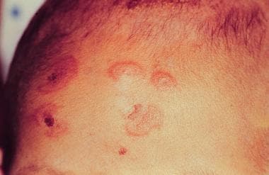
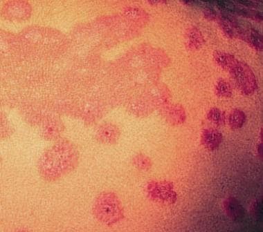
EM minor represents a localized eruption of the skin with minimal or no mucosal involvement. The papules evolve into pathognomonic target or iris lesions that appear within a 72-hour period and begin on the extremities (see the first image below). Lesions remain in a fixed location for at least 7 days and then begin to heal. An arcuate appearance may be present (see the second image below). Precipitating factors include herpes simplex virus (HSV), Epstein-Barr virus (EBV), and histoplasmosis. Because this condition may be related to recurrent HSV, recurrences of EM may follow, with many affected individuals experiencing several recurrences per year.
EM major is a more severe and potentially life-threatening disorder. One or more mucous membranes are involved, and as much as 10% of body area may have epidermal detachment. More than 50% of all cases are attributed to medications.
Erythema multiforme vs Stevens-Johnson syndrome and toxic epidermal necrolysis
Stevens-Johnson syndrome (SJS) and toxic epidermal necrolysis (TEN) have been considered severity variants of a single entity that has been divided into the following two broad categories: (1) EM, including both minor and major forms, and (2) SJS/TEN. Clinical descriptions are as follows:
-
EM minor - Typical targets or raised edematous papules distributed acrally
-
EM major - Typical targets or raised edematous papules distributed acrally with involvement of one or more mucous membranes; epidermal detachment involving less than 10% of total body surface area (TBSA)
-
SJS/TEN - Widespread blisters predominating on the trunk and face, presenting with erythematous or pruritic macules and one or more mucous membrane erosions; epidermal detachment involving less than 10% TBSA for SJS/TEN but 30% or more for TEN
More recent clinical data favor the view that EM and SJS, though sometimes confused with each other, are separate disorders. [1]
See also Dermatologic Manifestations of Stevens-Johnson Syndrome and Toxic Epidermal Necrolysis.
Pathophysiology
The pathophysiology of EM has not been fully elucidated, but it is probably immunologically mediated and appears to involve a hypersensitivity reaction that can be triggered by a variety of stimuli (particularly bacterial, viral, or chemical).
Cell-mediated immunity appears to be responsible for the destruction of epithelial cells. Early in the disease process, the epidermis becomes infiltrated with CD8 T lymphocytes and macrophages, whereas the dermis displays a slight influx of CD4 lymphocytes. These immunologically active cells are not present in sufficient numbers to be directly responsible for epithelial cell death. Instead, they release diffusable cytokines, which mediate the inflammatory reaction and resultant apoptosis of epithelial cells.
In some patients, circulating T cells transiently (for < 30 d) demonstrate a T-helper cell type 1 (Th1) cytokine response (interferon [IFN] gamma, tumor necrosis factor [TNF]-α, interleukin [IL]-2). Results of immunohistochemical analysis have also shown lesion blister fluid to contain TNF, an important proinflammatory cytokine.
Other evidence supports the hypothesis that the disease is the result of cell-mediated immune reactions. Individuals possessing human leukocyte antigen (HLA)-B12 are three times more likely to develop this disorder. The classic timing for a primary cell-mediated immune reaction is 9-14 days after the initiation of the offending drug. In recurrent exposure, the reaction occurs within several hours to 1-2 days, which is consistent with the timing of a secondary cell-mediated immune response.
Herpes simplex virus
HSV is a major cause of EM [2] ; in fact, recent or recurrent herpes has been reported as the principal risk factor for EM. Herpes-associated EM (HAEM) appears to represent the result of a cell-mediated immune reaction associated with HSV antigen. The immunologic reaction affects HSV-expressing keratinocytes. Cytotoxic effector cells, CD8+ T lymphocytes in the epidermis, induce apoptosis of scattered keratinocytes and lead to satellite cell necrosis. Neighboring epidermal cells are HLA-DR positive.
A relationship exists between HLA types A33, B35, B62 (B15), DR4, DQB1*0301, DQ3, and DR53 and recurrent EM. [3] In particular, HLA-DQ3 is specifically related to recurrent EM and may be a helpful marker for distinguishing HAEM from other cutaneous diseases. [4]
Drug hypersensitivity
The disease process also often involves an abnormal metabolism of a responsible drug. As noted above, the keratinocyte is the ultimate target of this disease process, with keratinocyte necrosis being the earliest pathologic finding.
Patients frequently display an altered metabolism of the responsible drug and are considered to be slow acetylators, both genotypically and phenotypically. This means that an increased proportion of drug metabolism is directed toward the alternative pathway of oxidation by the cytochrome P-450 system, resulting in increased production of reactive and potentially toxic metabolites. Affected individuals have a defect in the ability to detoxify these reactive metabolites, which may then behave as haptens by binding covalently to proteins on the surface of epithelial cells. This may then induce the immune response, leading to the severe skin reaction.
Etiology
Many suspected etiologic factors have been reported to cause EM. Both EM and SJS may be induced by medications, but infectious agents are also considered to be a major cause of EM. Approximately 50% of cases are idiopathic, with no precipitating factor identified. Other reported risk factors include male sex and a previous history of EM; pregnancy may contribute to the development of EM as well. Postvaccination causes include bacille Calmette-Guérin (BCG) vaccination, oral polio vaccine, vaccinia, and tetanus/diphtheria.
HSV and other infections
Infectious causes are more common in children and are implicated commonly in EM.
EM minor is regarded as being commonly triggered by HSV types 1 and 2, and HSV is the most common cause in young adults; in fact, many instances of idiopathic EM minor may be precipitated by subclinical HSV infection. Mycoplasmal infection is another common cause.
Bacterial
Bacterial infections that may trigger EM include the following:
-
Borreliosis
-
Catscratch disease
-
Diphtheria
-
Hemolytic streptococci
-
Legionellosis
-
Leprosy
-
Neisseria meningitidis
-
Mycobacterium avium complex
-
Pneumococci
-
Tuberculosis
-
Proteus/Pseudomonas/Salmonella/Staphylococcus/Yersinia species
-
Treponema pallidum [7]
-
Tularemia
-
Vibrio parahaemolyticus
-
Vincent disease
-
Rickettsial infections
Chlamydial infectious causes include lymphogranuloma venereum and psittacosis.
Viral
Viral infections that may trigger EM include the following:
-
Adenovirus
-
Coxsackievirus B5
-
Cytomegalovirus (CMV)
-
Echovirus
-
Enterovirus
-
EBV
-
Hepatitis A/B/C viruses (HAV/HBV/HCV)
-
HSV
-
Influenza [8]
-
Measles
-
Mumps
-
Paravaccinia
-
Parvovirus B19
-
Poliomyelitis
-
Varicella-zoster virus (VZV)
-
Variola
An association with severe acute respiratory syndrome coronavirus 2 (SARS-CoV-2) infection has been described. [9] It has also been suggested that EM may be a potential adverse consequence of COVID-19 vaccination. [10]
Other virus-related causes include the virus-drug interactions CMV infection–terbinafine [11] and EBV infection–amoxicillin. [12]
Other
EM may also be triggered by fungal infections (eg, coccidioidomycosis, dermatophytosis, and histoplasmosis) and some parasitic infections (eg, Trichomonas species and Toxoplasma gondii).
Drugs
More than 50% of cases of EM major are related to medication use, but no test has conclusively established the link between a single case and a specific drug.
The most common pharmacologic triggers are the sulfa drugs (30% of cases). The second most commonly involved agents are the anticonvulsants, including the following:
Causative antibiotics include the following:
-
Penicillin
-
Ampicillin
-
Tetracyclines
-
Amoxicillin
-
Cefotaxime
-
Cefaclor
-
Cephalexin
-
Ciprofloxacin [14]
-
Erythromycin
-
Minocycline
-
Sulfonamides
-
Trimethoprim-sulfamethoxazole
-
Vancomycin
Antituberculoid agents (eg, rifampin, isoniazid, thiacetazone, and pyrazinamide) are also known offenders. Antipyretic agents as triggers include analgesics (especially aspirin), as well as phenylbutazone, oxyphenbutazone, and phenazone.
Others agents that may cause EM include the following:
-
Acarbose
-
Albendazole
-
Alendronate [15]
-
Allopurinol [13]
-
Arsenic
-
Bromofluorene
-
Quinine
-
Cimetidine
-
Clofibrate
-
Corticosteroids
-
Diclofenac
-
Didanosine
-
Dideoxycytidine
-
Dihydrocodeine phosphate [16]
-
Diphosphonate
-
Estrogen
-
Etretinate
-
Fluconazole
-
Griseofulvin [17]
-
Gabapentin
-
Granulocyte-macrophage colony-stimulating factor (GM-CSF)
-
Hydralazine
-
Indapamide
-
Imiquimod [18]
-
Indinavir
-
Lamotrigine [13]
-
Methazolamide
-
Mefloquine
-
Methotrexate
-
Meprobamate
-
Mercurials
-
Minoxidil
-
Nifedipine
-
Nevirapine [13]
-
Nystatin
-
Nonsteroidal anti-inflammatory drugs (NSAIDs)
-
Phenolphthalein
-
Piroxicam [13]
-
Pyritinol
-
Progesterone
-
Potassium iodide
-
Secukinumab [21]
-
Sulindac
-
Suramin
-
Saquinavir
-
Thiabendazole
-
Thiouracil
-
Terbinafine
-
Theophylline
-
Tramadol [22]
-
Vandetanib [23]
-
Verapamil
Contact exposure
Contactants that may induce an EM-like eruption include the following:
-
Ammoniated mercury
-
Budesonide
-
Bufexamac
-
Capsicum
-
Chloromethylnaphthalene
-
Desoximetasone
-
Dinitrochlorobenzene (DNCB)
-
Disperse blue 124
-
Diphenylcyclopropenone
-
Fire sponge (Tedania ignis)
-
Herbal medicines (eg, Alpinia galanga) [24]
-
Isopropyl-p-phenylenediamine of rubber
-
Nickel
-
Nitrogen mustard
-
Oxybenzone
-
Phenylbutazone
-
Poison ivy [25]
-
Proflavin
-
Resin
-
Rosewood
-
Triamcinolone acetonide
Other etiologic factors
The following have also been reported as causes of EM:
-
Flavorings and preservatives (eg, benzoic acid and cinnamon [26] )
-
Immunologic disorders (eg, transient selective C4 deficiency of infancy, [27] collagen diseases, vasculitides, sarcoidosis, non-Hodgkin lymphoma, leukemia, multiple myeloma, myeloid metaplasia, and polycythemia)
-
Physical or mechanical factors (eg, tattooing, radiotherapy, cold, and sunlight)
-
Foods (eg, salmon berries and margarine)
-
Malignancy
-
Hormonal factors
Epidemiology
The exact incidence of EM in the United States is not defined; however, as many as 1% of dermatologic outpatient visits are for EM. Globally, the frequency of EM has been estimated to be in the range of 1.2-6 cases per million individuals per year.
Before the HIV epidemic among young males, EM showed a slight female predominance; however, it has since become more common in younger males (male-to-female ratio, 2-3:1), mainly seen in the second to fourth decades but sometimes also in children and adolescents (20%). [28] EM is rare in children younger than 3 years and in adults older than 50 years.
The following medical conditions seem to predispose individuals to a higher risk of developing EM:
-
HIV infection
-
Corticosteroid exposure
-
Bone marrow transplant
-
Systemic lupus erythematosus (SLE)
-
Graft-versus-host disease (GVHD)
-
Inflammatory bowel disease (IBD)
Individuals undergoing radiation therapy, chemotherapy, or neurosurgery for brain tumors are also at higher risk.
Prognosis
Most cases of EM are self-limited. In EM minor, the lesions evolve over 1-2 weeks and ultimately subside within 2-3 weeks without scarring. However, recurrence of EM minor is common, developing in as many as one third of cases, and is mostly preceded by apparent or subclinical HSV infection.
EM major has a mortality of less than 5%, which is directly proportional to the TBSA of sloughed epithelium. It usually has a more protracted course than EM minor, and clearing may require 3-6 weeks. Skin lesions usually heal with hyperpigmentation, hypopigmentation, or both. Scarring is usually absent, except after secondary infection. Sepsis secondary to loss of the cutaneous barrier is the principal cause of death.
Advanced age, visceral involvement, increased serum urea nitrogen level, and previous bone marrow transplantation are poor prognostic factors. Surprisingly, although people with HIV infection have an increased incidence of EM (approaching one case per 1000 individuals per year), they do not appear to have a higher mortality.
Continuous and persistent erythema multiforme
Two additional rare clinical forms of EM have been reported: continuous EM and persistent EM. Continuous EM manifests as a prolonged course with overlapping attacks and may be associated with systemic administration of glucocorticoids. Persistent EM has a protracted clinical course over months, is commonly associated with atypical skin lesions, and is commonly resistant to conventional treatment. It has been reported in association with IBD, occult renal carcinoma, persistent or reactivated EBV infection, and HSV infection.
Patient Education
Patients with EM should be educated regarding appropriate symptomatic treatment and reassured that the condition is usually self-limited. In addition, patients should be advised regarding the significant risk of recurrence and the possible need for suppressing recurrent HSV infection with appropriate antiviral therapy.
-
Target lesion of erythema multiforme.
-
Raised atypical targets and arcuate lesions.
-
Hemorrhagic crusts on the lips.
-
Interface dermatitis with prominent dyskeratotic cells in epidermis.
Tables

What would you like to print?
- Drug Overdose Deaths Involving Ketamine on the Rise
- FDA Investigates Tampons for Potential Lead and Metal Risks
- Hey Pharmacy, Do You Have… Thai Green Pit Viper Antivenom?
-
 A Futuristic Vision for Treating Myelofibrosis
-
Addressing Toxicities of Systemic Cancer Therapy and Improving the Cancer Survivorship Experience
-
EGFR Inhibitors in the Treatment of Stage IV CRC


 Poisoning Clues on the Skin: 8 Cases
