Practice Essentials
Mycetoma is a chronic, granulomatous disease of the skin and subcutaneous tissue, which sometimes involves muscle, bone, and neighboring organs. [1] It is characterized by tumefaction, abscess formation, and fistulae. It typically affects the lower extremities, but it can occur in almost any region of the body. Mycetoma predominately occurs in farm workers, but it can also appear in the general population. [2, 3, 4]
Mycetoma infection can be caused by fungi or bacteria. When caused by fungi, it is referred to as mycotic mycetoma or eumycetoma. When it is caused by bacteria, it usually involves infection by the actinomycetes group; such cases are called actinomycotic mycetoma or actinomycetoma. [5] The disease presentation, whether caused by fungi or bacteria, is quite similar.
Mycetoma is characterized by the formation of grains, which contain aggregates of the causative organisms that may be discharged onto the skin surface through multiple sinuses. [6] The characteristic color of the grains can assist in the identification of the specific etiologic agent.
Mycetoma due to actinomycetes should be differentiated from actinomycosis, which is an endogenous suppurative infection caused by Actinomyces israelii, other species of Actinomyces, or related bacteria, typically affecting the cervicofacial, thoracic, and pelvic sites (the latter is usually associated with the use of intrauterine devices). The branching bacteria that cause actinomycosis are non–acid-fast anaerobic or microaerophilic bacteria. These bacteria are smaller than 1 µm in diameter, smaller than eumycotic agents. Alternatively, the agents that cause actinomycetoma are always aerobic and are sometimes weakly acid-fast.
More than 56 different species of fungi and bacteria have been reported to cause mycetoma. Nocardia species, especially Nocardia brasiliensis, is the most commonly implicated actinomycetes. [7] The ratio of mycetoma cases caused by bacteria (actinomycetoma) to those caused by true fungi (eumycetoma) in Mexico has been reported to be 92:8. [8]
Mycetoma is usually painless; individuals who are affected seek medical attention mainly because of tumefaction and draining sinuses. In cases affecting the thorax or the head, mycetoma can be potentially fatal because of the spread of microorganisms to adjacent organs. Rarely, the disease spreads by hematogenous dissemination (Nocardia asteroides and N brasiliensis).
Actinomycetoma generally responds well to trimethoprim-sulfamethoxazole/amikacin (approximately 90% of cases). If the bacteria have become resistant to this treatment, antibiotic susceptibility testing should be performed to select the best antimicrobial agent or agents to be used. Linezolid and tedizolid, of the group of the oxazolidinones, have been proved useful in vitro, in vivo, and in some human clinical cases, [9] although the expensive price hinders their use in poor developing countries, where most cases are reported.
Eumycetoma tends to be a more chronic disease, and success with medical therapy is observed in only about 40% of cases. If the response to medical treatment is partial or negative, surgery of the affected area should be performed, and antifungal drugs should be continued until complete remission of the disease.
Background
Gill first described the disease in the Madura district of India in 1842, hence the term Madura foot. In 1860, Carter named the condition mycetoma, describing its fungal etiology. In 1913, Pinoy described the mycetoma produced by aerobic bacteria that belong to the actinomycete group and classified mycetomas as those produced by true fungi (eumycetoma) versus those due to aerobic bacteria (actinomycetoma). Both types have similar clinical findings. [10]
Pathophysiology
Mycetoma is produced by the introduction of microorganisms (bacteria or fungi) via localized trauma to the skin with thorns, wood splinters, or implantation with solid objects. Clinically, the disease begins as small, firm nodules that can persist (mini-mycetomas) or evolve to form extensive suppurative lesions that in some cases can reach more than 20 cm in diameter. Eumycetoma tends to be more localized than actinomycetoma.
Human-to-human or animal-to-human transmission has not been described for eumycetoma, but nosocomial transmission of Nocardia farcinica, one of the agents of actinomycetoma in postoperative surgical site infections, has been reported. [11]
The body parts affected most commonly in persons with mycetoma include the foot or lower leg, with infection of the dorsal aspect of the forefoot being typical. The hand is the next most common location; however, mycetoma lesions can occur anywhere on the body. Lesions on the chest and back are frequently caused by Nocardia species, whereas lesions on the head and neck are usually caused by Streptomyces somaliensis.
In experimentally induced N brasiliensis actinomycetoma in mice, production of granules (or grains) containing the bacterium can be observed 15 days after inoculation. The grains are surrounded by polymorphonuclear leukocytes, lymphocytes, plasma cells, and histiocytes. Murine infection can evolve into a chronic disease similar to the clinical manifestations observed in humans. Severe inflammation and deformity, abscesses, ulcers, and fistulae are present 28 days after infection.
The in situ production of cytokines in the microabscesses has been reported in murine infection. Tumor necrosis factor-alpha is produced in the first days of infection, decreasing later to nondetectable quantities at day 90. Interleukin (IL)–1-beta, interferon-gamma, transforming growth factor-beta, IL-10, IL-4, and IL-6 are produced constantly during the 90 days, but IL-6 is the only one with a significant increase once the mycetoma is fully established (90 d). [12]
The host immune response in humans and mice involves the production of high levels of anti–N brasiliensis immunoglobulin G antibodies. Quantitation of these antibodies is useful for diagnosis. [13] Immunoglobulin M anti–N brasiliensis antibodies can protect mice from an experimental infection. [14] Activation of cellular immunity and production of cytokines are involved in resistance and elimination of the N brasiliensis bacterial cells.
Salinas-Carmona et al (2012) have unveiled aspects of the physiopathogenic mechanisms of experimental actinomycetoma in mice. [15]
Etiology
Mycetoma occurs most often in farmers, shepherds, Bedouins, nomads, and people living in rural areas. Frequent exposure to penetrating wounds by thorns or splinters is a risk factor, especially in combination with contaminated soil material.
Eumycetomas can be produced by a variety of fungi (see Table 1 below); however, actinomycetomas are mainly produced by bacteria of four genera: Nocardia, Actinomadura, Streptomyces, and Nocardiopsis (see Table 2 below), the last of which is rare.
Eumycetoma is mainly caused by Madurella mycetomatis, which produces 70% of all eumycetoma cases globally. Less frequently, Trematosphaeria (Madurella) grisea, Scedosporium boydii, and Falciformispora (Leptosphaeria) senegalensis are isolated. [16] Other Madurella species have been identified in cattle dung in rural East Africa. [17]
The color of the grains is sometimes helpful in pinpointing the exact etiologic agent. For example, the grains of M mycetomatis or T grisea typically are black, while those of Pseudallescheria boydii (S apiospermum) and several actinomycetes are usually faint yellowish or white.
Although traditionally it has been considered that mycetoma is produced by the pathogenic characteristics of the causative agents, it has been observed that genetic polymorphisms involved in neutrophil function are related to either the production of human mycetoma or its size, in the case of M mycetomatis infection. IL-8 (CXCL8), its receptor CXCR2, thrombospondin-4, nitric oxide synthase, and complement receptor 1 have significant differences in mycetoma patients compared with geographically and ethnically matched controls. These findings open the possibility that certain individuals are predisposed to this infection. [18]
The use of molecular methods has permitted the identification of new species causing mycetoma, including Nocardia harenae, Nocardia wallacei, and Nocardia takedensis, and Actinomadura mexicana causing actinomycetoma. [19, 20, 21] Madurella fahalii, Aspergillus sydowi, and Microascus gracilis are identified as causing eumycetoma. [22, 23]
Table 1. Fungi Causing Mycetoma (Open Table in a new window)
White Grain |
Black Grain |
Acremonium falciforme |
Exophiala jeanselmei |
Acremonium kiliense |
T grisea |
Acremonium recifei |
M mycetomatis |
Cylindrocarpon destructans |
Madurella pseudomycetomatis |
Fusarium moniliforme |
Leptosphaeria tomkinsii |
Fusarium solani |
Leptosphaeria senegalensis |
Neotestudina rosatii |
Pyrenochaeta mackinnonii |
P boydii (S apiospermum) |
Pyrenochaeta romeroi |
---------------- |
Phlenodomus avramii |
Table 2. Microorganisms Causing Actinomycetoma in Humans (Open Table in a new window)
Etiologic Agent |
Grain |
Actinomadura madurae |
White, large, 1-5 mm in diameter |
Actinomadura pelletieri |
Red, hard, 1 mm in diameter |
N brasiliensis |
White to yellow, multilobed, soft, < 0.5 mm in diameter |
N asteroides |
Uncommon, white, soft, < 0.5 mm in diameter |
Nocardia otitidiscaviarum |
White to yellow, lobed, < 0.5 mm in diameter |
Nocardia transvalensis |
White to yellow, < 0.5 mm in diameter |
Nocardia veterana [24] |
-- |
Nocardia mexicana [25] |
-- |
N harenae |
-- |
N takedensis |
-- |
Nocardiopsis dassonvillei |
White to yellow, < 0.5 mm in diameter |
S somaliensis |
Yellow, hard, 2 mm in diameter |
Streptomyces sudanensis |
Yellow, hard, 2 mm in diameter |
Epidemiology
Mycetoma is endemic in Africa, in the known “mycetoma belt” that comprises countries like Sudan and Somalia through Mauritania and Senegal. Other endemic countries include Mexico and India. Mycetoma can also be found in natives of areas of Central and South America and the Middle or Far East between latitudes 15°S and 30°N. [26]
Eumycetoma is more common in areas where the average rainfall is scarce (ie, < 350 mm), whereas actinomycetoma tends to appear in areas with abundant rainfall (ie, >600 mm) [27] and has been described in Southeast Asia. [28]
In Sudanese hospitals, at least 300-400 patients are diagnosed with mycetoma every year.
Mycetoma is rare in the United States. Some cases are acquired during international travel, but cases acquired on US soil have also been reported. [29]
In general, traumatic inoculation of fungal elements into the skin or subcutaneous tissue by a thorn or splinter typically occur in those who walk bare-footed (eg, farmers, field workers), especially in developing countries.
Among the fungal pathogens responsible for mycetoma, M mycetomatis is the most common pathogen described in Africa. [30] T grisea is the most common etiologic pathogen in South America. P boydii (S apiospermum) is the most common etiologic agent in the United States. [30] In Mexico, which shares common climatic conditions with the African countries, most cases are found in rural areas and 98% are caused by actinomycetes, mainly N brasiliensis (86%) and A madurae (about 8%). [31] In India, 65% of cases are produced by actinomycetes and the rest by eumycetes, mostly M mycetomatis. [30]
Mycetoma has no apparent racial predilection. Mycetoma is more common in men than in women, with male-to-female ratios ranging from 3:1 to 5:1. It is most common in persons aged 20-50 years, with a mean of age 34 years.
Prognosis
Mycetoma carries a good prognosis if the disease is promptly diagnosed and treated. Mycetoma causes disfigurement but is rarely fatal in the absence of skull involvement. Although mycetoma carries a low risk of mortality, amputations or ankylosis can lessen the quality of life. [32] In late stages of mycetoma, the treatment response is limited.
The lesions are painless and slowly progressive; however, secondary bacterial infection or bone expansion may cause pain. When secondary bacterial infection occurs, Staphylococcus aureus is the most common etiologic agent. [33] In advanced cases, deformities or ankylosis and their corresponding disabilities can appear. Patients who are immunocompromised or who have undergone transplantation can develop invasive infection.
Complications of mycetoma result mainly from toxicity due to prolonged administration of antimicrobial or antifungal drugs. Disfigurement of the affected body parts may be a consequence of delayed treatment. Secondary bacterial infections can progress to full-blown bacteremia or septicemia, resulting in death. [6]
See the images below.
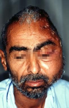
 Mycetoma in a 47-year-old shepherd from Mauritania who had a painless progressive swelling of the face for more than 20 years.
 Frontal view of mycetoma in a 47-year-old shepherd from Mauritania who had a painless progressive swelling of the face for more than 20 years.
-
Mycetoma in a 47-year-old shepherd from Mauritania who had a painless progressive swelling of the face for more than 20 years.
-
Frontal view of mycetoma in a 47-year-old shepherd from Mauritania who had a painless progressive swelling of the face for more than 20 years.
-
MRI coronal section of mycetoma in a 47-year-old shepherd from Mauritania who had a painless progressive swelling of the face for more than 20 years. On this T1-potentiated image, a large heterogenous mass surrounds the cranium. Bone invasion can be observed only in the area of the zygomatic fossa.
-
MRI with coronal view of mycetoma in a 47-year-old shepherd from Mauritania who had a painless progressive swelling of the face for more than 20 years. The mycetoma mass invades the left parapharyngeal space and almost reaches the lumen of the pharynx.
-
Actinomycetoma of the foot (left) and arm (center) caused by Nocardia brasiliensis. Multiple nodules and fistulae are present. Microscopic examination of the pus (right). Granules are observed, which are multilobulated and surrounded by abundant clubs.
-
Eumycetoma. Mycetoma of the hand (left). Microscopic features of a Madurella mycetomatis grain are observed (center). Notice the presence of brownish hyphae and intercellular cement (hematoxylin and eosin stain). Macrocolony of another eumycotic agent, Scedosporium apiospermum (Pseudallescheria boydii) (right).
Tables
White Grain |
Black Grain |
Acremonium falciforme |
Exophiala jeanselmei |
Acremonium kiliense |
T grisea |
Acremonium recifei |
M mycetomatis |
Cylindrocarpon destructans |
Madurella pseudomycetomatis |
Fusarium moniliforme |
Leptosphaeria tomkinsii |
Fusarium solani |
Leptosphaeria senegalensis |
Neotestudina rosatii |
Pyrenochaeta mackinnonii |
P boydii (S apiospermum) |
Pyrenochaeta romeroi |
---------------- |
Phlenodomus avramii |
Etiologic Agent |
Grain |
Actinomadura madurae |
White, large, 1-5 mm in diameter |
Actinomadura pelletieri |
Red, hard, 1 mm in diameter |
N brasiliensis |
White to yellow, multilobed, soft, < 0.5 mm in diameter |
N asteroides |
Uncommon, white, soft, < 0.5 mm in diameter |
Nocardia otitidiscaviarum |
White to yellow, lobed, < 0.5 mm in diameter |
Nocardia transvalensis |
White to yellow, < 0.5 mm in diameter |
Nocardia veterana [24] |
-- |
Nocardia mexicana [25] |
-- |
N harenae |
-- |
N takedensis |
-- |
Nocardiopsis dassonvillei |
White to yellow, < 0.5 mm in diameter |
S somaliensis |
Yellow, hard, 2 mm in diameter |
Streptomyces sudanensis |
Yellow, hard, 2 mm in diameter |
