Background
Alopecia mucinosa (or follicular mucinosis) was first reported by Pinkus in 1957. The dermatologic eruptions consist of follicular papules or indurated plaques that demonstrate distinct histologic changes in the hair follicles that lead to hair loss. The accumulation of mucinous material in the damaged hair follicles and sebaceous glands creates an inflammatory condition and a subsequent degenerative process. The face, the neck, and the scalp are the most frequently affected sites, though lesions may appear on any part of the body.
The presenting sign of alopecia mucinosa is hair loss in hair-bearing areas. Skin eruptions present as pruritic, pink–to–yellow-white follicular papules and plaques. Lesions may be isolated or multiple. Mycosis fungoides (MF) is recognized at the time of diagnosis in approximately 15-30% of patients with alopecia mucinosa.
Alopecia mucinosa represents various stages of follicular damage leading to hair loss. The reactive process is of unknown etiology. The role of circulating immune complexes and cell-mediated immunity has been considered.
Although the question of whether alopecia mucinosa is a transitional state evolving into MF is unresolved, it is known that alopecia mucinosa may precede the development of MF by several years. Thus, additional biopsy specimens and extremely close follow-up care are crucial in all variants of alopecia mucinosa.
Common treatments include topical, intralesional, and systemic corticosteroids; some success with other modalities and agents has been described.
Pathophysiology
Alopecia mucinosa is a disease process defined histopathologically by mucin deposition in hair follicles and sebaceous glands, which undergo epithelial reticular degeneration. The exact pathogenesis is unknown, though circulating immune complexes and cell-mediated immunity may play a role. The disease has three clinical variants, as follows:
-
Primary acute disorder of young persons
-
Primary chronic disorder of older persons
-
Secondary disorder associated with benign or malignant disease
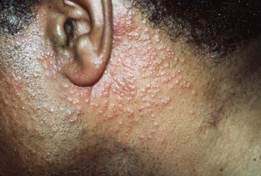
The primary acute disorder of young persons consists of focal cutaneous lesions with limited progression. Lesions are typically limited to the head, the neck, and the shoulders (see the image below). Most lesions spontaneously resolve between 2 months and 2 years. The majority of cases occur in pediatric patients, with the remainder occurring in patients younger than 40 years. Neonatal follicular mucinosis has also been reported. [1] Alopecia mucinosa may also manifest as acneiform lesions in young patients; this variant is typically benign and is not associated with cutaneous T-cell lymphoma (CTCL) or MF. [2]
Primary chronic alopecia mucinosa of older persons affects people older than 40 years. Lesions have a widespread distribution, and they may persist or recur indefinitely. No associated disorders have been identified.
Secondary alopecia mucinosa may be associated with either benign or malignant disease. It is usually seen between the ages of 40 and 70 years, and the lesions are widespread and numerous. Benign associations include the inflammatory conditions lupus erythematosus, lichen simplex chronicus, and angiolymphoid hyperplasia. Malignant associations include MF, Kaposi sarcoma, and Hodgkin disease, with MF being by far the most common association. [3, 4, 5, 6] When the secondary form of alopecia mucinosa appears in children, it typically shows a more benign progression than it does in adults. [7]
In most patients who exhibit both alopecia mucinosa and MF, the two conditions appear to develop concomitantly; however, the concern exists that individuals exhibiting only alopecia mucinosa may also be at risk for subsequent development of lymphoma.
Drug-induced alopecia mucinosa has been associated with the use of adalimumab [8] and imatinib. [9]
Epidemiology
Alopecia mucinosa is considered to be a rare condition, though precise data on its frequency are not available.
The three clinical variants of alopecia mucinosa typically affect different age groups, as follows:
-
Primary localized disease affects patients younger than 40 years and primarily occurs in the pediatric population [10]
-
Primary generalized disease affects people older than 40 years
-
Secondary disease with either a benign or a malignant association usually affects people in the fifth through eighth decades of life
Although both sexes are affected by alopecia mucinosa, the disorder is more frequent in males than in females. Alopecia mucinosa in pregnancy has been reported. [11] No racial predilection has been demonstrated.
Prognosis
The prognosis of alopecia mucinosa depends on the specific clinical variant, as follows:
-
Primary acute disease of young persons usually disappears within 2 years; however, childhood alopecia mucinosa is not always self-limited and may possibly be related to Hodgkin disease
-
Primary chronic disease of older persons usually lingers for several years, but it can fluctuate in the extent of skin involvement at any given time
-
Secondary alopecia mucinosa has the least favorable prognosis when associated with coexistent malignancy
In cases of primary alopecia mucinosa, morbidity is generally restricted to cosmesis, whereas in cases of secondary alopecia mucinosa, morbidity is related to the associated disease process.
Mortality is related to the coexistence of MF in secondary alopecia mucinosa. An estimated 15-40% of adults with alopecia mucinosa will eventually develop lymphoma, if they do not already have it. The malignant potential of alopecia mucinosa cannot be fully assessed, because of the enigmatic nature of this and other cutaneous T-cell abnormalities.
In a retrospective cohort study (N = 14) that evaluated long-term (20-year) outcomes for patients aged 22 years or younger who had a diagnosis of follicular mucinosis, Jin et al found that none of the patients subsequently developed MF or other hematologic malignancies. [12]
-
Alopecia mucinosa (follicular mucinosis). Image from Dirk M Elston, MD.
-
Alopecia mucinosa. Follicular degeneration with accumulation of mucin within follicles. Image from Dirk M Elston, MD.
-
Alopecia mucinosa. Image from San Antonio Uniformed Services Health Education Consortium (SAUSHEC) teaching files.
Tables

What would you like to print?
- Alopecia in Renal Disease: Common and Troublesome, but Treatable
- One-a-Day Pill for Alopecia Recommended for NHS Use
- FDA Approves JAK Inhibitor Deuruxolitinib for Alopecia Areata
-
 What's New in Psoriasis? Dermasphere Podcasters Weigh In
-
15 Top Doc Social Media Influencers 2025
-
American Academy of Dermatology (AAD) 2025 Annual Meeting