Background
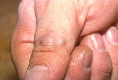
Glomus tumors are rare soft-tissue neoplasms originating from the glomus body (see the image below). [1] The glomus body is a neuromyoarterial apparatus composed of vascular structures, nerve cells, and smooth-muscle cells. The glomus body is responsible for thermoregulation.
Glomus bodies are found in the dermis throughout the body, with the highest concentrations in the hands and feet. Glomus tumors most frequently occur in those locations, including subungual regions of the fingers and the deep dermis of the hand (palm), forearm, and foot (sole). Most lesions are solitary.
Glomus tumors typically present in adults (ages 20-40 y) as small papules or nodules of the distal extremities, with most cases involving subungual sites. [2] The subcutaneous nodules may be red, purple, or blue depending on their depth.
Pain is the usual presenting symptom of a glomus tumor. Patients may report paroxysmal pain in response to temperature changes (especially cold) or pressure. However, the classic triad of point tenderness, pain, and sensitivity to cold is not always found.
Local soft-tissue tenderness and thickening may be present. A mass is sometimes detectable in the area of tenderness. On clinical examination, a positive result with the Love test and the Hildreth sign suggest the diagnosis. See Presentation for more detail.
Because the diagnosis of glomus tumor is primarily clinical, laboratory workup usually is not needed. If the diagnosis is uncertain in view of the patient's history and physical findings, imaging studies are warranted.
Surgical excision of the tumor is the mainstay of treatment. Pain relief should be provided until the procedure is performed. Excision should be limited to symptomatic lesions. Sclerotherapy and laser treatment have been used to destroy the tumor as therapeutic alternatives to surgical excision.
Pathophysiology
Glomus bodies play an important role in thermoregulation via arteriovenous shunting. The glomus body is composed of an afferent arteriole, anastomotic vessel (termed Sucquet-Hoyer canal), primary collecting vein, intraglomerular reticulum, and a capsular portion. These specialized arteriovenous anastomoses are particularly concentrated in the reticular dermis of the fingers.
Glomus tumors are thought to represent hamartomatous proliferations of modified smooth muscle cells originating from preexisting normal glomus cell populations. The three components of most glomus tumors include glomus cells, vasculature, and smooth muscle cells. The most common type of glomus tumor is the solid glomus tumor, characterized by a prominent smooth muscle cell component. [3] No evidence of mitosis is observed in the structure.
While glomus tumors predominate on the hands and fingers, these tumors can occur in a wide anatomic distribution, including sites not known to contain glomus cells, such as deep soft tissues, nerve, bone, and abdominal viscera. In fact, gastric glomus tumors account for approximately 2% of benign gastric tumors. [4] These tumors may arise from perivascular cells or pluripotent mesenchymal cells capable of differentiating into glomus cells.
Most glomus tumors are solitary and sporadic. However, glomus tumors have been reported in 5% of patients with neurofibromatosis type 1. In these cases the tumors are most often subungual and are often multiple; the presence of such tumors may be an important clue to the diagnosis of neurofibromatosis. [5]
A distinct but related condition known as glomuvenous malformations (GVMs) differ clinically from glomus tumors in that they occur more often in children and adolescents, are typically multifocal, and do not have a predilection for subungual sites. GVMs are more likely to be hereditary and painless. GVMs are thought to have different etiologies from glomus tumors, with GVMs resembling venous malformations and containing more dilated venous channels than glomus tumors.
Cases of GVM may be sporadic, but familial cases with autosomal dominant inheritance have also been described. The familial GVM trait has been mapped to 1p21-22 and is thought to result from loss-of-function mutations in the cytoplasmic protein glomulin. [6, 7, 8]
Rarely, glomus tumors can undergo malignant transformation or malignant glomus tumors can arise de novo. Malignant glomus tumors are termed glomangiosarcomas; they have a high local recurrence rate but very low rate of metastasis. Most reported cases have been in the abdominal viscera or lower extremity. [9, 10, 11, 12, 13, 14, 15, 16, 17, 18]
Findings in malignant glomus tumors have included the following [18] :
-
Large size
-
Deep location
-
Infiltrative growth
-
Mitotic activity
-
Nuclear pleomorphism
-
Necrosis
BRAF mutations have been found in glomus tumors with malignant histologic characteristics, suggesting that this may be a marker of malignant potential and/or a therapeutic target. However, further study is needed. [19]
Etiology
Glomus tumors are neoplasms caused by a proliferation of glomus cells, which make up a portion of the glomus body. The initiating event for glomus cell proliferation is unknown. Some authors have postulated that trauma induces solitary subungual glomus tumors, although this theory is not well studied.
Familial glomuvenous malformations (GVMs) are inherited in an autosomal dominant pattern with incomplete penetrance. Most hereditary GVMs are associated with defects in the glomulin gene (GLMN) located on chromosome 1. [6, 7, 8]
Epidemiology
Frequency
Glomus tumors account for 1-5% of all soft-tissue tumors of the upper extremity, occurring in most cases in the nail bed [2] ; however, the true incidence of glomus tumors could be even higher, likely as a result of misdiagnosis of many of these lesions as hemangiomas or venous malformations.
Glomuvenous malformations (GVMs) are much less common than glomus tumors. [8] GVMs are seen more frequently in children, with many patients reporting a positive family history.
Sex
Glomus tumors in general show no sex predilection; however, solitary subungual lesions are more commonly observed in women and multiple lesions are slightly more common in men. [20, 21, 22]
Age
Solitary glomus tumors can occur at any age. While previously thought to occur predominantly in young adults (ages 20-40 y), they have also been reported to be frequent in older adults (ages 40-70 y). [23] GVMs are often multifocal and typically are present at birth or early in life.
Prognosis
The prognosis for patients with glomus tumors is excellent. Excision of painful lesions most often results in cure, with a low recurrence rate for solitary lesions. [24, 25] With subungual glomus tumors, the most important complications are recurrence and nail deformity; recurrence requires repeat wide excision. [20] If the tumor extends into the germinal matrix of the nail bed, it may affect nail growth. Additionally, one case report describes infection due to rupture of a subungual glomus tumor. [26]
Malignant glomus tumors are usually locally infiltrative/aggressive. Their overall prognosis is good if they are treated with wide excision; otherwise, there is a risk of local recurrence. For example, a study of malignant glomus tumors of the head and neck that included a literature review and single-institution experience reported recurrence in 45% of patients. [13] While extremely rare, metastases have been described and are associated with a poor prognosis. [9, 10, 11, 12]
-
Glomus tumor.
-
Multiple glomus tumors.
-
Glomus tumor (4X). The tumor is composed of uniformly round, small, glomus cells with pale eosinophilic cytoplasm associated with conspicuous vasculature.
-
Glomus tumor (10X).
-
Glomangioma (2X). In this variant, blood vessels predominate.
-
Glomangioma (10X). Note the typical small, round glomus cells, often distributed in a monolayer or bilayer within the vessel walls.
