Practice Essentials
Idiopathic guttate hypomelanosis (IGH) is an acquired, benign leukoderma of unknown etiology. Idiopathic guttate hypomelanosis is most commonly a complaint of middle-aged, light-skinned women, but it is increasingly seen in both sexes and older dark-skinned people with a history of long-term sun exposure. See the image below.
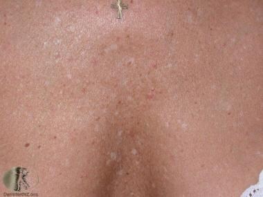 Idiopathic guttate hypomelanosis. Courtesy of DermNet New Zealand (https://www.dermnetnz.org/assets/Uploads/colour/igh1.jpg).
Idiopathic guttate hypomelanosis is a benign condition. The cause is not known, but it appears to be related to the effect of the sun on melanocytes, which makes them effete.
A variety of therapeutic methods, including topical steroids, topical retinoids, dermabrasion, cryotherapy, and minigrafting, have been used for idiopathic guttate hypomelanosis with variable success. [1]
Pathophysiology
Because pigmentation of the skin is due to an integration of melanocyte and keratinocyte function, an acquired defect of the epidermal melanin unit results in the observed hypopigmentation in idiopathic guttate hypomelanosis patients. Significantly fewer dopa oxidase-positive, KIT+, and melanocytes are seen in the lesions. [2, 3] In 1967, Hamada and Saito found a 50% reduction in melanocytes. [4]
There may be a genetic component to IGH, as families of IGH patients have a greater incidence of IGH when compared to controls. [5] Also, a study of 20 IGH patients found dermal fibrosis in 90% of patients. [6]
Prognosis
Idiopathic guttate hypomelanosis is cosmetic alone, albeit, it is indicative of cumulative sun exposure. Idiopathic guttate hypomelanosis progresses with increasing sun exposure and, to a lesser degree, with age.
Patient education
Progress in preventing idiopathic guttate hypomelanosis can be made by educating young women not to tan their legs.
Causes
The exact cause of idiopathic guttate hypomelanosis is not agreed upon; however, it is hypothesized that ultraviolet light plays an important role. [7] Another hypothesis is that microtrauma to the skin may contribute to the condition. [5]
Diagnostics
Laboratory studies
Idiopathic guttate hypomelanosis should be diagnosed on clinical grounds. A Wood light examination could be helpful in revealing unsuspected involvement. Dopa staining can be useful because dopa-positive melanocytes are decreased.
Procedures
In atypical cases of idiopathic guttate hypomelanosis, a biopsy may be indicated. Dermatoscopic examination may aid in diagnosis. Peripheral lesion findings include amoeboid, featherlike, and petaloid patterns described in lesions of idiopathic guttate hypomelanosis. [8, 9]
Histologic findings
Several light and electron microscopic studies have revealed a characteristic pathologic picture for idiopathic guttate hypomelanosis. Typical histologic findings are epidermal atrophy of the actinic type, a patchy decrease or absence of melanocytes and melanin, flat rete ridges, and basket weave hyperkeratosis. Skip areas of retained melanin can help histologically differentiate this condition from other disorders of pigmentation. [10]
Treatment
See Treatment and Medication.
Epidemiology
Frequency
United States
Idiopathic guttate hypomelanosis is a very common condition to the point of being almost universal in elderly fair-skinned individuals. In 2002, a case control study of 47 renal transplant patients demonstrated a significant positive association between HLA-DQ3 and the development of idiopathic guttate hypomelanosis and a significant negative association between HLA-DR8 and the development of idiopathic guttate hypomelanosis.
International
Idiopathic guttate hypomelanosis is most common in countries with fair-skinned populations having a high degree of sun exposure.
Race-, sex-, and age-related information
Idiopathic guttate hypomelanosis affects fair-skinned people at a younger age.
Idiopathic guttate hypomelanosis is seen far more frequently in women, beginning around the age of 30 years. However, with increasing age and sun exposure, it is found almost equally in elderly men and women. Why idiopathic guttate hypomelanosis occurs earlier in young women than in young men is unknown.
Idiopathic guttate hypomelanosis is related to the lack of pigmentary protection from the sun and sun exposure rather than to age. Fair-skinned women develop this condition first; later, with increasing age and exposure to sun, both sexes seem to be equally affected.
-
Idiopathic guttate hypomelanosis. Courtesy of DermNet New Zealand (https://www.dermnetnz.org/assets/Uploads/colour/igh1.jpg).
