Practice Essentials
Systemic lupus erythematosus (SLE) is an autoimmune disorder characterized by antibodies to nuclear and cytoplasmic antigens, multisystem inflammation, protean clinical manifestations, and a relapsing and remitting course. Approximately 90% of cases of SLE occur in women, frequently starting at childbearing age. [1] See the image below.
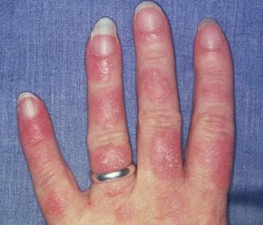 Photosensitive systemic lupus erythematosus (SLE) rashes typically occur on the face or extremities, which are sun-exposed regions. Although the interphalangeal spaces are affected, the metacarpophalangeal (MCP) and proximal interphalangeal (PIP) and distal interphalangeal (DIP) joints are spared. Photo courtesy of Dr. Erik Stratman, Marshfield Clinic.
Signs and symptoms of SLE
SLE is a chronic inflammatory disease that can affect almost any organ system, although it mainly involves the skin, joints, kidneys, blood cells, and nervous system. Its presentation and course are highly variable, ranging from indolent to fulminant.
The classic presentation of a triad of fever, joint pain, and rash in a woman of childbearing age should prompt investigation into the diagnosis of SLE. [2] However, male or female patients may present at any age with any of the following manifestations [3] :
-
Constitutional (eg, fatigue, fever, arthralgia, weight changes)
-
Musculoskeletal (eg, arthralgia, arthropathy, myalgia, frank arthritis, avascular necrosis)
-
Dermatologic (eg, malar rash, photosensitivity, discoid lupus)
-
Renal (eg, acute or chronic kidney failure, acute nephritic disease)
-
Neuropsychiatric (eg, seizure, psychosis)
-
Pulmonary (eg, pleurisy, pleural effusion, pneumonitis, pulmonary hypertension, interstitial lung disease)
-
Gastrointestinal (eg, nausea, dyspepsia, abdominal pain)
-
Cardiac (eg, pericarditis, myocarditis)
-
Hematologic (eg, cytopenias such as leukopenia, lymphopenia, anemia, or thrombocytopenia)
Clinical features tend to vary between children and adults with SLE: signs including malar rash, seizures, and fever are more common in children, while articular manifestations and pulmonary involvement more common in adults. [4]
In patients with suggestive clinical findings, a family history of autoimmune disease should raise further suspicion of SLE.
See Presentation for more detail.
Diagnosis of SLE
The diagnosis of SLE is based on a combination of clinical findings and laboratory evidence. Familiarity with the diagnostic criteria helps clinicians to recognize SLE and to subclassify this complex disease based on the pattern of target-organ manifestations.
The American College of Rheumatology (ACR) and the European League Against Rheumatism (EULAR) published updated criteria for the classification of SLE in 2019. [2] These criteria represent current concepts of SLE, and have excellent specificity and sensitivity. They replace the 1997 ACR criteria for SLE diagnosis. [5]
The ACR/EULAR classification requires an antinuclear antibody (ANA) titer of at least 1:80 on HEp-2 cells or an equivalent positive test at least once. If that is present, 22 "additive weighted" classification criteria are considered, comprising seven clinical domains (constitutional, hematologic, neuropsychiatric, mucocutaneous, serosal, musculoskeletal, renal) and three immunologic domains (antiphospholipid antibodies, complement proteins, SLE-specific antibodies).
Criteria are assigned points, ranging from 2 to 10. Patients with at least one clinical criterion and 10 or more points are classified as having SLE. See Workup.
Testing
Laboratory studies used in the diagnosis of SLE are as follows:
-
CBC with differential
-
Serum creatinine
-
Urinalysis with microscopy
-
ESR or CRP level
-
Complement levels
-
Liver function tests
-
Creatine kinase assay
-
Spot protein/spot creatinine ratio
-
Autoantibody tests
Imaging studies
The following imaging studies may be used to evaluate patients with suspected SLE:
-
Joint radiography
-
Chest radiography and chest CT scanning
-
Echocardiography
-
Brain MRI/MRA
-
Cardiac MRI
Procedures
Procedures that may be performed in patients with suspected SLE include the following:
-
Arthrocentesis
-
Lumbar puncture
-
Kidney biopsy
See Workup for more detail.
Management
Management of SLE often depends on the individual patient’s disease severity and disease manifestations, [6] although hydroxychloroquine has a central role for long-term treatment in all SLE patients.
Pharmacotherapy
Medications used to treat SLE manifestations include the following:
-
Antimalarials (eg, hydroxychloroquine)
-
Corticosteroids (eg, methylprednisolone, prednisone), short-term use recommended
-
Nonbiologic disease-modifying antirheumatic drugs (DMARDs): Cyclophosphamide, methotrexate, azathioprine, mycophenolate, cyclosporine
-
Nonsteroidal anti-inflammatory drugs (NSAIDS; eg, ibuprofen, naproxen, diclofenac)
-
Biologic DMARDs : Belimumab, rituximab, anifrolumab, and/or IV immune globulin
See Treatment and Medication for more detail.
See also the following Medscape articles:
Pathophysiology and Etiology
SLE is an autoimmune disorder characterized by multisystem inflammation with the generation of autoantibodies. Although the specific cause of SLE is unknown, multiple factors are associated with the development of the disease, including genetic, epigenetic, ethnic, immunoregulatory, hormonal, and environmental factors. [1, 7, 8] To date, about 100 SLE susceptibility genetic loci have been identified, mostly in European and Asian populations, explaining around 30% of the inheritability of lupus. [9] Many immune disturbances, both innate and acquired, occur in SLE.
Potential mechanisms
It is important to note that antibodies may be present for many years before the onset of the first symptoms of SLE. [10] One longstanding proposed mechanism for the development of autoantibodies involves a defect in apoptosis that causes increased cell death and a disturbance in immune tolerance. [11, 12, 13] The redistribution of cellular antigens during necrosis/apoptosis leads to a cell-surface display of plasma and nuclear antigens in the form of nucleosomes. Subsequently, dysregulated (intolerant) lymphocytes begin targeting normally protected intracellular antigens. The defective clearance of the apoptotic cell debris allows for the persistence of antigen and immune complex production. [14]
T cells have long been thought to play a central role in SLE pathogenesis, and T cells from patients with lupus show defects in both signaling and effector function. [15, 16] These T cells secrete less interleukin (IL)-2, and one defect in signaling seems to be linked to an increase in calcium influx, possibly due to changes in the CD3 signaling subunits. The following seem to be adversely affected in T cells from patients with SLE: effector activity such as CD8 cytotoxicity; T-regulatory, B-cell help; migration; and adhesion.
However, the method by which each of these deficits contributes to the exact clinical syndrome seen in an individual patient is still unknown. These T-cell abnormalities can be a target for therapy, as seen with the approval of belimumab, which targets the B-lymphocyte stimulator (BLys) signaling pathway. [15, 16]
Many clinical manifestations of SLE are mediated by circulating immune complexes that form with antigens in various tissues or the direct effects of antibodies on cell surface components. Immune complexes form in the microvasculature, leading to complement activation and inflammation. Moreover, antibody-antigen complexes deposit on the basement membranes of skin and kidneys. In active SLE, this process has been confirmed by demonstration of complexes of nuclear antigens such as DNA, immunoglobulins, and complement proteins at these sites.
Autoantibodies have been found to be biomarkers for future neuropsychiatric events in SLE. A prospective study of 1047 SLE patients demonstrated that individuals who had evidence of lupus anticoagulant (LA) had an increased future risk of intracranial thrombosis and that those with anti-ribosomal P antibodies had an increased future risk of lupus psychosis. [17]
A study of 35 SLE patients,16 of whom had overt neuropsychiatric symptoms, found that values of anti–double-stranded DNA antibodies, anti-nucleosome antibody, anti–cardiac phospholipid antibody (aCL-IgG), and anti-β2-glycoprotein I antibodies were significantly higher in the patients with neuropsychiatric symptoms. In addition, magnetic resonance imaging using 3D arterial spin labeling demonstrated a significantly higher incidence of decreased frontal lobe perfusion in the neuropsychiatric group. [18]
Serum antinuclear antibodies (ANAs) are found in nearly all individuals with active SLE. Antibodies to native double-stranded DNA (dsDNA) are relatively specific for the diagnosis of SLE. Whether polyclonal B-cell activation or a response to specific antigens exists is unclear, but much of the pathology involves B cells, T cells, and dendritic cells. Cytotoxic T cells and suppressor T cells (which would normally down-regulate immune responses) are decreased. The generation of polyclonal T-cell cytolytic activity is impaired. Helper (CD4+) T cells are increased. A lack of immune tolerance is observed in animal lupus models. Reports pointing to important roles of interferon-alpha, transcription factors, and signaling variations also point to a central role for neutrophils. [19]
A subset of SLE patients present with IgG autoantibodies to ribosomal P protein (anti-Rib-P antibodies). These antibodies are able to penetrate certain cells, binding to ribosomal proteins and blocking protein synthesis. In activated monocytes, anti-Rib-P antibodies enhance the production of tumor necrosis factor (TNF) and interleukin (IL)-6. The presence of anti-Rib-P antibodies has been associated with greater severity of SLE. [20]
Anti-Rib-P antibodies occur more often in juvenile-onset than in adult-onset SLE. In addition, the frequency of these antibodies tends to vary by ethnicity, ranging from 6% to 20% in various ethnic groups, but as high as 36% in Chinese patients with SLE. [21]
Genetics
There is a clear genetic component in SLE, with a sibling risk ratio 8-fold to 29-fold higher than that in the general population and a 10-fold increase in disease concordance in identical twins. [22]
A population-based study from Denmark found that the hazard ratio of SLE was 10.3 in first-degree relatives of patients with SLE and 3.6 in second- or third-degree relatives. [23] A population-based study from Taiwan estimated that the heritability of SLE was 43.9%; relative risks were 23.68 for siblings, 11.44 for parents, and 14.42 for offspring of patients. [24] In addition, SLE occurs in both twins in 30% of identical twins and 5-10% of nonidentical twins, which may be due to a combination of genetic and environmental factors. [8]
Some studies have synthesized what is known about the mechanisms of SLE disease and genetic associations. [25, 26] A genetic predisposition is supported by 40% concordance in monozygotic twins; if a mother has SLE, her daughter's risk of developing the disease has been estimated to be 1:40, and her son's risk, 1:250. [25, 26]
Although some single genes have been implicated to play a causative role in SLE, current knowledge points toward a large number of genes being involved in a multifactorial-type inheritance pattern in most patients. Genome-wide association studies have identified more than 60 risk loci for SLE susceptibility across populations, with most of the genetic risk shared across borders and ethnicities. [22]
Many of the loci with a strong association with SLE are involved in the immune and related biologic systems. [21] Genes previously associated with other autoimmune diseases have been associated with SLE (eg, PTPN22 and diabetes; STAT4 and rheumatoid arthritis).
A genome-wide study in a northern European population replicated the association of SLE with susceptibility genes related to B-cell receptor pathway signaling, and confirmed the association of SLE with genes at the interferon regulatory factor 5 (IRF5)-TNPO3 locus. The investigators also confirmed other loci associations with SLE (TNFAIP3, FAM167A-BLK, BANK1 and KIAA1542) with a lower contribution to individual risk for SLE. [27]
A meta-analysis of the association of interferon regulatory factor 5 (IRF5) with SLE found that a specific T allele, IRF5 rs2004640, is significantly associated with SLE in populations of European, Asian, and Latin American origins, whereas the A allele IRF5 rs10954213 is associated with SLE in patients of European origin but not in those of Asian origin. [28] Overall, the IRF5 gene polymorphism was found to be associated with SLE in multiple ethnic populations.
Studies of human leukocyte antigens (HLAs) reveal that HLA-A1, HLA-B8, and HLA-DR3 are more common in persons with SLE than in the general population. The presence of the null complement alleles and congenital deficiencies of complement (especially C4, C2, and other early components) are also associated with an increased risk of SLE.
Genetic studies point to disruptions in lymphocyte signaling, interferon response, clearance of complement and immune complexes, apoptosis, and DNA methylation. [25] Several genes associated with T-cell function and signaling have been associated with SLE, including PTPN22, TNFSF4, PDCD1, IL10, BCL6, IL16, TYK2, PRL, STAT4, and RASGRP3, as have immune-complex processing and innate immunity genes, including several complement genes (eg, C2, C4A, and C4B). [29] Hypomethylation (a form of epigenetic modification) of genes involved in osmotic lysis, apoptosis, inflammation, and cytokine pathways, among other immunologic functions, has also been associated with SLE. [28, 30]
The higher risk of developing SLE in women and in men with Klinefelter syndrome (ie, genotype XXY) may relate to enhanced expression of toll-like receptor 7 (TLR7), a key pathogenic factor in SLE that is encoded on an X chromosome locus. Souyris et al reported that in both women and males with Klinefelter syndrome, substantial fractions of primary B-lymphocytes, monocytes, and plasmacytoid dendritic cells express TLR7 on both X chromosomes, leading to greater immunoglobulin secretion. [31]
Brown et al identified a previously undescribed single-point missense gain-of-function TLR7 mutation, TLR7Y264H, in a child with severe SLE and subsequently found it in other patients with severe SLE. When introduced into mice, the TLR7Y264H variant caused lupus. [32]
Normally, one X chromosome in female cells is randomly selected for transcriptional silencing through X-chromosome inactivation. Pyfrom et al reported that abnormal epigenetic regulation of X-chromosome inactivation in B cells is a feature of SLE. Those epigenetic abnormalities predispose for aberrant expression of X-linked immunity genes (eg, TLR7) from the inactivated X chromosome, and likely contribute to the female bias in SLE. [33]
The frequencies of SLE risk alleles in the general population help to evaluate each individual genetic susceptibility by means of cumulative weighted genetic risk score (wGRS), which is defined as the sum of the number of risk alleles at various loci in an individual weighted by the natural logarithm of their odds ratios. [34] A wGRS analysis of five general populations found that high-wGRS populations carrying high frequencies of SLE risk alleles have higher prevalence of SLE. [35] The population order from the lowest to highest average of wGRS was as follows: Europeans < American Indian ≈ South Asians < East Asian < Africans.
In addition, high wGRS for SLE was found to be more frequent in patients with childhood-onset SLE, anti-dsDNA positivity, oral ulcers and immunologic, renal, and hematologic manifestations, suggesting that that the high genetic load on SLE risk is not only associated with susceptibility to SLE but also early onset and unfavorable prognosis of SLE. [35] However, a more detailed study of the increased prevalence of SLE in Asians and Africans would require extensive comparisons of genetic and environmental data, including generation of DNA sequence data to exclude European bias in genotyping arrays.
Infections and Microbes as Triggers
Numerous studies have investigated the role of infectious etiologies that may also perpetuate autoimmunity. [36] Patients with SLE have higher titers of antibodies to Epstein-Barr virus (EBV), have increased circulating EBV viral loads, and make antibodies to retroviruses, including antibodies to protein regions homologous to nuclear antigens. In patients with SLE and EBV infection, the B cells are not primarily defective; rather, the SLE/EBV phenomenon is due to a T-cell abnormality, which causes failure in normal immunoregulation of the B-cell response. [37] Viruses may stimulate specific cells in the immune network. Chronic infections may induce anti-DNA antibodies or even lupuslike symptoms, and acute lupus flares often follow bacterial infections.
A study by Manfredo Vieira et al found that in a mouse strain that is predisposed to autoimmunity, translocation of a gut pathobiont, Enterococcus gallinarum, to the liver and other systemic tissues (as might occur with loss of integrity of the gut barrier) triggers autoimmune responses. in a genetic background predisposing to autoimmunity. In this model, antibiotic treatment prevented mortality in this model, suppressed growth of E gallinarum in tissues, and eliminated pathogenic autoantibodies and T cells. [38]
Furthermore, these researchers recovered E gallinarum–specific DNA from liver biopsies of autoimmune patients, and found that cocultures of human hepatocytes with E gallinarum induced autoimmune-promoting factors, replicating the murine findings. Those results suggest that similar processes occur in susceptible humans. [38]
Other Risk Factors and Environmental Triggers
Pregnancy can be a time when lupus initially presents or flares, although more recent data suggests that pregnancy outcomes are favorable and flares are infrequent among patients with inactive or stable mild-moderated SLE. [39]
Vitamin D is involved in both in both innate and acquired immunity, and vitamin D deficiency has been implicated in autoimmunity and the development of rheumatic diseases, including SLE. [40] Young et al studied 436 individuals who reported having a relative with SLE but who did not have SLE themselves, and found that the combination of vitamin D deficiency and carriage of specific single-nucleotide polymorphisms was associated with significantly increased risk of transitioning to SLE. [41] Hu et al reported that in an Asian population, carriage of certain polymorphisms in the vitamin D receptor gene BsmI (Bb + BB genotype and B allele) can significantly increase risk for developing SLE. [42]
Other potential triggers include the following:
-
Silica dust exposure and cigarette smoking may increase the risk of developing SLE.
-
Cigarette smoking is also associated with higher risk of flares, predominantly cutaneous flares, and increase risk of poor outcomes, including chronic kidney disease and cardiovascular disease, in patients with lupus.
-
Estrogen use in postmenopausal women appears to increase the risk of developing SLE.
-
Photosensitivity is clearly a precipitant of skin disease. Ultraviolet light stimulates keratinocytes, which leads not only to overexpression of nuclear ribonucleoproteins (snRNPs) on their cell surfaces but also to the secretion of cytokines that simulate increased autoantibody production. [43]
-
Breastfeeding is associated with a decreased risk of developing SLE.
Possible early-life risk factors include the following [44] :
-
Low birthweight (< 2500 g)
-
Preterm birth (≥1 month early)
-
Childhood exposure to agricultural pesticides
Epidemiology
United States Statistics
Incidence
Estimates from the five national lupus registries funded by the Centers for Disease Control and Prevention (CDC) place the incidence of SLE at roughly 5.1 per 100,000 person-years (95% CI 4.6 to 5.6), higher in women than in men (8.7 vs 1.2 per 100,000 person-years), and highest among Black women (15.9 per 100,000 person-years). The American Indian/Alaska Native population had the second highest race-specific SLE estimates for women (10.4 per 100,000) and highest for men (3.8 per 100,000). Incidence in Hispanic women with SLE was 6.7 per 100,000. Based on those data and extrapolating age- and race-specific rates to the 2018 US Census data, it is estimated that 14,263 persons (95% CI 11,563 to 17,735) were newly diagnosed with SLE in the US. [45]
Prevalence
Older national prevalence estimates vary widely due to differences in case definitions, and study methods. The Lupus Foundation of America currently estimates prevalence to be at least 1.5 million cases, [46] which likely reflects inclusion of milder forms of the disease.
The pooled prevalence of SLE from the five national CDC-funded lupus registries was 72.8 cases per 100,000 people. In 2018, an estimated 204,295 individuals (95% confidence interval [CI] 160,902–261,725) in the US fulfilled the American College of Rheumatology (ACR) classification criteria for SLE. [45] The pooled prevalence was 9 times higher among women than men (128.7 vs 14.7 per 100,000). Pooled prevalence rates per 100,000 in women by ethnicity were as follows:
-
American Indian/Alaska Native: 270.6
-
Black: 230.9
-
Hispanic: 120.7
-
White: 84.7
-
Asian/Pacific Islander: 84.4
Rates among men were as follows:
-
American Indian/Alaska Native: 53.8
-
Black: 26.7
-
Hispanic: 18.0
-
Asian/Pacific Islander: 11.2
-
White: 8.9
Mortality and morbidity
A US study highlighted a marked improvement in the 10-year survival for SLE patients, with adjusted standardized mortality rates in 1968 vs 2013 of 4.5 (95% CI, 4.2 to 4.8) vs 3.4 (95% CI, 3.2 to 3.6) per million persons. However, the ratio of SLE to non-SLE mortality was about 35% higher in 2013 than in 1968. [47] Patients with lupus who are younger than age 40 years are at 52-fold higher risk of cardiovascular disease compared with age-matched healthy peers. [48] Lupus is a leading cause of mortality in young women, particularly those identified as Black or Hispanic. [49]
Disadvantaged populations carry a disproportionate burden of SLE complications and poor outcomes. The burden of cardiovascular disease was found to be 19-fold higher in people with lupus identified as Black than in those identified as White. [50] Mortality risk in people with SLE is 3-4 times higher in Blacks than in Whites, with the highest age-standardized mortality rate in Black women (3.38 per million persons). [47, 51] Additionally, death occurred at a much younger age in Black women with SLE than in White women with SLE (51 years vs 64 years). [51] Another cohort study highlighted that the mortality risk was four times higher in Hispanic and Asian women with SLE. [52]
International Statistics
The global incidence of SLE has been estimated to be 5.14 cases per 100,000 person-years. In women, the estimated incidence is 8.82 cases per 100,000 person-years. [53]
Worldwide, the prevalence of SLE varies. The highest rates have been reported in Italy, Spain, Martinique, and the United Kingdom Afro-Caribbean population. [54] Although the prevalence of SLE is high in Black persons in the United Kingdom, the disease is rarely reported in Africa, suggesting that there may be an environmental trigger, as well as a genetic basis, for disease in the UK population. [55]
A review of SLE across Asia-Pacific countries revealed considerable variation in prevalence and survival rates. [56] For example, overall prevalence rates ranged from 4.3 to 45.3 per 100,000, and the overall incidence ranged from 0.9 to 3.1 per 100,000 per year. Moreover, Asians with SLE had higher rates of kidney involvement than White persons did, and cardiovascular involvement was a leading cause of death in Asians. [56]
Thus, based on those studies, in general, women from the Black racial group have a higher rate of SLE than women of any other race, followed by women from the Asian racial group and then women from the White racial group. [54]
Gender and Age Disparities
Approximately 90% of cases of SLE occur in women, frequently starting at childbearing age. [1, 57] The association of lupus onset and flares with the use of exogenous hormones further suggests a role for hormonal factors in the pathogenesis of the disease. [58]
The risk of SLE development in men is similar to that in prepubertal or postmenopausal women. Interestingly, in men, SLE is more common in those with Klinefelter syndrome (ie, genotype XXY). In fact, a study by Dillon et al found that men with Klinefelter syndrome had a more severe course of SLE than women but a less severe course than other men. [59]
The female-to-male ratio peaks at 11:1 during the childbearing years. [60] A correlation between age and incidence of SLE mirrors peak years of female sex hormone production. Onset of SLE is usually after puberty, typically in the 20s and 30s, but SLE can begin during childhood; 20% of all cases are diagnosed during the first 2 decades of life. [61] A review of the worldwide literature (predominantly North America, Europe, and Asia) found that the incidence of pediatric-onset SLE ranged from 0.36 to 2.5 per 100,000 per year and the prevalence ranged from 1.89 to 25.7 per 100,000. [62]
The prevalence of SLE is highest in women aged 14 to 64 years. SLE does not have an age predilection in males, although it should be noted that in older adults, the female-to-male ratio falls. [63] This effect is likely due to loss of the estrogen effect in older women.
Prognosis
SLE carries a highly variable prognosis for individual patients. The natural history of SLE ranges from relatively benign disease to rapidly progressive and even fatal illness. SLE often waxes and wanes in affected individuals throughout life, and features of the disease vary greatly among individuals.
The disease course is milder and the survival rate higher in persons with isolated skin and musculoskeletal involvement than in those with kidney disease [64] and CNS disease. [65] A consortium report of 298 SLE patients followed for 5.5 years noted falls in SLE Disease Activity Index 2000 (SLEDAI-2K) scores after the first year of clinical follow-up and gradual increases in cumulative mean Systemic Lupus International Collaborating Clinics (SLICC) damage index scores. [66]
It is important to distinguish between the disease activity and the damage index (irreversible organ dysfunction). Although which instrument is the most effective for measuring SLE disease activity is still open to debate, there are several validated measures, including the following:
-
Systemic Lupus Activity Measure (SLAM)
-
SLEDAI
-
Lupus Activity Index (LAI)
-
European Consensus Lupus Activity Measurement (ECLAM)
-
British Isles Lupus Activity Group (BILAG) Index
Prognostic factors from the 2008 European League Against Rheumatism (EULAR) recommendations included the following [67] :
-
Clinical findings: Skin lesions, arthritis, serositis, neurologic manifestations such as seizures and psychosis, and kidney involvement
-
Diagnostic study results: Anemia, thrombocytopenia, leukopenia, increased serum creatinine levels
-
Immunologic test results: Serum C3 and C4 concentration (which may be low), as well as the presence of anti–double-stranded DNA (anti-dsDNA), anti-Ro/ Sjögren syndrome A (SSA), anti-La/Sjögren syndrome B (SSB), and antiphospholipid (aPL), and anti-ribonucleoprotein (anti-RNP)
Mortality
Although historically, SLE was associated with a reduced life expectancy, mortality in patients with SLE has decreased over the past few decades. [68] Prior to 1955, the 5-year survival rate in SLE was less than 50%; currently, the average 10-year survival rate exceeds 90%, [69, 65] and the 15-year survival rate is approximately 80%. [70] Previously, mortality was due to the disease itself; currently, mortality is often a result of medication adverse effects (eg, fatal infections in individuals receiving potent immunosuppressive medications) or cardiovascular disease (CVD) events.
A review of over 15,000 incident SLE patients by Li et al concluded that patients with high initial severity of SLE had elevated risk of all-cause mortality and CVD events compared with those who presented with milder disease. After multivariable adjustment, the CVD subdistribution hazard ratio (HRSD) for initially severe SLE versus mild SLE was 1.64 (95% confidence index [CI] 1.32, 2.04). The HR for mortality was 3.11 (95% CI 2.49, 3.89). [71]
Ten-year survival rates in Asia and Africa are significantly lower than those in the United States, ranging from 60-70%. [72, 73] However, that may reflect detection bias of severe cases only.
Decreased mortality rates associated with SLE can be attributed to earlier diagnosis (including milder cases), improvement in disease-specific treatments, and advances in general medical care. According to the Centers for Disease Control and Prevention (CDC), however, 35% of SLE-related deaths in the United States occur in patients younger than 45 years, making this a serious issue despite declining overall mortality rates. [74]
The EULAR task force also identified the following comorbidities as increasing the risk of morbidity and mortality in patients with SLE [75] :
-
Infections
-
Hypertension
-
Lipid disorders (dyslipidemia), atherosclerosis, and coronary heart disease
-
Diabetes mellitus
-
Bone-related conditions: Osteoporosis; avascular bone necrosis
-
Malignancies (eg, non-Hodgkin lymphoma, lung cancer, hepatobiliary cancer)
In 1976, Urowitz first reported bimodal mortality in early versus late SLE, noting that SLE-related deaths usually occurred within the first 5-10 years of symptom onset. [76] Mortality in the first few years of illness is typically from severe SLE disease (eg, CNS, kidney, or cardiovascular involvement) or infection related to immunosuppressive treatment. Infections account for 29% of all deaths in these patients. [77]
Late deaths (after age 35 years) are generally from myocardial infarction or stroke secondary to accelerated atherosclerosis. [68, 78, 69, 79] Inflammation is central to SLE pathogenesis and plays a major role in the development and accelerated progression of atherosclerosis. Manzi et al reported that women aged 35-44 years with SLE were 50 times more likely to develop myocardial ischemia than healthy Framingham study control women. [78] The presence of lupus nephritis may increase these risks. [80] The presence of traditional and nontraditional risk factors increases the risk of cardiovascular (CVD) disease in patients with SLE.
In a study by Petri et al that evaluated a large sample of SLE patients, the investigators reported that more than 50% of these patients had at least 3 classic cardiac risk factors, with the most common ones being a sedentary lifestyle, obesity, and hypercholesterolemia. [81] In another study, Salmon et al found that nontraditional CVD risk factors in SLE patients included having higher homocysteine levels, kidney impairment, enhanced LDL oxidation, and chronic inflammation. [82]
Causes of accelerated coronary artery disease in persons with SLE are likely multifactorial. They include endothelial dysfunction, inflammatory mediators, corticosteroid-induced atherogenesis, and dyslipidemia.
The influence of race on prognosis has been widely debated. The LUMINA study group reported that high disease activity and poverty predicted higher mortality in Black and Hispanic patients with lupus. [83] In the Michigan Lupus Epidemiology and Surveillance program, the proportion of patients with kidney disease was 2.2-fold higher, and that of progression to end-stage kidney disease was 3.4-fold higher, in Black patients than in White ones. [84] A 2022 study reported that patients from the Black racial group faced 19-fold higher risk of cardiovascular disease, with peak events occurring in the second year of lupus diagnosis. [50] This study highlighted discoid lupus as another unique predictor of cardiovascular disease in patients with SLE.
Patient Education
Stress the importance of adherence to medications and follow-up appointments for detection and control of SLE disease. Instruct patients with SLE to seek medical care for evaluation of new symptoms, including fever. Advise them regarding their heightened risks for infection and cardiovascular disease. Educate patients with SLE regarding aggressive lipid and blood pressure goals to minimize the risk of coronary artery disease.
Instruct patients with SLE to avoid exposure to sunlight and ultraviolet light. Also, encourage them to receive nonlive vaccines during stable periods of disease, to quit smoking, and to carefully plan pregnancies.
For patient education information, see Lupus (Systemic Lupus Erythematosus).
See also the American College of Rheumatology’s patient fact sheets for SLE, Systemic Lupus Erythematosus in Children and Teens, and Antiphospholipid Syndrome as well as the American Lupus Foundation's What is lupus?.
-
Systemic lupus erythematosus (SLE). The classic malar rash, also known as a butterfly rash, with distribution over the cheeks and nasal bridge. Note that the fixed erythema, sometimes with mild induration as seen here, characteristically spares the nasolabial folds (nasolabial fold sparing is indicated by black arrows).
-
Dermatomyositis. Acute onset of confluent macular erythema in a periorbital and malar distribution (involving the cheeks and extending over the nasal bridge), with extension to the chin in a female with juvenile dermatomyositis. Note the perioral sparing. In some patients, there may be more extensive involvement of the face, including the perioral region, forehead, lateral face, and ears. In contrast to SLE , in dermatomyositis with malar erythema, the nasolabial folds are often not spared.
-
Discoid lupus erythematosus.
-
Photosensitive systemic lupus erythematosus (SLE) rashes typically occur on the face or extremities, which are sun-exposed regions. Although the interphalangeal spaces are affected, the metacarpophalangeal (MCP) and proximal interphalangeal (PIP) and distal interphalangeal (DIP) joints are spared. Photo courtesy of Dr. Erik Stratman, Marshfield Clinic.
-
In systemic lupus erythematosus (SLE), many genetic-susceptibility factors, environmental triggers, antigen-antibody (Ab) responses, B-cell and T-cell interactions, and immune clearance processes interact to generate and perpetuate autoimmunity. HLA = human leukocyte antigen; UV = ultraviolet light.
-
This axial, T2-weighted brain magnetic resonance image (MRI) demonstrates an area of ischemia in the right periventricular white matter of a 41-year-old woman with long-standing systemic lupus erythematosus (SLE). She presented with headache and subtle cognitive impairments but no motor deficits. Faintly increased signal intensity was also seen on T1-weighted images, with a trace of enhancement following gadolinium that is too subtle to show on reproduced images. Distribution of the abnormality is consistent with occlusion of deep penetrating branches, such as may result from local vasculopathy, with no clinical or laboratory evidence of lupus anticoagulant or anticardiolipin antibody. Cardiac embolus from covert Libman-Sacks endocarditis remains less likely due to distribution.
-
Lupus band test. Microphotograph of a histologic section of human skin prepared for direct immunofluorescence using an anti-IgG antibody. The skin is from a patient with systemic lupus erythematosus and shows IgG deposit at 2 different places: the first is a band-like deposit along the epidermal basement membrane ("lupus band test" is positive); the second is within the nuclei of the epidermal cells (anti-nuclear antibodies).
-
Microphotograph of a fixed Hep-2 line cell prepared for indirect immunofluorescence. The preparation was exposed to a serum of a patient with systemic lupus erythematosus and labeled using a murine anti-human immunoglobulin G (IgG) antibody. It shows IgG deposit in the nucleus and nonspecific deposit in the cytoplasm.
-
Mesangial proliferative lupus nephritis with moderate mesangial hypercellularity. International Society of Nephrology/Renal Pathology Society 2003 class II (×200, hematoxylin-eosin).
-
Focal lupus nephritis. International Society of Nephrology/Renal Pathology Society 2003 class III (×200, immunofluorescence).
-
Membranous lupus nephritis showing thickened glomerular basement membrane. International Society of Nephrology/Renal Pathology Society 2003 class V (×200, silver stain).
-
The chest x-ray from a patient with lupus demonstrates a right-sided pleural effusion (yellow arrow) and atelectasis with scarring in the left lung base (blue arrow). In severe complications, a fibrothorax may develop.
-
Vasculitis, antiphospholipid antibodies, and renal failure are commonly found in patients with lupus; these conditions greatly increase the risk of developing pulmonary emboli. The diagnosis in a patient with shortness of breath, hemoptysis, and pleuritic chest pain is commonly made with ventilation-perfusion scans or computed tomography (CT) angiography. The CT angiogram demonstrates a filling defect in the left anterior segmental artery (arrow).
-
Libman-Sacks endocarditis is the most characteristic cardiac manifestation of lupus. It is characterized by clusters of verrucae on the ventricular surface of the mitral valve. These lesions consist of accumulation of immune complexes, platelets, and mononuclear cells. This can lead to heart failure, valvular dysfunction, emboli, and secondary infective endocarditis. Diagnosis is best made via echocardiography, which may reveal the characteristic valvular masses (arrows). IVS = interventricular septum; LA = left atrium; LV = left ventricle.
-
Histologic image of a normal renal cortex, including the glomerulus (1) and proximal (2) and distal (3) convoluted tubule. [Image from Wikipedia: https://en.wikipedia.org/wiki/File:Histology-kidney.jpg]
-
Discoid lupus erythematosus is a chronic scarring skin condition causing scaly plaques on the scalp, face, and ears.
-
Focal lupus nephritis. International Society of Nephrology/Renal Pathology Society 2003 class III (×100, hematoxylin-eosin).
Tables
Domain |
Criteria |
Points |
Constitutional |
Fever |
2 |
Hematologic |
Leukopenia Thrombocytopenia Autoimmune hemolysis |
3 4 4 |
Neuropsychiatric |
Delirium Psychosis Seizure |
2 3 5 |
Mucocutaneous |
Non-scarring alopecia Oral ulcers Subacute cutaneous or discoid lupus Acute cutaneous lupus |
2 2 4 6 |
Serosal |
Pleural or pericardial effusion Acute pericarditis |
5 6 |
Musculoskeletal |
Joint involvement |
6 |
Renal |
Proteinuria > 0.5 g/24 h Kidney biopsy class II or V lupus nephritis Kidney biopsy class III or IV lupus nephritis |
4 8 10 |
Domain |
Criteria |
Points |
Antiphospholipid antibodies |
Anti-cardiolipin antibodies or Anti-β2GP1 antibodies or Lupus anticoagulant |
2 |
Complement proteins |
Low C3 or low C4 Low C3 and low C4 |
3 4 |
SLE-specific antibodies |
Anti-dsDNA antibody or Anti-Smith antibody |
6 |
Test |
Description |
ANA |
Screening test; sensitivity 95%; not diagnostic without clinical features |
Anti-dsDNA |
High specificity; sensitivity only 70%; level is variable based on disease activity |
Anti-Sm |
Most specific antibody for SLE; only 30-40% sensitivity |
Anti-SSA (Ro) or Anti-SSB (La) |
Present in 15% of patients with SLE and other connective-tissue diseases such as Sjögren syndrome; associated with neonatal lupus |
Anti-ribosomal P |
Uncommon antibodies that may correlate with risk for CNS disease, including increased hazards of psychosis in a large inception cohort, although the exact role in clinical diagnosis is debated [111] |
Anti-RNP |
Included with anti-Sm, SSA, and SSB in the ENA profile; may indicate mixed connective-tissue disease with overlap SLE, scleroderma, and myositis |
Anticardiolipin |
IgG/IgM variants measured with ELISA are among the antiphospholipid antibodies used to screen for antiphospholipid antibody syndrome and pertinent in SLE diagnosis |
Lupus anticoagulant |
Multiple tests (eg, direct Russell viper venom test) to screen for inhibitors in the clotting cascade in antiphospholipid antibody syndrome |
Direct Coombs test |
Coombs test–positive anemia to denote antibodies on RBCs |
Anti-histone |
Drug-induced lupus ANA antibodies are often of this type (eg, with procainamide or hydralazine; p-ANCA–positive in minocycline-induced drug-induced lupus) |
ANA = antinuclear antibody; CNS = central nervous system; ds-DNA = double-stranded DNA; ELISA = enzyme-linked immunoassay; ENA = extractable nuclear antigen; Ig = immunoglobulin; p-ANCA = perinuclear antineutrophil cytoplasmic antibody; RBCs = red blood cells; RNP = ribonucleic protein; SLE = systemic lupus erythematosus; Sm = Smith; SSA = Sjögren syndrome A; SSB = Sjögren syndrome B. |
|
Class |
Classification |
Features |
Class I |
Minimal mesangial |
Normal light microscopy findings; abnormal electron microscopy findings |
Class II |
Mesangial proliferative |
Hypercellular on light microscopy |
Class III |
Focal proliferative |
< 50% of glomeruli involved Class III lupus nephritis is further subclassified as follows:
|
Class IV |
Diffuse proliferative |
=50% of glomeruli involved; classified segmental or global; treated aggressively Class IV lupus nephritis is also further subclassified, as follows:
Note: It remains to be determined whether further subcategories have a prognostic difference. [116] There are conflicting data from studies; some investigators report that class IV-G (A) has a better prognosis relative to class IV-S (A/C), which is less responsive to treatment. |
Class V |
Membranous |
Predominantly nephrotic disease Note: Class V may occur with class III or IV (then, both cases would be diagnosed) [109] |
Class VI |
Advanced sclerosing |
≥90% of glomeruli involved without residual activity [109] Chronic lesions and sclerosis |
Source (except as noted otherwise) : Weening JJ, D'Agati VD, Schwartz MM, et al. The classification of glomerulonephritis in systemic lupus erythematosus revisited. J Am Soc Nephrol. Feb 2004;15(2):241-50. [117] SLE = systemic lupus erythematosus. |
||
