Redondo P, Bastarrika G, Aguado L, et al. Foot or hand malformations related to deep venous system anomalies of the lower limb in Klippel-Trénaunay syndrome. J Am Acad Dermatol. 2009 Oct. 61(4):621-8. [QxMD MEDLINE Link].
Akcali C, Inaloz S, Kirtak N, Ozkur A, Inaloz S. A case of Klippel-Trenaunay syndrome involving only upper limbs. G Ital Dermatol Venereol. 2008 Aug. 143(4):267-9. [QxMD MEDLINE Link].
Qi HT, Wang XM, Zhang XD, Zhang MH, Li CM, Bao SG, et al. The role of colour Doppler sonography in the diagnosis of lower limb Klippel-Trénaunay syndrome. Clin Radiol. 2013 Jul. 68(7):716-20. [QxMD MEDLINE Link].
Li X, Tian J. Multidetector row computed tomography arteriography in the preoperative assessment of patients with Klippel-Trénaunay syndrome. J Am Acad Dermatol. 2009 Feb. 60(2):345-6; author reply 346. [QxMD MEDLINE Link].
Wen Z, Tong G, Liu Y. Potential Utilization of Lymphoscintigraphy in Patients With Klippel-Trenaunay Syndrome. Clin Nucl Med. 2021 Jan. 46 (1):25-30. [QxMD MEDLINE Link].
Bliznak J, Staple TW. Radiology of angiodysplasias of the limb. Radiology. 1974 Jan. 110(1):35-44. [QxMD MEDLINE Link].
Servelle M. Klippel and Trenaunay's syndrome. 768 operated cases. Ann Surg. 1985 Mar. 201(3):365-73. [QxMD MEDLINE Link].
Baskerville PA, Ackroyd JS, Browse NL. The etiology of the Klippel-Trenaunay syndrome. Ann Surg. 1985 Nov. 202(5):624-7. [QxMD MEDLINE Link].
McGrory BJ, Amadio PC. Klippel-Trenaunay syndrome: orthopaedic considerations. Orthop Rev. 1993 Jan. 22(1):41-50. [QxMD MEDLINE Link].
Ceballos-Quintal JM, Pinto-Escalante D, Castillo-Zapata I. A new case of Klippel-Trenaunay-Weber (KTW) syndrome: evidence of autosomal dominant inheritance. Am J Med Genet. 1996 Jun 14. 63(3):426-7. [QxMD MEDLINE Link].
Hofer T, Frank J, Itin PH. Klippel-Trenaunay syndrome in a monozygotic male twin: supportive evidence for the concept of paradominant inheritance. Eur J Dermatol. 2005 Sep-Oct. 15(5):341-3. [QxMD MEDLINE Link].
Hu Y, Li L, Seidelmann SB, et al. Identification of association of common AGGF1 variants with susceptibility for Klippel-Trenaunay syndrome using the structure association program. Ann Hum Genet. 2008 Sep. 72:636-43. [QxMD MEDLINE Link]. [Full Text].
Kihiczak GG, Meine JG, Schwartz RA, Janniger CK. Klippel-Trenaunay syndrome: a multisystem disorder possibly resulting from a pathogenic gene for vascular and tissue overgrowth. Int J Dermatol. 2006 Aug. 45(8):883-90. [QxMD MEDLINE Link].
Sung HM, Chung HY, Lee SJ, Lee JM, Huh S, Lee JW, et al. Clinical Experience of the Klippel-Trenaunay Syndrome. Arch Plast Surg. 2015 Sep. 42 (5):552-8. [QxMD MEDLINE Link].
Funayama E, Sasaki S, Oyama A, Furukawa H, Hayashi T, Yamamoto Y. How do the type and location of a vascular malformation influence growth in Klippel-Trénaunay syndrome?. Plast Reconstr Surg. 2011 Jan. 127(1):340-6. [QxMD MEDLINE Link].
Renard D, Larue A, Taieb G, Jeanjean L, Labauge P. Recurrent cerebral infarction in Klippel-Trenaunay-Weber syndrome. Clin Neurol Neurosurg. 2012 Feb 17. [QxMD MEDLINE Link].
Upadhyay H, Sherani K, Vakil A, Babury M. A case of recurrent massive pulmonary embolism in Klippel-Trenaunay-Weber syndrome treated with thrombolytics. Respir Med Case Rep. 2016. 17:68-70. [QxMD MEDLINE Link].
Chenbhanich J, Leelayuwatanakul N, Phowthongkum P. Klippel-Trenaunay-Weber syndrome as a cause of chronic thromboembolic pulmonary hypertension. BMJ Case Rep. 2018 Mar 22. 2018:[QxMD MEDLINE Link].
Yilmaz T, Cikla U, Kirst A, Baskaya MK. Glioblastoma multiforme in Klippel-Trenaunay-Weber syndrome: a case report. J Med Case Rep. 2015 Apr 17. 9:83. [QxMD MEDLINE Link].
Bhat L, Bisht S, Khanijo K. Klippel-Trenaunay-Weber Syndrome with Kasabach-Merritt Coagulopathy and Hydronephrosis. Indian Pediatr. 2015 Nov. 52 (11):987-8. [QxMD MEDLINE Link].
van der Loo LE, Beckervordersandforth J, Colon AJ, Schijns OE. Growing skull hemangioma: first and unique description in a patient with Klippel-Trénaunay-Weber syndrome. Acta Neurochir (Wien). 2017 Feb. 159 (2):397-400. [QxMD MEDLINE Link].
Opdenakker O, Renson T, Walle JV. Vesical Hemangioma in a Patient with Klippel-Trenaunay-Weber Syndrome. J Pediatr. 2019 May. 208:293-293.e2. [QxMD MEDLINE Link].
Furness PD 3rd, Barqawi AZ, Bisignani G, Decter RM. Klippel-Trénaunay syndrome: 2 case reports and a review of genitourinary manifestations. J Urol. 2001 Oct. 166(4):1418-20. [QxMD MEDLINE Link].
Ploegmakers MJ, Pruszczynski M, De Rooy J, Kusters B, Veth RP. Angiosarcoma with malignant peripheral nerve sheath tumour developing in a patient with klippel-trénaunay-weber syndrome. Sarcoma. 2005. 9(3-4):137-40. [QxMD MEDLINE Link]. [Full Text].
Fakir E, Roberts T, Stephen L, Beighton P. Klippel-Trenaunay-Weber syndrome: orodental manifestations and management considerations. Oral Surg Oral Med Oral Pathol Oral Radiol Endod. 2009 Jun. 107(6):754-8. [QxMD MEDLINE Link].
Liu NF, Lu Q, Yan ZX. Lymphatic malformation is a common component of Klippel-Trenaunay syndrome. J Vasc Surg. 2010 Dec. 52(6):1557-63. [QxMD MEDLINE Link].
Willis-Owen CA, Cobb JP. Total hip arthroplasty in Klippel-Trenaunay syndrome. Ann R Coll Surg Engl. 2008 Nov. 90(8):W6-8. [QxMD MEDLINE Link]. [Full Text].
Yaqub Y, Suarez J, Perez-Verdia A, Arvandi A, Nugent KM. Klippel-Trenaunay syndrome and radial artery coronary graft spasm. J Coll Physicians Surg Pak. 2009 Oct. 19(10):658-60. [QxMD MEDLINE Link].
Karunamurthy A, Pantanowitz L, Lepe JG, Reyes-Múgica M. Lethal outcomes in Klippel-Trenaunay-Weber syndrome (KTS). Pediatr Dev Pathol. 2013 Aug 5. [QxMD MEDLINE Link].
Reyes-Capó D, Cavuoto KM, Chang TC. Outcomes of Infantile-Onset Glaucoma Associated With Port Wine Birthmarks and Other Periocular Cutaneous Vascular Malformation. Asia Pac J Ophthalmol (Phila). 2018 Mar-Apr. 7 (2):95-98. [QxMD MEDLINE Link].
Garg A, Trent ME, Strouse JJ, Mitchell SE, Rowe PC. Delayed diagnosis of iliac vein thrombus in a sexually-active adolescent with Klippel-Trénaunay syndrome. J Pediatr Adolesc Gynecol. 2009 Jun. 22(3):e29-32. [QxMD MEDLINE Link].
Bouchard-Fortier G, El-Chaar D, Hawrylyshyn P, Kingdom J, Lyons E. Klippel-Trenaunay-Weber syndrome-associated arterial and venous malformations in the lower uterine segment. J Obstet Gynaecol Can. 2014 Aug. 36(8):665-6. [QxMD MEDLINE Link].
Rodriguez Peña M, Ovando E. [Klippel-Trenaunay-Weber syndrome with vesical and uterine involvement treated by endoscopic and endovascular routes]. Medicina (B Aires). 2020. 80 (1):84-86. [QxMD MEDLINE Link].
Zhang J, Wang K, Mei J. Late puerperal hemorrhage of a patient with Klippel-Trenaunay syndrome: A case report. Medicine (Baltimore). 2019 Dec. 98 (50):e18378. [QxMD MEDLINE Link].
Purkait R, Samanta T, Sinhamahapatra T, Chatterjee M. Overlap of sturge-weber syndrome and klippel-trenaunay syndrome. Indian J Dermatol. 2011 Nov. 56(6):755-7. [QxMD MEDLINE Link]. [Full Text].
Sfaihi L, Aissa K, Fourati H, Kamoun F, Mnif Z, Kamoun T, et al. Klippel Trenaunay syndrome in association with Sturge Weber syndrome about one case. Tunis Med. 2014 Feb. 92(2):173-4. [QxMD MEDLINE Link].
Lee A, Driscoll D, Gloviczki P, Clay R, Shaughnessy W, Stans A. Evaluation and management of pain in patients with Klippel-Trenaunay syndrome: a review. Pediatrics. 2005 Mar. 115(3):744-9. [QxMD MEDLINE Link].
van der Vleuten CJM, Zwerink LGJM, Klappe EM, et al. Is there a place for prophylaxis with DOACs in Klippel-Trenaunay syndrome and other low-flow vascular malformations with intravascular coagulopathy and thromboembolic events?. Thromb Res. 2022 May. 213:30-33. [QxMD MEDLINE Link]. [Full Text].
PIK3CA-Related Overgrowth Spectrum. National Organization for Rare Disorders (NORD). Available at https://rarediseases.org/rare-diseases/pik3ca-related-overgrowth-spectrum/. 2022; Accessed: April 7, 2022.
Canaud G, López Gutiérrez JC, Irvine A, Ankrah N, Papadimitriou A, Ridolfi A, et al. EPIK-1 Retrospective chart review study of patients with PIK3CA-related Overgrowth Spectrum (PROS) who have received alpelisib as part of a compassionate use programme. Annals of Oncology (2021) 32 (suppl_5): S1283-S1346.
Andreasen KR, Tabor A, Weber T. Klippel-Trenaunay-Weber syndrome in pregnancy and at delivery. J Obstet Gynaecol. 1999 Jan. 19(1):78-9. [QxMD MEDLINE Link].
Hergesell K, Kroger K, Petruschkat S, Santosa F, Herborn C, Rudofsky G. Klippel-Trenaunay syndrome and pregnancy. Int Angiol. 2003 Jun. 22(2):194-8. [QxMD MEDLINE Link].
Spicer MS, Goldberg DJ, Janniger CK. Lasers in pediatric dermatology. Cutis. 1995 May. 55(5):270-2, 278-80. [QxMD MEDLINE Link].
Onoda S, Komagoe S. Lymphaticovenular anastomosis for Klippel-Trenaunay-Weber syndrome. Int J Surg Case Rep. 2019 Apr 16. 58:67-69. [QxMD MEDLINE Link].
Yildiz F, Yilmaz M, Cengiz M, et al. Radiotherapy in the management of Klippel-Trénaunay-Weber syndrome: report of two cases. Ann Vasc Surg. 2005 Jul. 19(4):566-71. [QxMD MEDLINE Link].
Huang Y, Jiang M, Li W, Lu X, Huang X, Lu M. Endovenous laser treatment combined with a surgical strategy for treatment of venous insufficiency in lower extremity: a report of 208 cases. J Vasc Surg. 2005 Sep. 42(3):494-501; discussion 501. [QxMD MEDLINE Link].
Lambert G, Teplisky D, Cabezas M, Szhafir I, Silva M, Garriga M, et al. Mechanochemical Endovenous Ablation of Varicose Veins in Pediatric Patients with Klippel-Trénaunay Syndrome: Feasibility, Safety, and Initial Results. J Vasc Interv Radiol. 2021 Jan. 32 (1):80-86. [QxMD MEDLINE Link].
Meine JG, Schwartz RA, Janniger CK. Klippel-Trenaunay-Weber syndrome. Cutis. 1997 Sep. 60(3):127-32. [QxMD MEDLINE Link].
Spicer MS, Schwartz RA, Janniger CK. Nevus flammeus. Cutis. 1994 Nov. 54(5):315-20. [QxMD MEDLINE Link].
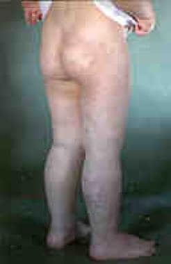 Klippel-Trenaunay syndrome in a young person. Note the port-wine stain extending to the buttocks. These lesions can be associated with venous malformations involving the rectum and bladder.
