Background
Cutaneous lupus erythematosus (CLE) can be divided into three main subtypes: acute, subacute, and chronic, all of which demonstrate photosensitivity.
Acute cutaneous lupus erythematosus (ACLE) most commonly presents as symmetric erythema overlying the malar cheeks and nasal bridge with sparing of the nasolabial folds (butterfly rash). However, it can also present as a diffuse morbilliform eruption with erythema and edema of the hands, with prominent sparing of the joints.
Subacute cutaneous lupus erythematosus (SCLE) characteristically presents as annular or psoriasiform plaques in a photodistribution.
Chronic cutaneous lupus erythematosus (CCLE) can be further divided into three main types: discoid lupus erythematosus (DLE), tumid lupus, and lupus panniculitis. Tumid lupus typically presents with juicy papules and plaques that lack scale and heal without scarring, whereas lupus panniculitis involves the subcutaneous tissue, leading to painful subcutaneous nodules that heal with depression and atrophy.
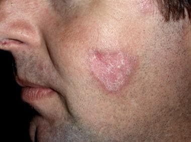
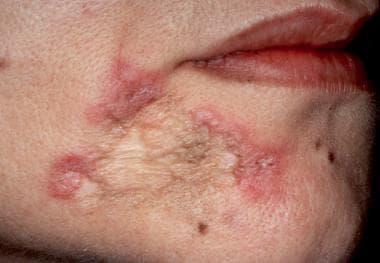
DLE classically presents with erythematous-to-violaceous, scaly plaques with prominent follicular plugging that often results in scarring and atrophy (see the images below). DLE may occur in the absence of systemic disease, or it may occur in association with systemic lupus erythematosus (SLE). DLE is the most common subtype of CLE. [1]
The risk of progression to SLE in patients with DLE has been demonstrated to be higher than previously reported.
Etiology
Lupus erythematosus is a polygenic autoimmune disease linked to various HLA subtypes, immune signaling, and environmental factors, which ultimately leads to autoantibody production and T-cell dysfunction. Although the exact etiology of discoid lupus erythematosus (DLE) is not well understood, genetic polymorphisms, mutations, and risk alleles have been identified in different populations of CLE patients, with most of them associated with innate and adaptive immunity pathways. Genes that act in apoptosis, leukocyte migration, type I interferon (IFN) pathway, complement cascade, antigen presentation, and antibody production are among the most frequently affected, including ITGAM, which has been found to portend high risk for DLE. It has also been determined that immunoglobulins play a role in the development of CLE, with immunoglobulin G (IgG), specifically, being responsible for the majority of immune complexes in DLE.
DLE likely occurs in genetically predisposed individuals, but the exact genetic connection has not been determined. In CLE, ultraviolet (UV) light exposure and/or stress are thought to trigger a cascade activating self-reactive B and T lymphocytes, which ultimately activates the innate and adaptive immune system and leads to tissue inflammation. [9]
Emerging research has shown a role for interferons, particularly IFN-1, and JAK-STAT (Janus kinase/signal transducers and activators of transcription) signaling in DLE, postulating the potential for treatment with JAK inhibitors. [10] Additionally, fibrosis in DLE has been linked to transforming growth factor (TGF)–β signaling, providing another molecular target for therapy. [11] The overexpression of two microRNAs (miR-31 and miR-485-p) has also been reported to contribute to DLE pathogenesis. [12]
Epidemiology
Worldwide, the prevalence of systemic lupus erythematosus (SLE) ranges from 17-48 cases per 100,000 population. The highest prevalence of SLE occurs in persons aged 40-60 years, with SLE onset most often occurring in patients in their 20s and 30s. SLE is approximately 8 times more common in women than in men. [13]
In Europe and the United States, the incidence of isolated cutaneous lupus erythematosus (CLE) has ranged from 4 to 4.3 cases per 100,000 population, slightly higher than the incidence of SLE (approximately 3 cases per 100,000 population). [2, 13, 14] In an epidemiological study from Olmstead County, Minnesota, Jarukitsopa et al demonstrated that the incidence of CLE rises steadily with age, peaking at age 60-69 years. [14]
Discoid lupus erythematosus (DLE) is responsible for 50-85% of cases of CLE and occurs 2-3 times more frequently in women than in men. DLE has also been reported to have a higher incidence in African Americans than in Whites. Although DLE may occur at any age, it most often develops in persons aged 20-40 years.
A limited number of population-based studies have estimated the incidence of DLE in the absence of SLE (“primary” DLE). [15, 16] In an epidemiological study using the Georgia Lupus Registry, Drenkard et al reported an overall age-adjusted DLE incidence of 3.7 cases per 100,000 person-years among Georgia residents. [15]
Prognosis
Although the prognosis of patients with discoid lupus erythematosus (DLE) is favorable regarding mortality, morbidity can be considerable. Patients may experience pain or burning of their lesions, or occasionally pruritus. Many patients with DLE experience disfigurement from the scars or atrophy that can develop. Scarring alopecia is particularly disturbing for patients. Prompt treatment of early lesions may help prevent or lessen the severity of scarring and atrophy.
DLE can negatively impact patient quality of life. [17] In fact, a study demonstrated that over one third of patients with cutaneous lupus met criteria for depression or anxiety with need for psychiatric intervention. [18] Furthermore, a significant correlation has been found between skin disease activity in cutaneous lupus and quality of life. [19]
Exacerbation is common with increased sun exposure, particularly in the spring and summer. Serious systemic disease is rare, but when it occurs, patients may develop life-altering sequelae. Malignant degeneration within DLE lesions is uncommon, but may occur. Hence, prompt biopsy of suggestive lesions developing within chronic DLE lesions is warranted. [20, 21]
Patient Education
Instruct patients in sun-avoidance techniques and the proper use of sunscreens, wide-brimmed hats, and protective clothing. Advise patients to quit smoking. Discuss the possibility of systemic involvement with patients.

-
Discoid lupus erythematosus on the face.
-
Chronic scarred lesion of discoid lupus erythematosus.
-
Lesions of discoid lupus erythematosus in the conchal bowl demonstrate patulous follicles with follicular plugging.
-
Palmar lesions of discoid lupus erythematosus.
-
Scarring alopecia of discoid lupus erythematosus.
-
Widespread scarring alopecia.
-
Hypertrophic lesions of chronic cutaneous lupus erythematosus on the dorsal hands. Characteristic lesions were observed elsewhere.
Tables

What would you like to print?
- Ultraprocessed Foods Increase Risk for Systemic Lupus Erythematosus in Women
- Rituximab’s Role in Systemic Lupus Erythematosus Set for Further Randomized Controlled Trials
- Does Cutaneous Lupus Erythematosus Raise Risk for Atherosclerotic Cardiovascular Disease?
-
 Lupus and Lifestyle: What Patients Can Control
-
In Lupus, How to Spot Hidden Heart Risk
-
Lupus by the Numbers: What the Data Reveals


 Identifying Lesions on Skin of Color