Practice Essentials
Onycholysis is characterized by a spontaneous separation of the nail plate starting at the distal free margin and progressing proximally. (See the images below.) In onycholysis, the nail plate is separated from the underlying and/or lateral supporting structures. Less often, separation of the nail plate begins at the proximal nail and extends to the free edge, which is seen most often in psoriasis of the nails (termed onychomadesis). Rare cases of onycholysis are confined to the nail's lateral borders.
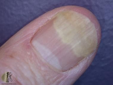 Thumb onycholysis. Courtesy of DermNet New Zealand (https://www.dermnetnz.org/assets/Uploads/hair-nails-sweat/olysis1.jpg).
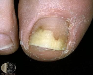 Great toe onycholysis. Courtesy of Professor Raimo Suhonen and DermNet New Zealand (https://www.dermnetnz.org/assets/Uploads/hair-nails-sweat/s/olysis7.jpg).
Signs and symptoms
Nails are smooth, firm, and without inflammatory reaction.
Discoloration under the nail may occur as a result of secondary infection.
See Presentation for more detail.
Diagnosis
Laboratory studies may include the following:
-
Mycologic studies to exclude onychomycosis
-
Nail biopsy if mycologic studies do not yield a positive result
See Workup for more detail.
Management
Treatments may include topical or intralesional corticosteroids, pulsed-dye laser, or psoralen plus ultraviolet A (PUVA) light.
Severe cases of onycholysis that are left untreated may result in nail bed scarring.
See Treatment and Medication for more detail.
Pathophysiology
Nails with onycholysis usually are smooth, firm, and without inflammatory reaction. Onycholysis is not a disease of the nail matrix, but nail discoloration may appear underneath the nail as a result of secondary infection. When onycholysis occurs, a coexistent yeast infection is suggested. Treating primary and secondary factors that exacerbate onycholysis is important. Left untreated, severe cases of onycholysis may result in nail bed scarring.
Etiology of Onycholysis
Endogenous, exogenous, hereditary, and idiopathic factors can cause onycholysis. Contact irritants, trauma, and moisture are the most common causes of onycholysis, but other associations exist.
Endogenous factors in onycholysis
Systemic diseases and states in onycholysis are as follows:
-
Amyloid and multiple myeloma
-
Anemia (iron deficient)
-
Bronchiectasis
-
Diabetes mellitus
-
Erythropoietic porphyria
-
Histiocytosis X
-
Hyperthyroidism [1]
-
Hypothyroidism
-
Ischemia (peripheral, impaired circulation)
-
Leprosy
-
Lupus erythematosus
-
Neuritis
-
Pellagra
-
Pemphigus vulgaris
-
Pleural effusion
-
Porphyria cutanea tarda
-
Pregnancy
-
Psoriatic arthritis [2]
-
Reiter syndrome
-
Sarcoidosis
-
Scleroderma
-
Shell nail syndrome
-
Syphilis
-
Yellow nail syndrome
Dermatologic diseases in onycholysis are as follows:
-
Pachonychia congenita
-
Congenital ectodermal defect
-
Pemphigus vegetans
-
Congenital abnormalities of the nail
-
Neoplastic disorders in onycholysis are as follows:
-
Squamous cell carcinoma (of nail bed)
-
Carcinoma (lung)
Exogenous factors in onycholysis
Nonmicrobial factors in onycholysis (may be encountered at the job site, ie, as occupational onycholysis) are as follows:
-
Mechanical - Mechanical force (trauma), repetitive minor trauma, or maceration
-
Chemical - Allergic or irritant contact dermatitis from various nail cosmetics (methyl methacrylate monomer, formaldehyde 1-2%, nail base coat/hardeners, polymerized 2-ethylcyanoacrylate adhesive used in artificial nails, nail lacquer), [3] gasoline, paint removers, dicyanodiamide, thioglycolate, solvents, and hydroxylamine sulphate in color developer
-
Chemical - Irritant contact dermatitis from prolonged immersion of nails in water, sugar onycholysis in confectioners/bakers, and exposure to highly destructive toxins (eg, hydrofluoric acid)
Biologic/microbial factors are as follows:
-
Dermatophytosis (ie, Trichophyton rubrum, Trichophyton mentagrophytes infection)
-
Yeast (Candida infection)
-
Bacteria (Pseudomonas infection [4] )
-
Virus (herpes simplex infection)
Drug associations with onycholysis
Photo-induced associations, with medication and subsequent exposure to sunlight, are as follows:
-
Tetracycline and its derivatives
-
Psoralens
-
Fluoroquinolones
-
Chloramphenicol
-
Benoxaprofen
-
Chlorpromazine
-
Chlortetracycline
-
Demethylchlortetracycline
-
Minocycline [7]
-
Oral contraceptives
-
5-Methoxypsoralen (Psoraderm 5)
-
Aminolevulinic acid (from phototherapy) [8]
-
Olanzapine [9]
-
Aripiprazole [9]
-
Griseofulvin [10]
Photo-induced associations with onycholysis, without medication and exposure to sunlight, are as follows:
-
Spontaneous photo-onycholysis [11]
-
Bullous photo-onycholysis (during pseudoporphyria resulting from hemodialysis)
-
Non–photo-induced associations with onycholysis [12] are as follows:
-
Doxorubicin
-
Mitoxantrone
-
Captopril
-
Bleomycin
-
Retinoids
-
Tetracycline
-
Etoposide
-
Hydroxylamine
Other factors in onycholysis
These include the following:
-
Congenital onycholysis
-
Hereditary partial onycholysis
-
Idiopathic acquired onycholysis
-
Hereditary distal onycholysis [18]
-
Foreign body implantation
Epidemiology
The worldwide incidence of onycholysis is unknown.
Race-, sex-, and age-related demographics
Distribution of onycholysis by race is unknown; however, onycholysis has been observed in persons of all races.
Individuals of either sex can have onycholysis; however, studies demonstrate an overwhelmingly female predilection.
People of any age can present with onycholysis, although it primarily is a disease of adulthood.
Patient Education
Educate patients with onycholysis about avoiding possible contact irritants, trauma, and moisture.
In addition, recommend that patients take the following measures:
-
Keep nail beds dry
-
Keep nails short; clip affected portions
-
Wear light cotton gloves under vinyl gloves for wet work
For patient education resources, see the Skin Conditions & Beauty Center and the patient education articles Nail Psoriasis and Onychomycosis.
-
Thumb onycholysis. Courtesy of DermNet New Zealand (https://www.dermnetnz.org/assets/Uploads/hair-nails-sweat/olysis1.jpg).
-
Great toe onycholysis. Courtesy of Professor Raimo Suhonen and DermNet New Zealand (https://www.dermnetnz.org/assets/Uploads/hair-nails-sweat/s/olysis7.jpg).
