Background
Phytophotodermatitis (PPD) is a cutaneous phototoxic inflammatory eruption resulting from contact with light-sensitizing botanical substances (also known as furanocoumarins) and long-wave (320-400 nm) ultraviolet A (UVA) radiation. [1] Furanocoumarins (or furocoumarins), present in some plants (eg, parsley, celery, carrots, and limes), react with UVA, [2, 3] and this reaction leads to the formation of eruptions on the skin. [4]
The eruption usually begins approximately 24 hours after exposure and peaks at 48-72 hours. [5] The occurrence of the rash requires exposure to both furanocoumarins and sunlight, and it is a direct phototoxic reaction that is entirely independent of the immune system. [6] The phototoxic result may be intensified by wet skin, sweating, and heat. The onset of the rash may be delayed and may not occur immediately after exposure to all of the elements. [7] Once the rash does occur, it may take weeks to resolve. [7]
PPD typically manifests as a burning erythema that may subsequently blister (see the image below). Postinflammatory hyperpigmentation lasting weeks to months may ensue. [8] In some patients, the preceding inflammatory reaction may be mild and may go unrecognized; in such cases, patients present with only pigmentary changes. Pain may be associated with the blister, resulting from necrosis of the affected epidermis. [6] The rash typically is nonpruritic; if pruritus occurs, further investigation into alternative diagnoses should be considered. [2] (See also Berloque Dermatitis and Drug-Induced Photosensitivity.)
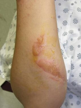 26-year-old female airline flight attendant exposed to lime while serving drinks en route to Caribbean. During Caribbean layover, she had significant sun exposure. Combination of lime juice and sun exposure led to drip-pattern blister formation on dorsal forearm consistent with phytophotodermatitis. Image clearly depicts potential severity of phytophotodermatitis with extensive blister formation.
Pathophysiology
Cutaneous inflammation produced by plants can be separated into the following four groups on the basis of their specific mechanism of action:
-
Urticarial dermatitis
-
Phototoxic dermatitis
PPD is a phototoxic reaction that is entirely independent of the immune system. In other words, it can occur in any individual, and prior sensitization or an intact immune system is not required. For PPD to develop, temporal exposure to both a photosensitizing substance (eg, a psoralen) and UV radiation. Furanocoumarins are photosensitizing chemical components produced by certain plants; they consist of psoralens, 5-methoxypsoralens, 8-methoxypsoralens, angelicin, bergaptol, and xanthotal.
The natural sunlight emission spectrum reaching the earth ranges from approximately 270 nm to 5000 nm. This electromagnetic radiation consists of photons, and there is a reciprocal relation between the wavelength and the energy of the photons. Only light that is absorbed into the skin can cause a photochemical reaction.
Within the light spectra, UVA (320-400 nm) produces PPD most efficiently, with peak activity at 335 nm, and is responsible for the vast majority of photoreactions resulting in PPD. When a photon with the appropriate wavelength strikes a furanocoumarin, the energy is absorbed, raising this chemical to a triple excited state from the ground state. Upon return to the ground state, energy is released in the form of heat, fluorescence, or phosphorescence, and a photoproduct may form.
Two distinct photochemical reactions have been described in PPD, each occurring independently of the other:
-
Type I reaction occurs in the absence of oxygen
-
Type II reaction occurs in the presence of oxygen
These photochemical reactions damage cell membranes and DNA and result in DNA interstrand crosslinking between the psoralen furan ring and the thymines or the cytosines of DNA.
During the type I oxygen-independent reaction, the RNA and nuclear DNA become fastened to the exposed ultraviolet-activated furocoumarins. Likewise, the type II oxygen-dependent reaction results in cell membrane damage and edema from activated furocoumarins. This results in activation of arachidonic acid metabolic pathways and in cell death (sunburn cells and apoptotic keratinocytes). Clinically, erythema, blistering, epidermal necrosis, and eventual epidermal desquamation occur.
A postinflammatory pigment alteration may follow the acute phase of this phototoxic reaction. This alteration occurs primarily via the following two mechanisms:
-
First, melanin, which is normally found in the epidermis, "falls" into the dermis and is ingested by melanophages
-
Second, an increased number of functional melanocytes and melanosomes are distributed in the epidermis following PPD and also account for the hyperpigmentation; this hyperpigmentation may serve as a protective mechanism against further UV injury; clinically, it corresponds with irregular hyperpigmentation (or occasionally hypopigmentation resulting in dyschromia) seen as the end stage of the phototoxic reaction
Etiology
The plants that most commonly cause PPD come from the Umbelliferae (Apiaceae) family. (See the images below.)
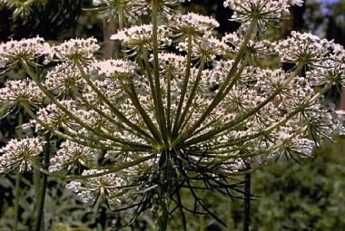 Queen Anne's lace, from Umbelliferae (Apiaceae) family, is well known to produce furanocoumarin-induced phototoxic eruption.
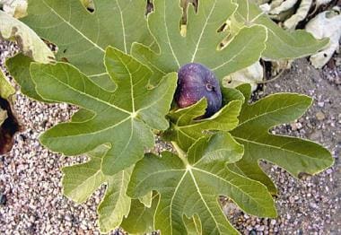 Common fig (ficus) contains furanocoumarins and should be considered among potential offending agents that cause phytophotodermatitis.
PPD is most commonly caused by ingestion of or topical exposure to psoralens (furanocoumarins). [9] Psoralens have been isolated from at least four different plant families: Umbelliferae (Apiaceae), [10] Rutaceae, [11, 12] Moraceae, and Leguminosae (Fabaceae). (See Table 1 below.)
Table 1. Common Causes of Phytophotodermatitis (Open Table in a new window)
Family |
Genus |
Species |
Common Names |
Main Compounds |
Umbelliferae (Apiaceae) |
Ammi |
majus |
Queen Anne's lace, bishop's weed |
8-methoxypsoralen (8-MOP), 5-methoxypsoralen (5-MOP), imperatorin |
|
Heracleum |
maximum |
Cow parsnip [13] |
8-MOP, 5-MOP, imperatorin, phellopterin |
|
Heracleum |
mantegazzianum |
Giant hogweed, [14] cartwheel flower |
8-MOP, 5-MOP, imperatorin, phellopterin |
|
Pastinaca |
sativa |
Parsnip |
8-MOP, 5-MOP, imperatorin, isopimpinellin |
|
Apium |
graveolens |
Celery |
Psoralens, 8-MOP, 5-MOP |
Rutaceae |
Citrus |
bergamia |
Bergamot lime |
5-MOP |
|
Citrus |
maxima |
Zabon [15] |
5-MOP |
|
Dictamnus |
albus |
Gas plant, “burning bush of Moses” |
8-MOP, 5-MOP |
Moraceae |
Ficus |
carica |
Psoralens, 5-MOP |
|
Leguminosae (Fabaceae) |
Psoralea (Cullen) |
corylifolia (corylifolium) |
Babchi, bakuchi, [19] scurf pea |
Psoralens |
Chart modified from Plants and the Skin. 1993:70-71. [20]
Epidemiology
US and international statistics
The overall incidence of PPD is unknown, but it undoubtedly varies according to the population studied and is based on the risk of exposure to psoralen compounds. Because furanocoumarins are found in a wide range of wild and domestic plants, numerous different patient groups may become exposed. An example of an international greenery known to produce PPD is the fig tree, Ficus carica, which is often sought for the fruit it produces, as well as for analgesic folk medicine applications. Ficus pumila is native to China, Japan, and Taiwan but can be found worldwide.
Age-, sex-, and race-related demographics
PPD may occur at any age. It should be noted, however, that PPD occurring on a child may be mistaken for child abuse. One classic example is a handprint pattern seen on a child who has been touched by a parent cooking with lime juice; another is a linear drip pattern seen on the hands and arms of a child who has been eating ice pops made with real juice.
Both sexes may be affected.
Individuals of any racial group may be affected, but PPD is most easily recognized in lighter-skinned patients.
Prognosis
With identification and avoidance of the offending agent, the prognosis for patients with PPD is good. Most commonly, PPD is a localized cutaneous phenomenon that initially gives rise to a burning sensation, which may be followed acutely by erythema and blistering. Eventually, the affected sites may desquamate and show permanent hyperpigmentation or hypopigmentation. Scarring, however, is rare.
Patient Education
It is important to reassure patients that PPD is a self-limited problem that resolves with removal of the offending agent.
-
37-year-old White woman presented to clinic complaining of rash on medial part of right thigh and left arm that was acquired after clearing some weeds in her yard. Phototoxic combination of sunlight and psoralen-containing plant produced this bizarre linear vesicular eruption.
-
Closer clinical view of bizarre angulated vesicular streaks, which occurred after contact with plant and ultraviolet light exposure.
-
26-year-old female airline flight attendant exposed to lime while serving drinks en route to Caribbean. During Caribbean layover, she had significant sun exposure. Combination of lime juice and sun exposure led to drip-pattern blister formation on dorsal forearm consistent with phytophotodermatitis. Image clearly depicts potential severity of phytophotodermatitis with extensive blister formation.
-
Two-month follow-up picture of patient with drip-pattern blister formation on dorsal forearm illustrates potential postinflammatory pigmentation changes and scarring that may occur with severe blistering of phytophotodermatitis.
-
Close-up view of vesicular linear streaks with morphology suggestive of scattered foci of epidermal necrosis.
-
Queen Anne's lace, from Umbelliferae (Apiaceae) family, is well known to produce furanocoumarin-induced phototoxic eruption.
-
Common fig (ficus) contains furanocoumarins and should be considered among potential offending agents that cause phytophotodermatitis.
Tables
Family |
Genus |
Species |
Common Names |
Main Compounds |
Umbelliferae (Apiaceae) |
Ammi |
majus |
Queen Anne's lace, bishop's weed |
8-methoxypsoralen (8-MOP), 5-methoxypsoralen (5-MOP), imperatorin |
|
Heracleum |
maximum |
Cow parsnip [13] |
8-MOP, 5-MOP, imperatorin, phellopterin |
|
Heracleum |
mantegazzianum |
Giant hogweed, [14] cartwheel flower |
8-MOP, 5-MOP, imperatorin, phellopterin |
|
Pastinaca |
sativa |
Parsnip |
8-MOP, 5-MOP, imperatorin, isopimpinellin |
|
Apium |
graveolens |
Celery |
Psoralens, 8-MOP, 5-MOP |
Rutaceae |
Citrus |
bergamia |
Bergamot lime |
5-MOP |
|
Citrus |
maxima |
Zabon [15] |
5-MOP |
|
Dictamnus |
albus |
Gas plant, “burning bush of Moses” |
8-MOP, 5-MOP |
Moraceae |
Ficus |
carica |
Psoralens, 5-MOP |
|
Leguminosae (Fabaceae) |
Psoralea (Cullen) |
corylifolia (corylifolium) |
Babchi, bakuchi, [19] scurf pea |
Psoralens |

What would you like to print?
- Nemolizumab Effective in Atopic Dermatitis
- Allergic Contact Dermatitis: New Culprits
- Inflammation Now the Key Target for Seborrheic Dermatitis
-
 Lupus Rashes: What to Spot, What to Shield
-
What's New in Psoriasis? Dermasphere Podcasters Weigh In
-
American Academy of Dermatology (AAD) 2025 Annual Meeting