Continually Updated Clinical Reference
 
 
  All Sources     eMedicine     Medscape     Drug Reference     MEDLINE
 
eMedicine - Tinea Barbae : Article by

Quick Find
Authors & Editors
Introduction
Clinical
Differentials
Workup
Treatment
Medication
Follow-up
Miscellaneous
Multimedia
References

Related Articles
Acne Vulgaris

Actinomycosis

Candidiasis, Cutaneous

Contact Dermatitis, Allergic

Contact Dermatitis, Irritant

Folliculitis

Rosacea

Syphilis


Patient Education
Skin, Hair, and Nails Center

Ringworm on Body Overview

Ringworm on Body Causes

Ringworm on Body Symptoms

Ringworm on Body Treatment


Author: Jacek C Szepietowski, MD, PhD, Professor and Vice-Head, Department of Dermatology, Venereology and Allergology, Wroclaw Medical University, Poland

Coauthor(s): Robert A Schwartz, MD, MPH, Professor and Head of Dermatology, Professor of Medicine, Professor of Pediatrics, Professor of Pathology, Professor of Preventive Medicine and Community Health, UMDNJ-New Jersey Medical School

Editors: Franklin Flowers, MD, Chief, Division of Dermatology, Professor, Department of Medicine and Otolaryngology, University of Florida College of Medicine; Richard P Vinson, MD, Assistant Clinical Professor, Department of Dermatology, Texas Tech University School of Medicine; Consulting Staff, Mountain View Dermatology, PA; Paul Krusinski, MD, Director of Dermatology, Professor, Department of Internal Medicine, Fletcher Allen Health Care, University of Vermont; Joel M Gelfand, MD, MSCE, Medical Director, Clinical Studies Unit, Assistant Professor, Department of Dermatology, Associate Scholar, Center for Clinical Epidemiology and Biostatistics, University of Pennsylvania; William D James, MD, Paul R Gross Professor of Dermatology, University of Pennsylvania School of Medicine; Vice-Chair, Program Director, Department of Dermatology, University of Pennsylvania Health System

Author and Editor Disclosure

Synonyms and related keywords: ringworm of the beard, barber's itch, trichophytosis barbae, tinea sycosis, sycosis

Background

Tinea barbae is a superficial dermatophyte infection that is limited to the bearded areas of the face and neck and occurs almost exclusively in older adolescent and adult males. The clinical presentation of tinea barbae includes inflammatory, deep, kerionlike plaques and noninflammatory superficial patches resembling tinea corporis or bacterial folliculitis.

Pathophysiology

Tinea barbae is caused by the keratinophilic fungi (dermatophytes) that are responsible for most superficial fungal skin infections. They infect the stratum corneum of the epidermis, hair, and nails. Several enzymes, including keratinases, are released by dermatophytes, which help them invade the epidermis. The mechanism that causes tinea barbae is similar to that of tinea capitis. In both diseases, hair and hair follicles are invaded by fungi, producing an inflammatory response. Tinea barbae is caused by both zoophilic and anthropophilic dermatophytes.

Infection caused by zoophilic dermatophytes usually is of greater severity than that produced by anthropophilic organisms. Thus, zoophilic dermatophytes are the primary cause of inflammatory kerionlike plaques, which most likely result from a more intense host reaction. Recently, kerion formation has been described as resulting from Trichophyton rubrum infection. T rubrum, an anthropophilic dermatophyte, can invade hair shafts and deeper tissues (although rarely), resulting in an inflammatory reaction. Usually, infection involving hair is more severe; therefore, tinea barbae caused by anthropophilic dermatophytes often has a more severe course than tinea corporis caused by the same pathogen.

The formation of kerion is postulated by 2 theories. The first theory suggests that it results from diffusion of metabolites and/or toxins from the fungus; however, kerion formation most likely results from an immunologic response to dermatophyte antigens.

Frequency

United States

Tinea barbae is uncommon in the United States.

International

Currently, tinea barbae is infrequent around the world. As with other dermatophytoses, it is more common in countries in which weather is characterized by high temperatures and humidity. Tinea barbae was observed more frequently in the past before single-use razors became available, and infection frequently was transmitted by barbers who used unsanitary razors. Therefore, it is not surprising that tinea barbae once was termed barber's itch. Now that habits and equipment have changed, this source of infection has been all but eliminated. Currently, tinea barbae is more common among rural inhabitants, and zoophilic dermatophytes constitute its primary pathogens.

Mortality/Morbidity

Permanent alopecia and scarring frequently follow spontaneous resolution of inflammatory plaques and nodules. In superficial chronic tinea barbae, alopecia may occur in the center of the lesions; however, this is not common.

Sex

Men are affected almost exclusively because the disease involves the bearded areas of the face and neck. Involvement of the same areas in healthy women and children is classified as tinea faciei.

Age

Hair appears on the face at puberty; therefore, tinea barbae may occur almost exclusively in older adolescent and adult males.


History

  • Infection often begins on the chin or neck, but in severely affected patients, it may cover the entire bearded area of the face and neck, occasionally resulting in indurated verrucous plaques or nodules.
  • Tinea barbae may be asymptomatic; however, mild pruritus is characteristic.
  • Spontaneous resolution may occur, especially in inflammatory tinea barbae.
  • Lupoid sycosis, a deep form of tinea barbae, is so named because it may resemble lupus vulgaris.

Physical

Clinical manifestations of tinea barbae relate to the causative pathogen. Two clinical varieties of the disease are identified as follows:

  • Inflammatory deeper tinea barbae is caused primarily by zoophilic dermatophytes. This variety, termed a kerion, is the most common clinical presentation. Most patients show solitary plaques or nodules; however, multiple plaques are relatively common. Usually localized on the chin, cheeks, or neck, involvement of the upper lip is rare. The characteristic lesion is an inflammatory reddish nodule with pustules and draining sinuses on the surface. Hairs are loose or broken, and depilation is easy and painless. Pus-filled whitish masses involve the hair root and follicle. Over time, the surface of the indurated nodule is covered by exudate and crust. This variety of tinea barbae usually is associated with generalized symptoms, such as regional lymphadenopathy, malaise, and fever.
  • Noninflammatory superficial tinea barbae is caused by anthropophilic dermatophytes. This variety of barbae is less common and resembles common tinea corporis or bacterial folliculitis (sycosiform variety). Typically, erythematous patches show an active border composed of papules, vesicles, and/or crusts. Hairs are broken next to the skin, or they plug the hair follicle. In the sycosiform variety, small follicular pustules are observed. Hairs are broken or loose. This variety represents a chronic variant of tinea barbae.

Causes

Tinea barbae is caused by several dermatophytes, including zoophilic and anthropophilic organisms; however, zoophilic dermatophyte infection occurs more commonly. Frequently, animals (eg, cattle, horses, cats, dogs) constitute the source of infection. Trichophyton species are most common, thus the term trichophytosis barbae also is used. Among zoophilic dermatophytes, Trichophyton mentagrophytes var granulosum and Trichophyton verrucosum are the most common causative agents. Microsporum canis and Trichophyton mentagrophytes var erinacei may cause tinea barbae but are rare.

T rubrum and Trichophyton violaceum are the most common anthropophilic dermatophytes responsible for tinea barbae; however, infections from Trichophyton megninii (endemic in Sardinia, Sicily, Portugal) and Trichophyton schoenleinii (endemic in Eurasia, Africa, Brazil) also may occur, especially in endemic regions. Infection of bearded skin by anthropophilic dermatophytes may be the result of autoinoculation from tinea pedis or onychomycosis.


Acne Vulgaris
Actinomycosis
Candidiasis, Cutaneous
Contact Dermatitis, Allergic
Contact Dermatitis, Irritant
Folliculitis
Rosacea
Syphilis

Other Problems to be Considered

Bacterial folliculitis
Candidal folliculitis
Carbuncles
Pustular syphilis
Nodule-cystic acne
Iododerma
Bromoderma
Lupoid sycosis may resemble lupus vulgaris (cutaneous tuberculosis)


Other Tests

  • Mycologic features form the basis of diagnosis. Procedures include direct microscopy, culture, and a Wood lamp examination showing fluorescence when M canis infection causes tinea barbae.
  • Specimen selection and collection is important. Usually, the material consists of infected hairs (depilated with forceps) and pustular masses. In the superficial variety, which resembles tinea corporis, collect scrapings from the border of the lesion where the inflammatory reaction is more severe. Taking material from the lesion's border increases the possibility of detecting fungi on direct microscopy and culture. Send the specimen for culture, or examine it for fungus.
  • Direct microscopic examination is performed rapidly and easily; however, it requires experience. Place the material on a slide and add a solution of 10-20% potassium hydroxide, with or without dimethyl sulfoxide. This solution provides visualization of fungal elements. Gently warm the slide, especially if no dimethyl sulfoxide is added. Some recommend using special stains, such as chlorazol black E stain or Parker blue-black ink. Direct microscopy usually shows hyphae and/or arthroconidia. Wait for some minutes before evaluating the preparation under microscopy, or reexamine the slide after one-half hour, since fungal elements may be difficult to see just after addition of potassium hydroxide solution.
  • Culture identifies the causative fungus and usually is performed on Sabouraud agar with the addition of cycloheximide and chloramphenicol. These 2 substances inhibit bacterial and other fungal growth to obtain pure dermatophyte colonies. Cultures take approximately 3 weeks to become positive, and final fungal identification is based primarily on morphology and microscopy of the colonies. Occasionally, additional tests are required. Special media for rapid dermatophyte identification that include a color indicator currently are available. In the presence of dermatophytes, the color changes from yellow to bright red.

Histologic Findings

Biopsy specimens occasionally may be required to diagnose tinea barbae. Biopsy shows folliculitis and perifolliculitis with a mixed cellular infiltrate and spongiotic alterations within the follicular epithelium. Lymphocytes or neutrophils also may be evident within follicular epithelium. Neutrophils also may be seen within follicular keratin as microabscesses.

Since fungal elements often are difficult to visualize with hematoxylin and eosin stain, periodic acid-Schiff stain is recommended. Arthroconidia and/or hyphae may be evident within the hair shaft and in the hair follicle. An inflammatory infiltrate is present in the dermis, which in chronic lesions may contain giant cells.


Medical Care

Since hairs are infected in tinea barbae, the therapeutic procedure is similar to that of tinea capitis. Shaving or hair depilation is recommended with warm compresses to remove crusts and debris. Topical formulations with antifungal compounds (eg, shampoo, lotion, cream) can be applied, but tinea barbae requires oral antifungal therapy. Details of therapeutic modalities for tinea barbae are found in Medication, below.

Griseofulvin remains in wide use. Newer antifungals, especially terbinafine, also can be used. At the author's institution, experience with terbinafine in tinea barbae has been excellent, with all cures achieved within 4 weeks of initiating therapy. Itraconazole can be recommended as one-pulse therapy at a dosage of 400 mg/d divided into 2 doses for 1 week. Some prefer to use a second pulse after 3 weeks. Itraconazole also may be used continuously for 4 weeks at a dose of 200 mg/d. Fluconazole treatment is not well documented, but data from tinea capitis studies indicate that a dose of 150 mg once per week for up to 6 weeks also may be effective. All of these new antifungal drugs generally are well tolerated.


The goals of pharmacotherapy are to eradicate the infection, to reduce morbidity, and to prevent complications.

Drug Category: Antifungals

Mechanism of action usually involves inhibiting pathways (enzymes, substrates, transport) necessary for sterol/cell membrane synthesis or altering the permeability of the cell membrane (polyenes) of the fungal cell.

Drug NameGriseofulvin (Fulvicin P/G, Grifulvin V)
DescriptionFungistatic activity. Fungal cell division is impaired by interfering with microtubule. Binds to keratin precursor cells. Keratin is gradually replaced by noninfected tissue, which is highly resistant to fungal invasions.
Adult Dose500 mg microsize (330-375 mg ultramicrosize) PO qd or bid, continue for 2 wk after clinical lesions resolve
Pediatric DoseSuggested dose: 20 mg microsize/kg/d (5 mg/lb/d) PO or 7.3 mg ultramicrosize/kg/d (3.3 mg/lb/d) PO
ContraindicationsDocumented hypersensitivity; hepatic injury
InteractionsReduced effects of cyclosporine, salicylates, and warfarin (decreased hypoprothrombinemic activity); avoid alcohol use, since disulfiramlike reaction may occur; intense UV light exposure may result in phototoxic reaction; contraceptives may lose their effectiveness
PregnancyC - Safety for use during pregnancy has not been established.
PrecautionsBetter absorption when taken with fatty food; adverse effects may include abdominal pain, nausea, diarrhea, headache, and rarely, Stevens-Johnson syndrome and photodermatitis; 20% of patients experience adverse effects with griseofulvin; in prolonged therapy, observe patients closely; monitor renal, hepatic, and hematopoietic function regularly

Drug NameTerbinafine (Lamisil)
DescriptionMember of allylamine family and a fungicidal agent that inhibits ergosterol synthesis via squalene epoxidase, which results in decreased ergosterol level and increased concentration of squalene; leads to cell death.
Use medication until symptoms significantly improve.
Adult Dose250 mg/d PO for 4 wk
Pediatric DoseNot established
ContraindicationsDocumented hypersensitivity
InteractionsCimetidine, rifampin, and cyclosporine rarely may interact; toxicity may increase with coadministration of rifampin and cimetidine; coadministration with cyclosporine may decrease cyclosporine level
PregnancyB - Usually safe but benefits must outweigh the risks.
PrecautionsGenerally well tolerated; adverse effects are primarily GI, including hepatobiliary dysfunction; transient loss of sense of taste is rare but specific adverse effect; blood dyscrasias, Stevens-Johnson syndrome, and ocular disturbances are described; minimally inhibits cytochrome P450 enzyme pathway

Drug NameItraconazole (Sporanox)
DescriptionFungistatic activity. Synthetic triazole antifungal agent that slows fungal cell growth by inhibiting cytochrome P-450-dependent synthesis of ergosterol, a vital component of fungal cell membranes.
Adult Dose400 mg/d PO divided bid for 1 wk or 200 mg/d PO divided bid for 4 wk; not to exceed 400 mg/d; increase in 100-mg increments if no improvement
Pediatric DoseNot established
ContraindicationsDocumented hypersensitivity; coadministration with lovastatin, simvastatin, cisapride, triazolam, or midazolam; coadministration with cisapride may cause arrhythmia; coadministration with triazolam and midazolam may increase plasma levels; rhabdomyolysis may occur with coadministration of HMG-CoA reductase inhibitors
InteractionsFrequent: increased level of cyclosporine, tacrolimus, and digoxin may occur after coadministration; phenytoin and rifampin may decrease effect; amlodipine and nifedipine taken concurrently may cause edema; hypoglycemia was observed after coadministration with sulfonylureas; avoid alcohol use, since disulfiramlike reactions may occur; antacids may reduce absorption; coadministration with terfenadine (recalled from US market), astemizole (recalled from US market), lovastatin, simvastatin, cisapride, triazolam, and midazolam; serious cardiovascular events, including death, were reported in patients treated with itraconazole and H1-receptor inhibitors (terfenadine, astemizole); coadministration with cisapride may cause arrhythmia; coadministration with triazolam and midazolam may increase their plasma levels; rhabdomyolysis may occur with coadministration of HMG-CoA reductase inhibitors (lovastatin, simvastatin)
PregnancyC - Safety for use during pregnancy has not been established.
PrecautionsGenerally well tolerated, but approximately 12% of patients may have adverse effects including GI abnormalities; hepatobiliary dysfunction has been observed; recommended to be taken with fatty food, which increases absorption from alimentary tract

Drug NameFluconazole (Diflucan)
DescriptionFungistatic activity. Synthetic oral antifungal (broad-spectrum bistriazole) that selectively inhibits fungal cytochrome P-450 and sterol C-14 alpha-demethylation, which prevents conversion of lanosterol to ergosterol, thereby disrupting cellular membranes.
Adult Dose150 mg PO qwk for up to 6 wk
Pediatric DoseNot established
ContraindicationsDocumented hypersensitivity
InteractionsLevels may increase with hydrochlorothiazide; fluconazole levels may decrease with chronic coadministration of rifampin; coadministration of fluconazole may decrease phenytoin clearance; may increase concentrations of theophylline, tolbutamide, glyburide, and glipizide; effects of anticoagulants may increase with fluconazole coadministration; increases in cyclosporine concentrations may occur when administered concurrently
PregnancyC - Safety for use during pregnancy has not been established.
PrecautionsGenerally well tolerated, but approximately 12% of patients show adverse effects, which include nausea, vomiting, abdominal discomfort, and hepatotoxicity; adjust dose for renal insufficiency; monitor closely if rashes develop and discontinue drug if lesions progress; may cause clinical hepatitis, cholestasis and fulminant hepatic failure (including death), with underlying medical conditions (eg, AIDS or a malignancy) and while taking multiple concomitant medications; not recommended in breastfeeding


Deterrence/Prevention

  • Eliminating the source of infection is of great importance.
  • If farm workers become infected, examine all animals for the presence of fungal skin lesions.
  • Treatment of other fungal skin infections, such as tinea pedis or onychomycosis, may prevent the infection's spread by autoinoculation.

Prognosis

  • Prognosis usually is good.
  • Inflammatory lesions undergo spontaneous remission within a few months; however, if untreated, they leave scarring alopecia.
  • Noninflammatory lesions are more likely to be chronic and may not tend to resolve spontaneously.

Patient Education


Medical/Legal Pitfalls

  • Failure to recognize and adequately treat tinea barbae, since untreated severe forms of tinea barbae may result in scarring alopecia. A potential for litigation exists if permanent cosmetic disfigurement occurs that may have been prevented.


Media file 1:  
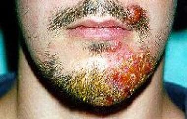
Inflammatory tinea barbae resulting from Trichophyton mentagrophytes var granulosum infection.
Click to see larger pictureClick to see detailView Full Size Image
Media type:  Photo

Media file 2:  Wax model of kerionlike tinea barbae. Courtesy of the Museum of the Department of Dermatology, University of Medicine, Wroclaw, Poland.
Click to see larger pictureClick to see detailView Full Size Image
Media type:  Photo


  • Baran W, Szepietowski JC, Schwartz RA. Tinea barbae. Acta Dermatoven APA. 2004;13:91-4. [Full Text].
  • Beswick SJ, Das S, Lawrence CM, Tan BB. Kerion formation due to Trichophyton rubrum. Br J Dermatol. Nov 1999;141(5):953-4. [Medline].
  • Bihari A, Simon G. Tinea barbae profunda. Borgyogyaszati Venerol Szemle (Budapest). 1998;74:283-6.
  • Bonifaz A, Ramírez-Tamayo T, Saúl A. Tinea barbae (tinea sycosis): experience with nine cases. J Dermatol. Dec 2003;30(12):898-903. [Medline].
  • Ceburkovas O, Schwartz RA, Janniger CK. Tinea capitis: current concepts. J Dermatol. Mar 2000;27(3):144-8. [Medline].
  • De Lacerda MH, Caldeira JB, Delfino JP, et al. [Sycosis of the beard (tinea barbae). Analysis of 42 cases]. Med Cutan Ibero Lat Am. 1981;9(3):161-78. [Medline].
  • Foti C, Diaferio A, Daddabbo M, Angelini G. Tinea barbae associated with erythema nodosum in immunocompetent man. J Eur Acad Dermatol Venereol. 2001;15:250-251. [Medline].
  • Glaser DA, Riordan AT. Images in clinical medicine. Tinea barbae: man and beast. N Engl J Med. Mar 12 1998;338(11):735. [Medline].
  • Gupta G, Burden AD, Roberts DT. Acute suppurative ringworm (kerion) caused by Trichophyton rubrum. Br J Dermatol. Feb 1999;140(2):369-70. [Medline].
  • Kapdagli H, Ozturk G, Dereli T, et al. Candida folliculitis mimicking tinea barbae. Int J Dermatol. Apr 1997;36(4):295-7. [Medline].
  • Kawada A, Aragane Y, Maeda A, et al. Tinea barbae due to Trichophyton rubrum with possible involvement of autoinoculation. Br J Dermatol. May 2000;142(5):1064-5. [Medline].
  • Kick G, Korting HC. Tinea barbae due to Trichophyton mentagrophytes related to persistent child infection. Mycoses. Nov 1998;41(9-10):439-41. [Medline].
  • Kiska DL, Cynamon MH. Photo quiz. Tinea barbae caused by Trichophyton verrucosum. Clin Infect Dis. Oct 1997;25(4):805, 871. [Medline].
  • Krajewska-Kulak E, Niczyporuk W, Lukaszuk C, et al. Difficulties in diagnosis and treating tinea in adults at the Department of Dermatology in Bialystok (Poland). Dermatol Nurs. 2003;15:527-533. [Medline].
  • Kurita M, Kishimoto S, Kibe Y, et al. Candida folliculitis mimicking tinea barbae. Acta Derm Venereol. Mar-Apr 2000;80(2):153-4. [Medline].
  • Kwasniewska J. Current antifungal agents in dermatology. Postepy Dermatol (Poznan). 1997;14:129-35.
  • Macura AB. Resistance to antimycotic drugs. Postepy Dermatol (Poznan). 1997;14:137-40.
  • Maeda M, Nakashima T, Satho M, et al. Tinea barbae due to Trichophyton verrucosum. Eur J Dermatol. 2002;12:272-274. [Medline].
  • Michalowski R, Kuczynska L. [Initial experiences with a natamycin-, neomycin-, and hydrocortisone- containing lotion in tinea barbae]. Mykosen. Mar 1984;27(3):153-6. [Medline].
  • Ratka P, Slusarczyk E, Sloboda T, Kusmierski W. [Case of tinea barbae profunda caused by Microsporum nanum]. Przegl Dermatol. Sep-Dec 1983;70(5-6):549-52. [Medline].
  • Soyer HP, Cerroni L. The significance of histopathology in the diagnosis of dermatomycoses. Acta Derm Venerol (Ljubljana). 1992;1:84-7.
  • Szepietowski J. Dermatomycoses and onychomycosis. Medycyna Rodzinna. 1999;2:29-35.
  • Szepietowski J. The contemporary therapy of superficial fungal infections. Terapia i Leki. 2000;28:18-23.
  • Szepietowski J. Dermatomycoses and onychomycosis. A practical guide. Medycyna Praktyczna, Krakow. 2001.
  • Szepietowski JC, Bielicka E, Maj J. Inflammatory tinea barbae due to Trichophyton rubrum infection - autoinnoculation from fingernail onychomycosis?. Case Rep Clin Pract Rev. 2002;3:254-6. [Full Text].
  • Tanuma H, Doi M, Nishiyama S, Katsuoka K. A case of tinea barbae successfully treated with terbinafine. Mycoses. Jan-Feb 1998;41(1-2):77-81. [Medline].
  • Trotha R, Gräser Y, Platt J. Tinea barbae caused by a zoophilic strain of Trichophyton interdigitale. Mycoses. Feb 2003;46(1-2):60-3. [Medline].

Tinea Barbae excerpt

Article Last Updated: Feb 27, 2007