Practice Essentials
Tinea corporis is a superficial dermatophyte infection characterized by either inflammatory or noninflammatory lesions on the glabrous skin (ie, skin regions other than the scalp, groin, palms, and soles). [1] Three anamorphic (asexual or imperfect) genera cause dermatophytoses: Trichophyton, Microsporum, and Epidermophyton. Dermatophytes may infect humans (anthropophilic) or nonhuman mammals (zoophilic), or they may reside primarily in the soil (geophilic). [2, 3]
Signs and symptoms
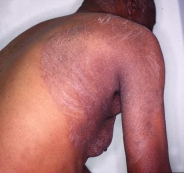
The lesion typically begins as an erythematous, scaly plaque that may rapidly worsen (see the image below). These scaly plaques typically have a raised border with peripheral scale. This scale is usually trailing scale, with the free edge of the scale pointed inward.
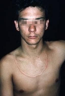
Following central resolution, the lesion may become annular in shape (see the image below).
The inflammation can cause scale, crust, papules, vesicles, and even bullae to develop, especially in the advancing border. Rarely, tinea corporis can present as purpuric macules. Infections due to zoophilic or geophilic dermatophytes may produce a more intense inflammatory response than those caused by anthropophilic microbes.
Patients who are immunocompromised or infected with the human immunodeficiency virus (HIV) often have atypical presentations, including deep abscesses or a disseminated skin infection.
Majocchi granuloma
This variant of tinea corporis is a fungal infection of the hair, hair follicles, and, often, surrounding dermis.
Pathophysiology
Dermatophytes preferentially inhabit the nonliving, cornified layers of the skin, hair, and nail, which are attractive for the warm, moist environment conducive to fungal proliferation. Fungi may release keratinases and other enzymes to invade deeper into the stratum corneum, though typically the depth of infection is limited to the epidermis and, at times, its appendages. They generally do not invade deeply, owing to nonspecific host defense mechanisms that can include the activation of serum inhibitory factor, complement, and polymorphonuclear leukocytes.
After the incubation period of 1-3 weeks, dermatophytes invade peripherally in a centrifugal pattern. In response to the infection, the active border has an increased epidermal cell proliferation with resultant scaling. This creates a partial defense by shedding the infected skin and leaving new, healthy skin central to the advancing lesion. Elimination of dermatophytes is achieved by cell-mediated immunity.
T rubrum is a common dermatophyte and, because of its cell wall, is resistant to eradication. This protective barrier contains mannan, which may inhibit cell-mediated immunity, hinder the proliferation of keratinocytes, and enhance the organism's resistance to the skin's natural defenses. [13]
Etiology
Tinea corporis can be caused by a variety of dermatophytes, though prevalence and patient history are very helpful in identifying the most likely organism. Internationally, the most commonly reported cause has been T rubrum.Trichophyton tonsurans, Trichophyton mentagrophytes, [7, 14] Trichophyton interdigitale, Trichophyton verrucosum, [15] Microsporum canis,Microsporum gypseum, [6] and Microsporum langeronii [16] are also known to produce infection. Tinea imbricata is caused by T concentricum.
The organism Trichophyton indotineae (previously classified as type VIII T mentagrophytes) is a pathogen of growing concern as a cause of tinea corporis and cruris. [17] It is often resistant to commonly used agents (eg, terbinafine).
Dermatophytoses may be acquired from different sources (eg, people, animals, or soil). Infected humans are the most common source of tinea corporis in the United States. Contact with contaminated household pets, farm animals, and fomites (eg, in hair brushes or towels) can spread infection. T verrucosum causes 98% of dermatophyte infections in cattle and is showing increasing prevalence of infection in human contacts. T mentagrophytes is spread by rabbits, guinea pigs, and small rodents. [14] Infection with M gypseum, a geophilic organism, can mimic tinea imbricata in presentation.
Because fungal arthroconidia can survive in the environment, outbreaks may recur.
Epidemiology
United States and international statistics
Dermatophyte infections are the most common fungal infections in the United States and among the most common sources of skin disease worldwide. Over the period from 2005 to 2014, these infections accounted for 4,981,444 outpatient visits in the United States, and in 2019, the direct medical cost of managing them was approximately $845 million. [18]
Tinea corporis is a common infection more often seen in typically hot, humid climates. Previous studies cited T rubrum as the source of nearly 50% of tinea corporis cases in the United States. [19] In a 2024 study using data from a major US commercial laboratory (Labcorp), Zarzeka et al reported 15,563 cases of tinea corporis for the period from March 1, 2019, to March 1, 2023 [20] ; the most common causative organism was T tonsurans, followed by T rubrum and then by yeasts (with Candida species accounting for the majority), nondermatophyte molds, and unspecified fungus.
T tonsurans is the dermatophyte that most commonly causes tinea capitis, and people with an anthropophilic tinea capitis infection are more likely to develop associated tinea corporis. Consequently, the prevalence of tinea corporis caused by T tonsurans has been increasing. [21, 20]
M canis has been associated with 14% of tinea corporis infections. Seyfarth et al reported a rare case of Microsporum fulvum skin infection (forearm), identified by ITS sequencing and mass spectrometry. [22]
A 5-year study (N = 2370) from Kuwait reported that fungal skin infections remained prevalent in that country, specifically in the capital area. [23] In patients with dermatophytes, six species were isolated: T mentagrophytes (39%), M canis (16%), T rubrum (10%), Epidermophyton floccosum (6.2%), Trichophyton violaceum (2.4%), and T verrucosum (0.4%). [23] There is evidence to suggest that the T mentagrophytes variant T interdigitale can be found on all body sites, not just the feet. [24]
Age- and sex-related demographics
Tinea corporis affects persons of all age groups, but prevalence is highest in preadolescents. Tinea corporis acquired from animals is more common in children. Tinea corporis secondary to tinea capitis typically occurs in children because tinea capitis is more common in this population.
Tinea corporis occurs in both men and women. Women of childbearing age are more likely to develop tinea corporis as a result of their greater frequency of contact with infected children.
Prognosis
For localized tinea corporis, the prognosis is excellent, with cure rates of 70-100% after treatment with topical azoles or allylamines or short-term or pulse systemic antifungals. Dermatophyte infections do not result in significant mortality, but they can greatly affect quality of life.

-
Annular plaque.
-
Large, erythematous, scaly plaque.
-
Tinea corporis. Courtesy of Wikimedia Commons (https://commons.wikimedia.org/wiki/File:Tinea_corporis.png).
-
Dermatophytosis or ringworm with mildly raised boarder and central clearing. Courtesy of Wikimedia Commons [James Heilman, MD] (https://commons.wikimedia.org/wiki/File:Yeartinfection.JPG).
Tables

What would you like to print?
- Could a Fungal Infection Cause a Future Pandemic?
- New Sexually Transmitted Fungal Infection Emerges in MSM
- Emerging Fungal Infections Demand All-Out Approach
-
 Immunoglobulin A Nephropathy (IgAN): 5 Differential Diagnoses to Know
-
Adele's Fungal Infection Has People Talking About Jock Itch
- Increased Hospitalizations Involving Fungal Infections During COVID-19 Pandemic, United States, January 2020–December 2021


