Practice Essentials
A systematic approach to the evaluation of alopecia allows for more effective management. Below is a step-by-step approach that can be used in scarring alopecia:
-
Initial classification of alopecia type
-
Applying scarring alopecia terminology based on clinical features
-
Using histology findings to further support/define the diagnosis
-
Providing an overall evaluation/diagnosis with treatment options
Initial classification
The initial determination of alopecia type usually begins with the establishment of alopecia as either a scarring (cicatricial) alopecia or nonscarring alopecia. Nonscarring alopecias tend to have preserved follicular ostia. No clinically visible inflammation is noted in most presentations, although histologic inflammation may be present. The most common nonscarring alopecias include alopecia areata and telogen effluvium.
Scarring alopecias have loss of follicular ostia, or atrophy. Clinical inflammation is frequently, but not always, present. Histologic inflammation may be present. Ultimately, histologic confirmation is the best method to confirm the presence of a fibrosing/scarring process with loss of hair follicles.
Many alopecia types are biphasic. For example, androgenetic alopecia eventually results in loss of ostia and thus may appear like a scarring alopecia. This article focuses on the alopecia types that are believed to be due to an inflammatory response with rapid secondary scarring if not controlled.
Scarring alopecia terminology based on clinical features
Once the patient is determined to have scarring alopecia, establishing and clarifying the diagnostic options and terminology is important to assist in confirming the diagnosis, initiating treatment, and suggesting a prognosis. Several manuscripts have examined and attempted to clarify the literature findings, [1, 2, 3] as summarized below.
Also important to note is that examination alone cannot be the final step; the literature is filled with examples of misdiagnosis and diagnostic mimics in alopecia. [4, 5, 6, 7] This includes patients presenting with nonscarring, noninflammatory alopecia that is later found to be scarring and inflammatory. [8] Even amongst the scarring alopecia presentation, it is sometimes debated as to the role of immune alteration versus pure external factors that cause the scarring alopecia. Much of the confusion is often based in the similarities of clinical presentation, which emphasizes again the importance of not basing diagnosis purely on clinical examination findings alone. [9]
Diagnoses in which lymphocytes predominate are as follows:
Lichen planopilaris
The diagnosis of lichen planopilaris (LPP) is confirmed by a combination of clinical and histologic features. The following subtypes are recognized [10] :
-
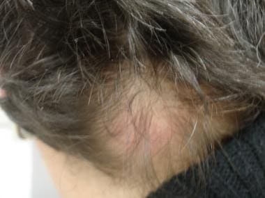
Frontal fibrosing alopecia (FFA): This is a potential clinical variant of lichen planopilaris, with similar histologic features. Frontal fibrosing alopecia has been associated with the postmenopausal state (ie, postmenopausal frontal fibrosing alopecia), although not all patients with this diagnosis are postmenopausal. Note the image below.
 End-stage scarring alopecia (ESSA) with prior history of itching and burning, along with a receding hairline. Started as the lichen planopilaris variant, frontal fibrosing alopecia. Used with permission, rights retained, courtesy of Rashid M. Rashid, MD, PhD, Morzak Research Collaborative.
-
Fibrosing alopecia in pattern distribution (FAPD): This also is a potential clinical variant of lichen planopilaris, with similar histologic features. Additionally, fibrosing alopecia in pattern distribution has some histologic/clinical overlap with androgenetic alopecia. Fibrosing alopecia in pattern distribution may also be an overlap of lichen planopilaris with androgenetic alopecia.
-
Graham-Little syndrome: This entity is also a possible overlapping condition, with features of lichen planopilaris and other alopecia types.

Central centrifugal cicatricial alopecia (CCCA)
CCCA is a diagnostic category adopted by the North American Hair Research Society to encompass terms such as hot comb alopecia, follicular degeneration syndrome, pseudopelade in African Americans, and central elliptical pseudopelade in whites. [10] Despite the many attempts to clarify and unify the terminology of central CCCA patterns of scarring alopecia, CCCA is not clearly a diagnostic entity in and of itself.
Pseudopelade
Pseudopelade is a term used to describe a clinically noninflammatory patchy alopecia. The most common condition to produce this appearance is lichen planopilaris. The term “idiopathic pseudopelade” refers to a distinct fibrosing alopecia characterized by a thin dermis, with dense eosinophilic dermal collagen and thick recoiled elastic fibers. The fibrous tracts are broad and may contain granulomas, but the surrounding elastic sheath is preserved.
Traction alopecia
As the name implies, traction alopecia is alopecia secondary to physical traction. Variant presentations exist with terminology that is optional for differentiating purposes. This includes alopecia linearis frontalis (ALF), more commonly known as marginal alopecia, and chignon alopecia, which is when a tight hair bun causes the traction changes.
Secondary systemic scarring alopecia
Secondary systemic scarring alopecia is a term used when the alopecia is a feature of a systemic disease (eg, scleroderma, discoid lupus erythematosus). Discoid lupus erythematosus is often a cause of alopecia. In the classic lupus classifications scheme, discoid lupus erythematosus is one of the chronic cutaneous lupus types (along with tumid lupus and lupus panniculitis).
Trichotillomania
Trichotillomania is a nonscarring psychiatric alopecia that can have a scarring clinical presentation. Trichotillosis is an alternative term that some physicians believe is more acceptable to patients. [11]
Chemotherapy alopecia
Chemotherapy alopecia is often thought of as nonscarring alopecia; however, reports of permanent alopecia suggest that hair stem cells can be permanently destroyed. Note the image below.
 Alopecia due to primary cutaneous follicular center cell lymphoma. Used with permission, rights retained, courtesy of Rashid M. Rashid, MD, PhD, Morzak Research Collaborative.
Alopecia mucinosa
Follicular mucinosis can exist by itself or can be a manifestation of mycosis fungoides.
Keratosis pilaris atrophicans
Keratosis pilaris atrophicans involves a spectrum of conditions that resemble keratosis pilaris, but result in hair loss.
Diagnoses in which neutrophils predominate are as follows:
-
Folliculitis decalvans: Folliculitis decalvans presents with recurrent crops of follicular pustules that result in permanent epilation. Staphylococci are often cultured from pustules, but this condition does not respond the standard antibiotic regimens. The best results are obtained by long-term use of topical corticosteroids together with an oral tetracycline.
-
Perifolliculitis capitis abscedens et suffodiens: Other terms used for perifolliculitis capitis abscedens et suffodiens include dissecting cellulitis and Hoffman disease. It resembles hidradenitis suppurativa and the 2 often occur together.
-
Tufted hair folliculitis: Tufted hair folliculitis often contains Staphylococcus aureus, but it is unclear if a preexisting abnormality predisposes to staphylococcal infection. Clinically, 5-20 hairs emerge from a common dilated follicular orifice, producing a clinical resemblance to doll hair. This is often seen as a reactive pattern present in multiple types of scarring alopecia and even as a reaction to medications. [12]
Diagnoses in which a mix of cell types predominate are as follows:
-
Acne keloidalis: Others terms used for this condition include acne keloidalis nuchae, dermatitis papillaris capillitii, sycosis nuchae, and folliculitis keloidalis. Patients present with pustules, alopecia, and hypertrophic scarring on the posterior neck. Many plasma cells are typically present in the infiltrate.
-
Acne necrotica: Acne necrotica is sometimes also called folliculitis necrotica. This condition is rare and exists in 2 variants. The first is acne necrotica varioliformis, which is a distinctive necrotizing disorder of the hair follicle that heals with varioliform scars. The second is acne necrotica miliaris, which is a nonscarring folliculitis.
-
Erosive pustular dermatosis of the scalp: This condition often follows surgery or other trauma. It presents with an expanding patch of crusts and pustules. [13] It has been reported to respond to potent topical corticosteroids, tacrolimus, and light therapy.
Diagnoses for which the least evidence-based characterization is available are as follows:
-
Pressure alopecia: Pressure alopecia is due to constant pressure to the region. Often described as a nonscarring reversible alopecia, it has also been reported to result in permanent scarring. This may be more accurately termed externally traumatic alopecia or iatrogenic alopecia. [14] As in chemotherapy alopecia, the rapidly growing hair follicle and hair cell source (bulge and associated regions) may be especially susceptible to antimetabolic insult, such as lack of blood flow.
-
Lipedematous alopecia: Lipedematous alopecia is a rare condition with unknown scarring potential. It is also referred to as lipedematous scalp.
-
Senescent alopecia: Senescent alopecia is also known as senile alopecia, a diffuse thinning due to decreased terminal hairs but without increased miniaturization. [15] It is thought to affect people aged 40-50 years or older with no family history or evidence of pattern balding. Senescent alopecia is not a primary diagnosis in all clinics and often is debated as a diagnosis as actually being late-onset androgenetic alopecia. [16]
-
Keratosis follicularis atrophicans
-
Possible variants include keratosis pilaris decalvans, an X-linked and sporadic condition, and folliculitis spinulosa decalvans, which is autosomal dominant.
Prognosis
The natural history of scarring alopecia has not been extensively validated in the literature. The morbidity of alopecia has been reviewed. [17] In addition is psychiatric morbidity; the correlation of scarring alopecia with systemic disease may be important and is becoming a larger focus of research.
Patient education
Instruct patients to discontinue hairstyling practices that cause traction alopecia.
Evaluation and follow up should be emphasized, even if patient is disappointed that the physician does not have a quick fix for the problem or cannot reverse its course. At the very least, halting progression is often possible and a point to highlight.
Signs and symptoms
See Presentation.
Diagnostics
Depending on the presentation, it may be prudent to (1) evaluate the patient for potential mimics (eg, syphilis), (2) draw bacterial/fungal cultures if infection is suspected, (3) rule out overlap conditions that may be autoimmune or stress based (thyroid, anemia), and (4) consider problems with systemic correlation. [17]
Studies have also suggested a role for evaluating zinc and vitamin D levels in alopecia patients. Although the correlation may exist, it has not yet been established if they are directly associated. Nevertheless, such testing is relatively inexpensive. [18, 19]
Also see Procedures and Histologic Findings.
Management
See Treatment and Medication.
Pathophysiology
The etiology for most forms of scarring alopecia is largely unknown and represents a fertile area for research.
One study suggested an autosomal dominant pattern of inheritance with CCCA, with hairstyling and gender as strong contributing factors. [20] A population study of environmental and medical risk factors for CCCA development described a possible correlation between the clinical presentation of CCCA and the finding of diabetes. The study also reminds us that traction makes alopecia, including CCCA worse; hot comb use does not necessarily play a role. [21] The major limitations of this study should be acknowledged and include the lack of biopsies and the reliance on self-reporting. Clinical CCCA does not always translate into histological CCCA because clinical mimics are common in alopecia.
For lichen planopilaris, related entities include frontal fibrosing alopecia and fibrosing alopecia in pattern distribution. Research has suggested that dysfunction in the PPAR receptor plays a role in the pathophysiology of lichen planopilaris. The dysfunction results in toxic fatty acid buildup. [22]
Studies have also show a significantly higher ratio of Langerhans cells to T lymphocytes in lichen planopilaris compared with that seen in traction alopecia. [9]
Sebaceous gland damage is also believed to be more common in scarring alopecia and can be a histologic clue to scarring. [23] Although it is not clear if a difference exists between external trauma–induced scarring alopecia and internal dysfunction, such as in lichen planopilaris. Nevertheless, it can be a good clue to differentiate from clinical mimics that can be seen in nonscarring alopecia.
Scarring alopecia can also result from rather unexpected causes. In particular, epidermal growth factor receptor inhibitors and chemotherapy-induced scarring/permanent alopecia have been reported. [24]
Traction alopecia is believed to be related directly to external factors, such as traction or trauma resulting from activities such as break dancing. [25]
Epidemiology
Frequency
United States
Epidemiology studies are mostly from clinics dedicated to alopecia. Within the alopecia population, the prevalence of scarring alopecia is believed to be approximately 7%. [26] The relative prevalence of each type of scarring alopecia varies by different reports and is highly clinic dependent. [26]
No large evidence-based medicine studies are available to report the epidemiology of lichen planopilaris, central centrifugal cicatricial alopecia, or pseudopelade in the general population.
Traction alopecia is most commonly seen in the African American population because of the practice of styling the hair in tight braids or the use of chemical hair straighteners. Female athletes who pull their hair back tightly have been found to develop from this problem. Traction alopecia is also reported in nurses who secure their nurse's caps to their scalp with bobby pins. [14, 15] The exact frequency of traction alopecia in the United States has yet to be documented.
International
Traction alopecia is seen worldwide. Its frequency usually depends on cultural customs. Japanese women who wear a traditional hairdo, Sikh men in India, and others who wear ponytails are examples of individuals who may be affected.
Population studies show a prevalence of 17.1% in African schoolgirls (6-21 y) and of 31.7% in women (18-86 y). [27] This is by far one of the most frequent types of scarring alopecia presentations.
Scarring alopecia prevalence studies are otherwise still lacking. Partial hindrance of such data production is based on the lack of well-established diagnostic criteria. Prevalence from specialty clinics has been estimated, although extrapolation to the general population is still difficult. [28]
Race-,sex-, and age-related information
Large race-based studies are not yet available. Central centrifugal cicatricial alopecia is believed to be predominant in African Americans, while lichen planopilaris occurs mostly in lighter-skinned patients.
The sex distribution of scarring alopecia is anecdotally believed to be predominantly favoring the female population.
Traction alopecia is initially seen in children and young adults. Traction alopecia is an uncommon overall cause of hair loss in adults. However, in the African American population, this entity is a significant cause of alopecia. The exact frequency has yet to be documented in children, young adults, and adults.
Age distribution on the other scarring alopecia types has not been well studied. The majority of case reports suggest these conditions present in persons older than 20 years.
-
Traction alopecia.
-
Alopecia due to primary cutaneous follicular center cell lymphoma. Used with permission, rights retained, courtesy of Rashid M. Rashid, MD, PhD, Morzak Research Collaborative.
-
Recalcitrant scarring pressure alopecia several years after an ICU stay. Used with permission, rights retained, courtesy of Rashid M. Rashid, MD, PhD, Morzak Research Collaborative.
-
End-stage scarring alopecia (ESSA) with prior history of itching and burning, along with a receding hairline. Started as the lichen planopilaris variant, frontal fibrosing alopecia. Used with permission, rights retained, courtesy of Rashid M. Rashid, MD, PhD, Morzak Research Collaborative.
-
Dissecting scalp cellulitis. Used with permission, rights retained, courtesy of Rashid M. Rashid, MD, PhD, Morzak Research Collaborative.
-
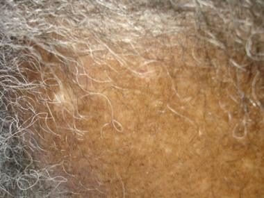
Armpit scarring alopecia in lichen planopilaris variant. Patient presented with frontal fibrosing alopecia. Used with permission, rights retained, courtesy of Rashid M. Rashid, MD, PhD, Morzak Research Collaborative.
-
Acne keloidalis. A misnomer term based on clinical examination findings. Used with permission, rights retained, courtesy of Rashid M. Rashid, MD, PhD, Morzak Research Collaborative.
-
Lichen planopilaris in its active stage presenting in a lighter-skinned patient. Courtesy of Rashid M Rashid, MD, PhD, and Ronald Rapini, MD.
-
Alopecic and aseptic nodules of the scalp (AANS) is a new entity reported first in Japan as "pseudocyst of the scalp." The main location of the nodules was the occiput. The associated alopecia was nonscarring. Histology is nonspecific but often shows deep granulomas. AANS reponds to tetracyclines. Photo courtesy of Sami Abdennader, MD (rights retained).