Background
Aphthous stomatitis (also known as recurrent aphthous ulcers or canker sores) is among the most common oral mucosal lesions observed by physicians and dentists. It is a disorder of unknown etiology that may cause significant morbidity. One or several discrete, shallow, painful ulcers are visible on the unattached oral mucous membranes. These ulcers may be broadly classified as minor, major (see the image below), or herpetiform (see Pathophysiology). Individual ulcers typically last 7-10 days and heal without scarring. Larger ulcers may last several weeks to months and can scar when healing.
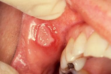 Major aphthous ulcer. Large oval ulcer with white pseudomembrane and raised red border is located on right upper labial mucosa adjacent to buccal commissure. Note irregular margin typical of major aphthae.
Selection of treatment from among the many therapeutic options that have been described should be guided by the severity of disease, the level of supporting evidence available, the cost, and the adverse effect profile. Treatment of recurrent aphthous ulcers focuses on palliating symptoms, shortening the healing time, and preventing future episodes. (See Treatment.)
Pathophysiology
Although the process in idiopathic recurrent aphthous ulcers is usually self-limiting, ulcer activity can be almost continuous in some individuals. Similar ulcers can be noted in the genital region. Behçet syndrome, systemic lupus erythematosus (SLE), and inflammatory bowel disease (IBD) are systemic diseases associated with oral recurrent aphthous ulcers.
Recurrent aphthous ulcers occur on nonkeratinized or poorly keratinized surfaces of the mucosa, such as the following:
-
Labial and buccal mucosa
-
Maxillary and mandibular sulci
-
Unattached gingiva
-
Soft palate
-
Tonsillar fauces
-
Floor of the mouth
-
Ventral surface of the tongue
-
Inferior lateral surface of the tongue
The clinical presentation of aphthous ulcers (see Presentation) is defined by the number of recurrences and the severity of disease. Clinically, the number and size of the lesions are the two main criteria used to divide ulcers into the following three forms:
-
Minor
-
Major
-
Herpetiform
Simple aphthae are common and considered mild, with one to four episodes per year. In general, there are few lesions of the minor or herpetiform form. In contrast, complex aphthosis has a severe clinical course, with an almost continuous presence of minor or major ulcers. These may be debilitating and may also involve the genitalia of both men and women. It is imperative that these patients be evaluated to rule out Behçet disease, as well as IBD.
Recurrent aphthous ulcer minor (Mikulicz ulcer)
This is the most common form, accounting for 80-85% of cases. Discrete, painful, shallow, recurrent ulcers smaller than 1 cm in diameter characterize this form. At any time, one or multiple ulcers may be manifest. These ulcers heal within 7-14 days without scarring. The periodicity varies among individuals, with some having long ulcer-free episodes and some never being free of ulcers.
Recurrent aphthous ulcer major (Sutton ulcer, periadenitis mucosa necrotica recurrens)
This form accounts for about 5-10% of cases and presents as round or oval ulcers that range in size from 2-3 cm in diameter. Major aphthae typically present as a single ulcer, but multiple ulcers may occur. The ulcers present on the soft palate, lips, or oropharynx. They may be deep with smooth or irregular borders. The ulcers may coalesce. Healing, which may take 6 weeks or even months, results in scarring; severe distortion of oral and pharyngeal mucosa may occur. These are more common in patients with HIV disease.
Herpetiform recurrent aphthous ulcer
In this rare form (< 5% of cases), ulcers are typically about 1-2 mm in diameter. The aphthae tend to occur in clusters or crops consisting of 10-100 ulcers. Clusters may be small and localized, or they may be distributed throughout the soft mucosa of the oral cavity. These too occur predominantly on unkeratinized mucosa. It is important to differentiate these ulcers from those caused by herpes simplex virus (HSV), which also may appear as recurrent crops. HSV infection often presents with vesicles that quickly ulcerate and involve the keratinized mucosa of the hard palate, dorsal tongue, and attached gingiva.
Etiology
Although the clinical characteristics of recurrent aphthous ulcer have been well defined, the precise etiology and the pathogenesis remain unclear. Many possibilities have been investigated. Recurrent aphthous ulcer is a multifactorial condition, and it is likely that immune-mediated destruction of the epithelium is the common factor in its pathogenesis. Host risk factors associated with recurrent aphthous ulcer are described below.
Genetics
A family history of recurrent aphthous ulcers is evident in some patients. A familial connection includes a young age of onset and symptoms of increased severity. Recurrent aphthous ulcer is highly correlated in identical twins. [1]
Associations between specific human leukocyte antigen (HLA) haplotypes (eg, HLA-B51) and recurrent aphthous ulcer have been investigated. No consistent association has been demonstrated. However, in two Iranian studies, polymorphisms in the inflammasome-related gene NLRP3 and in the promoter region for the IL6 gene were found to be significantly associated with recurrent aphthous ulcers in a small cohort of patients. [2, 3] Still, host susceptibility is clearly variable, with a polygenic inheritance pattern, and penetrance likely depends on other factors. [4]
Hematinic deficiency
In several studies, hematinic deficiencies (ie, deficiencies of iron, folic acid, or vitamin B6 or B12) were twice as common in patients with recurrent aphthous ulcers as in control subjects. As many as 20% of patients with recurrent aphthous ulcer have a hematinic deficiency, with some studies also reporting notable elevations of blood homocysteine. [5] Lower dietary intake of folate and vitamin B12 is more common among persons with aphthous ulcers. [6] Treatment with vitamin B12 1000 μg/day has shown benefit in some individuals, regardless of serum B12 levels. [7, 8]
A small group of adolescents were shown to have reduced incidence and pain from recurrent aphthous stomatitis when given 2000 mg/day of ascorbic acid. [9] Vitamin D deficiency is also more prevalent in patients with recurrent aphthous stomatitis, though no correlation has been established between vitamin D levels and a patient’s clinical course, including number of ulcers, duration of ulcers, or frequency of ulcer development. [10]
Thus, serologic workup is warranted. Hemoglobin and red blood cell (RBC) indices are not sufficient in all cases.
Immune dysregulation
Although no unifying theory of the immunopathogenesis of recurrent aphthous ulcer has been established, immune dysregulation appears to play a significant role. Cytotoxic action of lymphocytes and monocytes on the oral epithelium may cause the ulceration, but the trigger remains unclear. Upon histologic analysis, recurrent aphthous ulcer consists of mucosal ulcerations with mixed inflammatory cell infiltrates. Helper T (Th) cells predominate in the preulcerative and healing phases, whereas suppressor T cells predominate in the ulcerative phase.
Other findings associated with immune dysregulation include the following:
-
Reduced response of patients' lymphocytes to mitogens
-
Circulating immune complexes
-
Alterations in the activity of natural killer (NK) cells in various stages of disease
-
Increased adherence of neutrophils
-
Reduced quantities and functionality of regulatory T cells in lesional tissue
-
Increased expression of proinflammatory Th1 genes [11]
-
Release of tumor necrosis factor (TNF)-α
-
Significant involvement of mast cells in the pathogenesis of recurrent aphthous ulcer
-
Reduced cellular expression of heat-shock protein 27 and interleukin (IL)-10 in aphthous lesions [12]
-
Elevated levels of salivary and serum cortisol, as well as increased anxiety [13]
-
Increased Toll-like receptor activity [14]
-
Increased levels of autoimmune thyroid-related problems and antithyroid antibodies (though the significance of this is unclear) [17]
Microbial infection
Researchers have disagreed about the role of microbes in the development of recurrent aphthous ulcers. The emphasis has been on a microbial agent as a primary pathogen or an antigenic stimulus. Numerous studies have failed to provide strong evidence to support the role of HSV, human herpesvirus (HHV), varicella-zoster virus (VZV), or cytomegalovirus (CMV) in the development of aphthous ulcers. [18]
Recurrent aphthous ulcer formation may be a T-cell–mediated response to antigens of Streptococcus sanguis that cross-react with the mitochondrial heat-shock proteins and induce damage to oral mucosa. Helicobacter pylori has been detected in lesional tissue of oral ulcers, and a 2014 meta-analysis found H pylori infection to be associated with an increased risk of recurrent aphthous stomatitis. [19] Still, the frequency of serum immunoglobulin G (igG) antibodies to H pylori has not been found to be increased in recurrent aphthous ulcers, and this organism has not been proved to be causative. [20, 21, 22]
A study by Hijazi et al identified imbalances in the oral mucosal microbiome in patients with recurrent aphthous stomatitis. [23] Although no phylum-level differences were appreciated between nonulcerated sites in these patients and healthy controls, patients with recurrent aphthous stomatitis did exhibit an increased abundance of Bacteroidales species. Further investigation would be required to determine whether these microbiome imbalances play a causative role in aphthous stomatitis and to define how diet and oral hygiene influence the oral mucosal microbiome.
Epidemiology
US and international statistics
In North America, recurrent aphthous ulcers are the most common oral mucosal disease. The incidence is approximately 20% overall, rising to more than 50% in certain groups of students in professional schools. Children from higher socioeconomic groups may be affected more than those from lower socioeconomic groups. A 2004 study cited the following point prevalence and lifetime prevalence rates [24] :
-
Point prevalence in the pediatric population in the United States: 1.2-1.5%
-
Lifetime prevalence in the pediatric population in the United States: 40.18%
Internationally, recurrent aphthous ulcers have been reported on every populated continent, with frequencies ranging from 2% to 66%. Epidemiologic studies have been conducted in various subpopulations and have provided data on both point prevalence and lifetime prevalence, as follows:
-
Lifetime prevalence in the adult population in the United States and Canada: 46.4-69.4% [25]
-
Europe lifetime prevalence: 36-37% [25]
-
Jordan lifetime prevalence: 78% [30]
-
Iran lifetime prevalence: 25.2% [31]
-
Sulaimani City, Iraq lifetime prevalence: 28.2% [32]
-
India point prevalence: 1.5% in Northern India [33]
-
India lifetime prevalence: 50.3% [34]
Age- and sex-related demographics
Recurrent aphthous ulcer minor is the most common form of childhood recurrent aphthous ulcer. Approximately 1% of American children may have recurrent aphthous ulcers, with onset before age 5 years. The percentage of patients who are affected decreases after the third decade. [35]
Recurrent aphthous ulcer major has a typical onset after puberty and can persist for the remainder of an individual's life, although after late adulthood episodes become much less common. [35]
Herpetiform recurrent aphthous ulcer typically occurs first in the second decade of life; in the majority of cases, onset comes before age 30 years. The frequency and the severity of episodes may increase during the third and fourth decades and then decrease with advancing age. [35]
In children and in some adult communities who are affected, the incidence of recurrent aphthous ulcer is higher in women and girls than in men or boys. [25]
-
Minor aphthous ulcer. Small superficial oval erosions with yellow pseudomembrane and erythematous border are evident on labial aspect of left lower lip.
-
Major aphthous ulcer. Large oval ulcer with white pseudomembrane and raised red border is located on right upper labial mucosa adjacent to buccal commissure. Note irregular margin typical of major aphthae.
-
Herpetiform aphthous ulcer. Grouped and single tiny white-to-yellow ulcers are scattered on labial mucosa and on ventral aspect of tongue.
Tables

What would you like to print?
- Overview
- Presentation
- DDx
- Workup
- Treatment
- Medication
- Corticosteroids
- Corticosteroids, Topical
- Anesthetics, Topical
- Salivary Stimulants
- Immunosuppressants
- Gastrointestinal Agents, Other
- H pylori Agents
- Oral Rinses
- Wound Care
- Hemorheologic Agents
- Antigout Agents
- Leukotriene Receptor Antagonists
- Antitubercular Agents
- Anti-inflammatory, Topicals
- Calcineurin Inhibitors
- Acne Agents, Topical
- Acne Agents, Systemic
- Ear, Eye, Nose & Throat, Herbals
- Show All
- Media Gallery
- References