Practice Essentials
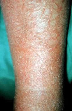
Asteatotic eczema, also known as eczema craquelé and xerotic eczema, is characterized by pruritic, dry, cracked, and polygonally fissured skin with irregular scaling and the appearance of cracked porcelain. The pattern of cracking has been likened to a crazy pavement pattern or a dried-up riverbed and most commonly involves the pretibial areas, but it may also occur on the thighs, on the hands, and on the trunk (see image below).
Superficial bleeding and fissures can occur as the epidermis loses water, as it splits, and as it cracks deeply enough to disrupt papillary dermal capillaries. The inflammation can be associated with asymmetric leg edema. Eczema with increased lichenification occasionally supervenes as patients rub and scratch the pruritic areas. The eruption can be generalized or localized. Generalized asteatosis is a distinct entity and should provoke a search for possible associated diseases.
It most commonly occurs in elderly patients with decreased sebaceous and sweat gland activity during the winter months, especially in areas where indoor humidity is decreased by heating. [1] In addition patients on antiandrogen therapy, people using degreasing agents, and people bathing without replacing natural skin emollients lost to bath water are at risk for asteatotic eczema. Areas of hypoesthetic skin, such as scar tissue, may be predisposed to developing asteatotic eczema. [2]
Asteatotic eczema is a clinical diagnosis; appropriate laboratory studies are indicated for identified or suspected associated diseases.
Asteatotic eczema treatment is aimed at restoring the skin barrier. Patients should be advised to use fragrance-free foaming soap alternatives and humidifiers. Topical emollients and topical steroids are the first-line therapies. [3] Cannabinoid formulations have shown some efficacy in relieving chronic pruritus in several dermatologic conditions, including asteatotic eczema. Where legal, cannabinoid formulations may prove to be an effective adjuvant therapy.
Asteatotic eczema responds well to therapy; however, if the causative factors are not eliminated, recurrences are common. Although most cases resolve without ill effects, asteatotic eczema can be chronic with relapses frequent during the winter months and during times of low humidity.
For patient education resources, visit the Skin Conditions & Beauty Center. Also, see the patient education article Eczema.
Pathophysiology
Initially, excess water loss from the epidermis results in dehydration of the stratum corneum with upward curling of corneocytes. The outer keratin layers require 10-20% water concentration to maintain their integrity. A significant decrease in free fatty acids in the stratum corneum is present in people with asteatotic dermatitis. Stratum corneum lipids act as water modulators, and cutaneous loss of these lipids can increase transepidermal water loss to 75 times that of healthy skin. [4] Elderly persons with decreased sebaceous and sweat gland activity, patients on antiandrogen therapy, people using degreasing agents, and people bathing without replacing natural skin emollients lost to bath water are at risk for asteatotic eczema. Areas of hypoesthetic skin, such as scar tissue, may be predisposed to developing asteatotic eczema. [2]
When the stratum corneum loses water, the cells shrink. A significantly decreased cellular volume can stress the skin's elasticity, creating fissures. Edema in the dermis leads to additional stretch on the overlying epidermis. Fissures rupture dermal capillaries, causing clinical bleeding. The disruption of cutaneous integrity can result in inflammation with risk of infection. Transepidermal absorption of allergens and irritants is increased as the epidermis is damaged, increasing susceptibility to allergic contact dermatitis and irritant contact dermatitis. [5] Allergic contact dermatitis and irritant contact dermatitis may cause a persistent and possibly more extensive dermatitis despite therapy. Furthermore, low environmental humidity contributes to xerosis, creating a clinical picture of asteatotic dermatitis in some dermatologic conditions, such as atopic dermatitis.
Etiology
Overwhelmingly the following are the most frequent contributors to asteatotic eczema:
-
Xerosis and friction
-
Frequent or prolonged bathing in hot water, use of soap on the involved site, and infrequent use of emollients for water retention in the stratum corneum
-
Degreasing agents - Solvents, cleansers
-
Decreased sebaceous and sweat gland activity in elderly persons
-
Low environmental humidity and cold winds that increase the loss of water by convection
-
Atopy
Multiple more uncommon etiologic factors may coexist to cause asteatotic eczema, including the following:
-
Decreased keratin synthesis in elderly persons
-
Radiation
-
Long-term malabsorption of essential fatty acids, including linoleic acid and linolenic acid
-
Thyroid disease - Myxedema and other thyroid diseases with diminished sweat and sebaceous gland activity [8]
-
Neurologic disorders - Decreased sweating in denervated areas
Epidemiology
Frequency
Seasonality is prominent, and most patients present in the winter months, especially in areas where indoor humidity is decreased by heating. The frequency of asteatotic eczema is increased in the northern United States, particularly during the winter season.
In China, asteatotic eczema was the second most common type of dermatitis among patients ≥60 years old with a prevalence of 8.1%. In patients < 60 years old the prevalence was 2.8%. [16]
Sex
Men older than 60 years develop asteatotic eczema more commonly than women.
Age
The median patient age at presentation is 69 years. Asteatosis can also occur in young people.
Prognosis
Asteatotic eczema responds well to therapy; however, if the causative factors are not eliminated, recurrences are common. Although most cases resolve without ill effects, asteatotic eczema can be chronic with relapses frequent during the winter months and during times of low humidity.
-
Asteatotic dermatitis on the lower extremity.
-
Asteatotic dermatitis on the lower extremity.
-
Asteatotic dermatitis on the lower extremity.