Background
First described in 1913 by Snow, [1] acute hemorrhagic edema of infancy (AHEI) was originally thought to be a purely cutaneous variant of Henoch-Schönlein purpura (HSP). Over the years, many others have described AHEI, including Del Carril, Diaz Sobillo, and Vidal [2] in Argentina in 1936 and Finkelstein in Europe 1938. [3] AHEI has also continued to be known by other names, including Seidlmayer cockade purpura, [4] purpura en cocarde avec oedema, urticarial vasculitis of infancy, and acute benign cutaneous leukocytoclastic vasculitis of infancy. [5]
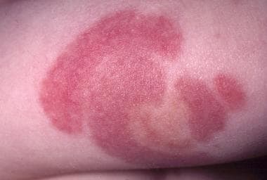
AHEI is now considered a separate entity from HSP because of the infrequency of both visceral involvement and immunoglobulin A (IgA) skin depositions, [6, 5, 7, 8] as well a better prognosis. The cutaneous findings are dramatic both in appearance and rapidity of onset. AHEI is characterized by the triad of fever; edema; and rosette-, annular-, or targetoid-shaped purpura primarily over the face, ears, extremities, feet and hands in a nontoxic infant or young child (see the image below). [9, 10, 11, 12] In a single-center retrospective study of infants diagnosed with AHEI, 95% of cases presented with prodromal symptoms. [13]
 Large cockade (rosette or knot of ribbons), annular, or targetoid purpuric lesions found primarily on the face, ears, and extremities are characteristic of acute hemorrhagic edema of infancy.
HSP, in comparison, usually presents with palpable purpura or petechiae associated with one or more symptoms, including abdominal pain, arthritis/arthralgias, and nephritis; however, any of these symptoms may be absent, which often leads to confusion in diagnosing the condition. The diagnosis may be particularly difficult to make when a patient presents with isolated symptoms, such as abdominal pain, without the typical rash. Scalp edema and/or scrotal swelling also may be seen in patients with HSP. A skin biopsy may be needed to distinguish AHEI from HSP. [14]
Pathophysiology
The specific etiology of acute hemorrhagic edema of infancy (AHEI) is unknown, although some consider the disease to be an immune complex–mediated vasculitis. [15] Eighty-four percent of reported cases were preceded by viral infections (acute upper respiratory tract infection, gastroenteritis), medication use (ie, antibiotics), and immunizations. [16, 17] In one review of a large referral center in Israel, various infectious conditions were identified in patients with AHEI, including Escherichia coli urinary tract infection, rota virus gastroenteritis, adenovirus upper respiratory tract infection, Streptococcus pyogenes tonsillitis, and herpes simplex gingivostomatitis. [18] In addition, reports have also described an association with Coxsackie virus infection. [19, 20] A possible association with COVID-19 has also been reported. [21, 22]
Etiology
The cause of acute hemorrhagic edema of infancy (AHEI) is unknown. AHEI is considered to be an immune complex disorder; however, immune complexes have been demonstrated in only some cases. [23, 24]
Epidemiology
Frequency
Over 300 cases of AHEI have been published in medical literature worldwide. [23] However, AHEI is often misdiagnosed. In a retrospective review medical records, 69 cases of possible AHEI were identified with 40 classified as probable. Of the 40 probable cases, only 24 (60%) received an initial diagnosis of AHEI. [25]
United States
Acute hemorrhagic edema of infancy (AHEI) is uncommon in the United States. Specific frequency data have not been reported.
International
Acute hemorrhagic edema of infancy (AHEI) has been reported in countries throughout the world, although incidence is unknown.
Race
No racial predilection has been described for acute hemorrhagic edema of infancy (AHEI).
Sex
Acute hemorrhagic edema of infancy (AHEI) is more common among male infants than among female infants [23] ; the male-to-female ratio is approximately 4.6:1.
Age
The age range of onset for acute hemorrhagic edema of infancy (AHEI) is 2-60 months (median, 11 mo; mean, 13.75 mo). [23, 26] However, a congenital case of AHEI has been reported. [27]
Prognosis
Acute hemorrhagic edema of infancy (AHEI) is usually benign, self-limited, and without sequelae, with spontaneous recovery occurring within 1-3 weeks. AHEI may recur, but this is uncommon. One case report describes an AHEI patient whose eruption resolved with unusual scarring. [17] Rare reports have described complications such as arthritis, nephritis, [28, 29] abdominal pain, gastrointestinal tract bleeding, intussusception, [30] scrotal pain, compartment syndrome, [31] and testicular torsion. [32]
Patient Education
Educate parents about the benign self-limited nature of acute hemorrhagic edema of infancy (AHEI) and the fact that recurrences, although uncommon, can occur.
-
Large cockade (rosette or knot of ribbons), annular, or targetoid purpuric lesions found primarily on the face, ears, and extremities are characteristic of acute hemorrhagic edema of infancy.
-
The left leg in this patient with acute hemorrhagic edema of infancy is markedly more edematous than the right leg.
-
Leukocytoclastic vasculitis and fibrinoid necrosis is seen in patients with acute hemorrhagic edema of infancy. This histologic pattern also is seen in Henoch-Schönlein purpura, although patients with Henoch-Schönlein purpura usually have immunoglobulin A deposition, and immunoglobulin A deposition is demonstrable in only approximately one third of patients with acute hemorrhagic edema of infancy (hematoxylin and eosin, magnification X40).
-
This toddler with acute hemorrhagic edema of infancy has a discoloration in the area of the umbilicus similar to that described as Cullen sign.
-
Note the concentric arcs of purpura on the patient's arm.
-
Despite the frightening appearance of purpura in these patients, they usually are in no significant distress.

