Background
Acneiform eruptions are dermatoses that resemble acne vulgaris. Such eruptions may develop as a result of infections, hormonal or metabolic abnormalities, genetic disorders, or drug reactions. Lymphoma has also been reported as a cause of acneiform eruptions. [1]
Patients with acneiform eruptions present with acnelike lesions such as papulonodules, pustules, and cysts. (See the image below.) Unlike patients with acne vulgaris, they typically do not present with comedones. The physical location may be outside of the area in which acne vulgaris occurs. Acneiform eruptions can be distinguished from acne vulgaris by a history of sudden onset, monotonous lesion morphology, and development of the eruption at an age outside the range typical of acne vulgaris. In the case of drug-induced acneiform eruptions, the eruption resolves with discontinuance of the medication.
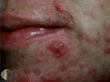 Acneiform eruption. Image from DermNet New Zealand (https://www.dermnetnz.org/assets/Uploads/doctors/quizzes/48/case6.jpg).
The workup of acneiform eruptions varies greatly, reflecting the wide variety of diseases. This can include skin biopsies, cultures and sensitivities, serologic tests, and empiric trials of drug withdrawal. Depending on the type of eruption present, biopsies may demonstrate suppurative folliculitis, eosinophilic folliculitis, keratosis pilaris, or granulomatous dermatitis. [2]
Treatment varies with the particular disease suspected and consists of a wide range of methods, including excision, laser ablation, topical or oral antibiotics, topical or oral retinoids, and drug withdrawal. Oral tetracyclines represent an efficacious prophylactic option for acneiform eruptions due to epidermal growth factor receptor (EGFR) inhibitors. They can be recommended to suitable patients beginning treatment.3 Rifampin and clindamycin may also be utilized for EGFR inhibitor–related acneiform eruptions. [3]
Although oral tetracyclines are commonly recommended for EGFR inhibitor–related acneiform eruptions, there is good evidence favoring the use of systemic retinoids instead. [4] Low-dose isotretinoin for refractory grade II/III EGFR inhibitor–related acneiform eruptions has been recommended, as has doxycycline 100 or 200 mg/day as a prophylactic option. [5]
Nevus Comedonicus
Nevus comedonicus (also known as comedone nevus and nevus acneiformis unilateralis) is an infrequent developmental anomaly manifesting as aggregated open comedones and consisting of dilated follicular or eccrine orifices plugged with keratin. Although the majority of cases are isolated, nevus comedonicus may also occur as part of nevus comedonicus syndrome in association with the following abnormalities:
-
Central nervous system - Epilepsy, electroencephalographic (ECG) abnormalities, transverse myelitis, microcephaly
-
Skeletal system - Scoliosis, hemivertebrae, spina bifida occulta, foot deformities, absent fifth finger, syndactyly, supernumerary digits
-
Skin - Ichthyosis, trichilemmal cysts, leukoderma, white hairs, Sturge-Weber syndrome, hemangiomas, linear basal cell nevus
-
Eye - Congenital cataracts (unilateral and bilateral) [6]
-
Other - Bilateral oligodontia, [7] multiple basal cell carcinomas, rare systemic malignancies
Nevus comedonicus appears as a collection of discrete, dilated follicular ostia plugged with pigmented keratinaceous material. Dermatoscopy displays multiple, well-defined structureless brown homogenous circular pores with keratin plugs. [8] They can be single or multiple, usually unilateral, and range in size from a few centimeters to half of the entire body surface. (See the image below.)
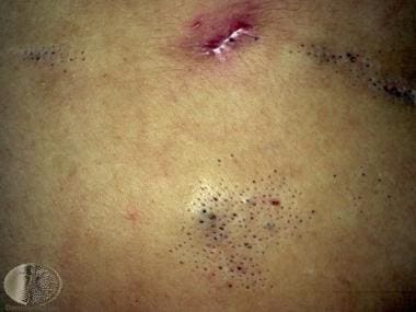 Nevus comedonicus. Image from DermNet New Zealand (https://www.dermnetnz.org/assets/Uploads/lesions/com-naev1.jpg).
Nevus comedonicus is typically found on the face, trunk, neck, and upper extremities. [9] Rarely, it has been described on the palms [10] and soles, scalp, [11, 12] or penis. [13] When it occurs on the elbows and knees, it can appear as verrucous nodules. When it occurs in the intertriginous zones, the mechanical stress can produce hidradenitis suppurativa–like lesions. [14] Nevus comedonicus may be linear, interrupted, unilateral, bilateral, present in a dermatomal distribution, following the lines of Blaschko, or segmental. [15, 16]
The lesions are typically present at birth or develop in early childhood. They are usually asymptomatic, and help is generally sought for cosmetic reasons. The lesions grow as the patient does and often grow faster at puberty.
Uncommonly, the lesions may become repeatedly inflamed and infected, potentially leading to repeated bacterial infections, drainage, cysts, fistula and abscess formation, and scarring. Nevus comedonicus syndrome complicated by hidradenitis suppurativa‒like lesions has been described. [17, 18] Cases of nevus comedonicus on the scalp with late-onset presentation have been reported. [19, 20]
The differential diagnosis of nevus comedonicus includes familial dyskeratotic comedones and linear comedone formations usually linked with acne vulgaris or chronically sun-damaged skin (Favre-Racouchot disease). Infrequently, multiple comedones in other unusual contexts may raise nevus comedonicus as a possible consideration.
Because nevus comedonicus is typically an asymptomatic benign condition, most cases require no treatment, though therapy may often be implemented for cosmetic concerns. Treatment is generally surgical, through excision or laser ablation of the involved skin. Surgical excision of small lesions can be curative and should be considered. Incomplete excision may result in recurrence of the lesion. Laser ablation is effective in eradicating extensive nevus comedonicus but can cause significant scarring and may necessitate the use of grafts if the treatment area is large. [21, 22]
Some lesions have been found to improve when treated with topical retinoic acid, salicylic acid, or ammonium lactate lotion.
Eruptive Vellus Hair Cyst
Eruptive vellus hair cysts (EVHCs) manifest as flesh-colored papules found usually on the face, chest, neck, thighs, groin, buttocks, and axillae. [23] They represent an anomaly of the vellus hair follicles and may be hereditary. Histopathologic evaluation reveals a middermal epithelial cyst containing vellus hairs and keratinous material. These cysts may undergo spontaneous regression, form a connection to the epidermis, or undergo degradation with a resultant foreign body granulomatous formation.
Treatment of EVHCs is often difficult. Incision and drainage of individual lesions carries the risk of subsequent scarring, and modalities such as carbon dioxide laser ablation are difficult to use over large surface areas. Topical retinoids and 12% lactic acid preparations have proved useful in some instances.
Steroid Acne
Steroid acne [24, 25] is observed as monomorphous papulopustules located predominantly on the trunk and extremities, with less involvement of the face. Characteristically, it appears after the administration of systemic corticosteroids, including intravenous (IV) therapy. Topical or inhaled corticosteroids may cause an acneiform eruption of the area of skin under which the topical preparation is applied or in around the nose or mouth in the case of inhaled steroids.
The eruption usually resolves after discontinuance of the steroid and, in addition, may respond to the usual treatments for acne vulgaris.
Chloracne
Exposure to halogenated aromatic hydrocarbon compounds (eg, chlorinated dioxins and dibenzofurans) through inhalation, ingestion, or direct contact of contaminated compounds or foods induces a cutaneous eruption of polymorphous comedones and cysts referred to as chloracne. Other associated skin findings may include xerosis and pigmentary changes. Internal changes involving the ophthalmic, nervous, and hepatic systems may also occur, and some chloracnegens can be oncogenic.
Treatment is difficult because chloracne may persist for years, even without further exposure.
Chemicals that contain iodides, bromides, and other halogens can also induce an acneiform eruption resembling that of steroid acne; however, the iodide-induced eruption may be more extreme.
Medication-Related Eruption
Antibiotics may induce an acute generalized pustular eruption. Penicillins and macrolides are the greatest offenders. Patients usually are febrile with leukocytosis, and the eruption does not usually involve comedones. Other implicated antibiotics include trimethoprim-sulfamethoxazole, doxycycline, ofloxacin, and chloramphenicol.
Additional medications that can also produce an acnelike eruption include corticotropin, nystatin, isoniazid, itraconazole, hydroxychloroquine, naproxen, mercury, amineptine, [26, 27, 28] the antipsychotics olanzapine and lithium, chemotherapeutic agents, and epidermal growth factor receptor (EGFR) inhibitors. [29, 30, 31, 32, 33, 34, 35, 36, 37] (For more information, see Drug Eruptions.)
Acneiform eruptions may be associated with tyrosine kinase 2 (TYK2) inhibitors such as deucravacitinib, [38] with HER2 inhibitors such as trastuzumab, [39] and with mitogen-activated protein kinase (MEK) inhibitors such as binimetinib and selumetinib. [40, 41] Vitamin B12 may induce an acneiform eruption. [42] Corticosteroids and Janus kinase (JAK) inhibitors employed for inflammatory bowel disease tend to exacerbate a tendency for acne or trigger new acneiform eruptions. [43] The JAK inhibitor upadacitinib employed for atopic dermatitis may produce an acneiform eruption. [44]
Infection-Related Eruption
Various infections may display an acneiform pattern. Gram-negative folliculitis, a persistent papulopustular eruption, may be a complication in patients receiving prolonged treatment with oral antibiotics for acne vulgaris or rosacea. Use of antibiotics (eg, tetracyclines) can alter the normal skin flora of the skin and thereby allow growth of gram-negative organisms in the nares. These gram-negative organisms typically spread to the skin of the upper lip, chin, and jawline, where they cause a folliculitis. Culture of the papulopustules grows gram-negative bacilli and gram-negative rods, including Escherichia coli and Klebsiella, Enterobacter, and Proteus species.
The typical history is of a patient with a sudden acne flare despite no change in treatment or a patient unresponsive to traditional therapies. Oral isotretinoin is considered standard of care.
Malassezia (Pityrosporum) is another infectious folliculitis that is presumably caused by a host reaction to the yeast Malassezia furfur (previously named Pityrosporum ovale) a normal human skin commensal organism. It appears primarily on the trunk and upper extremities of late adolescents and young adults. Unlike acne vulgaris, it is pruritic, does not contain comedones, and responds to empiric antifungal therapy rather than antibiotics.
The diagnosis is typically made clinically, though the yeast and hyphae can be observed in biopsy specimens in the widened follicular ostia along with keratinous material, and occasionally, rupture of the follicular wall may occur. Patients may be treated with topical leave-on, wash-off, or systemic antifungal therapy.
Eosinophilic pustular folliculitis (EPF) is a disease of unclear etiology, thought to be an allergic hypersensitivity. It appears as a recurrent pruritic papulopustular eruption on the face, trunk, and extremities. Histopathology reveals a predominantly perifollicular infiltration of eosinophils with some mononuclear cells and subcorneal pustules composed of eosinophils. Three main types of EPF exist: (1) infantile EPF, (2) HIV-associated EPF, and (3) classic Ofuji disease in immunocompetent patients (typically, Japanese patients). Patients may also demonstrate blood eosinophilia and leukocytosis.
Treatment modalities and results vary greatly. Options include topical or systemic corticosteroids, oral antibiotics, dapsone, isotretinoin, and pulsed ultraviolet (UV) A phototherapy (PUVA). Indomethacin is the treatment of choice for classic Ofuji disease.
Several other infectious diseases may also result in acneiform eruptions, including the following:
-
In secondary syphilis, [45, 46] papulopustules and nodules, some crusted, may occur on the face, trunk, and extremities; the causative agent, Treponema pallidum, may be easily observed in biopsy specimens with the Warthin-Starry stain; in addition, serologic tests and the presence of spirochetes on darkfield microscopy may reveal the diagnosis
-
Mycotic infections may also manifest cutaneously with papules and nodules that may ulcerate and crust
-
Sporothrix schenckii, the agent responsible for sporotrichosis, [47] commonly induces a lymphocutaneous reaction but can also produce a persistent fixed localized cutaneous papulonodular eruption that may involve the face; the organism can be demonstrated histologically, by peripheral blood smear, and by fungal culture
-
Cutaneous coccidioidomycosis, usually caused by inhalation and dissemination of Coccidioides immitis, may rarely occur by primary inoculation and appear as papulopustules, nodules, or plaques that can eventually ulcerate and crust
Rosacea
Rosacea appears similarly to acne vulgaris, with papulopustules on the face; however, rosacea patients may also have facial flushing and telangiectasias, and they do not have comedones. Four subtypes of rosacea exist: (1) erythematotelangiectatic, (2) papulopustular, (3) phymatous, and (4) ocular.
Rosacea is more common in lighter-skinned individuals and in women in the third or fourth decade of life. Men, however, more commonly develop sebaceous hyperplasia of the nose (rhinophyma). Associated ocular findings are variable but include blepharitis, conjunctivitis, iritis, iridocyclitis, hypopyon iritis, and even keratitis. Although the definitive etiology is unknown, weather extremes, hot or spicy foods, alcohol, and Demodex folliculorum mites can trigger and exacerbate this condition. Acne rosacea has also been associated with the ingestion of a high-dose vitamin B6 supplement. [48]
Histopathologic evaluation of rosacea skin reveals granulomatous inflammation. Treatment primarily includes skin-barrier sunscreens and topical antibiotics such as metronidazole, retinoids, and oral tetracyclines.
Perioral and Periocular Dermatitis
Perioral dermatitis, also a disorder of unclear etiology, is mainly observed in the young White female population in the form of papulopustules with erythematous base. The eruption is predominantly perioral in location, characteristically sparing the vermilion border of the lip, but it may also include the perinasal and periorbital areas. A variant known as periocular dermatitis affects the skin around the eyes. The eruption is thought to be a variant of rosacea, as biopsies show changes similar to those of rosacea.
Prior use of topical corticosteroids has been theorized to cause this condition, but neither duration of use nor steroid strength has been shown to be clearly related. Demodex mites, [49] moisturizers, fluorinated compounds, and contact irritants or allergens have also been implicated as causes of eruption.
Therapy typically includes cessation of topical steroids or other offending agents and administration of topical anti-inflammatory treatments (eg, topical metronidazole, topical pimecrolimus cream, and azelaic acid), as well as oral anti-inflammatory dose antibiotics such as doxycycline.
-
Acneiform eruption. Image from DermNet New Zealand (https://www.dermnetnz.org/assets/Uploads/doctors/quizzes/48/case6.jpg).
-
Nevus comedonicus. Image from DermNet New Zealand (https://www.dermnetnz.org/assets/Uploads/lesions/com-naev1.jpg).
Tables

What would you like to print?
- Heritability Strongly Linked to Acne Fulminans
- Acne in Primary Care: The Best of Times?
- Consider Emergency Contraception for Teens With Acne
-
 FDA Approves Triple Combination Topical Treatment for Acne
-
Severe, Atypical Skin Reactions With Amivantamab in NSCLC
- Antibiotic Resistance in Dermatology Part 2: Combating Resistance


 Identifying Lesions on Skin of Color
