Background
Oral pigmentation is a relatively common condition that may involve any portion of the oral cavity. Multiple causes are known, and they may range from simple iatrogenic mechanisms, such as implantation of dental amalgam, to complex medical disorders, such as Peutz-Jeghers syndrome (PJS) and Addison disease. [1] Local irritants, such as smoking, may also result in melanosis of varying degrees. [2] In general, oral pigmented lesions most commonly result from implantation of exogenous materials or endogenous, excessive melanin deposition. [3] Occasionally, however, these lesions may result from an increase in the number of cells, which can range from benign nevi to fatal oral melanoma.
As previously mentioned, pigmented entities may arise from intrinsic or extrinsic sources. The color may range from light brown to blue-black. The color depends on the source, quantity, and depth of the pigment from which the color is derived. [4] Melanin is brown, yet it imparts a blue, gray, brown, or black color to the eye. [5] This effect is due to the physical properties of light absorption and reflection described by the Tyndall light phenomenon or effect. Amalgam and graphite tattoos typically appear as black, blue, or gray discolorations. [5]
Oral conditions associated with increased melanin production are common; however, those due to a true increase in the number of melanocytes are rare. Clinicians must visually inspect the oral cavity, obtain clinical histories, and be willing to perform a biopsy for any oral pigmented lesion that is not readily diagnosed. Early biopsy of focal pigmentations of undetermined etiology is extremely important in order to detect oral melanomas at an early stage. Patients with oral melanoma often recall having a previous pigmentation in the same area months to years before the melanoma diagnosis, and the condition may even have elicited a prior comment from physicians or dentists. [6]
Pathophysiology
The four major mechanisms leading to increased oral pigmentation are discussed in detail: physiologic pigmentation, systemic diseases (eg, Peutz-Jeghers syndrome [PJS]), oral mucosal insults (eg, amalgam tattoo), and neoplastic processes (eg, melanoma).
Physiologic pigmentation
Physiologic pigmentation, also known as racial or ethnic pigmentation, is an increased production of melanin pigment by melanocytes in dark-skinned individuals. [5] The pigmentation is due to an increased melanocytic activity rather than an increase in the number of melanocytes. [7] The tendency to develop pigmentation appears to be genetically determined, but color intensity might be influenced by smoking, hormones, systemic medications, and physical factors. [5, 8] African Americans and other dark-skinned individuals such as Greeks, Syrians, Indians, and Italians are more commonly affected. [5] This type of pigmentation appears as flat, light-to-dark–brown lesions, which are evenly pigmented and bilaterally symmetrical. It most commonly involves the attached gingiva, especially in the facial regions of the anterior maxilla and mandible. [8] In this particular site, the pigmentation appears as a well-demarcated, ribbonlike, dark-brown band that usually spares the marginal gingiva (see image below). [5, 9] It does not alter the normal architecture of the tissue such as gingival stippling. When it involves the tongue, it may appear as pinpoint pigmentations at the tips of the fungiform papillae on the dorsal surface. [8]
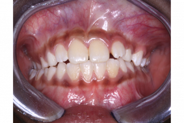 Physiologic pigmentation; bilaterally symmetrical, ribbon-like, brown band involving maxillary and mandibular facial gingiva.
Systemic diseases
Peutz-Jeghers syndrome
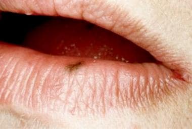
PJS is an autosomal dominant disorder characterized by intestinal hamartomatous polyps in association with mucocutaneous melanocytic macules. Pigmented macules, often confluent and varying in size and shades of brown, appear in almost all cases periorally and on the lips and buccal mucosae. Any oral site may be involved, and the degree of pigmentation and oral involvement vary among affected individuals. Besides the complications related to the intestinal polyps, such as small intestine intussusception at a young age, PJS also causes an increased cancer risk. The relative risk of developing cancer in this syndrome is increased 15-fold compared with that of the general population. [10, 11] The malignant tumor primarily involves the GI tract (esophagus, stomach, small intestine, colon, and pancreas), but extragastrointestinal sites such as lung, breast, and female reproductive organs may also be involved. [11]
Addison disease
Addison disease is the insufficient production of adrenocortical hormones caused by the destruction of the adrenal cortex (primary hypoadrenocorticism) and leading to increased production of adrenocorticotropic hormone (ACTH). [12] As an increased amount of glandular tissue becomes destroyed, the patient starts developing symptoms related to decreased levels of cortisol and, often, aldosterone. These symptoms include weakness, unexplained weight loss, salt craving, unexplained isotonic or hyponatremic dehydration, hyperkalemia, anorexia, nausea, hypoglycemia, episodes of shock during a severe illness, and behavioral changes. [12, 13, 14] Furthermore, there is an increase in melanin production in the skin and possibly mucous membranes due to the melanocyte-stimulating activity of high ACTH plasma levels. [15]
Oral mucosal insults
Most reactive pigmented lesions in the oral cavity are due to iatrogenic implantation of exogenous material (eg, amalgam tattoo) or to excessive production of melanin, which is produced by melanocytes. These cells are specialized dendritic cells present in the basal cell layer of the mucous membrane. The clinically visible pigmentation in the oral cavity depends on the number of melanocytes or the degree of melanin produced by these cells. The range of color pigmentation varies from gray to brown to black to dark blue. The closer the pigmentation to the surface, the darker the color (black).
Amalgam tattoo
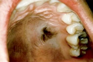
The amalgam tattoo is the most common localized, pigmented lesion of the oral mucosa, affecting 3.3% of the US adult population. [16] This lesion is the result of iatrogenically embedded amalgam particles during dental procedures. [17, 18] It appears as a soft, painless, nonulcerated, blue, gray, or black macule with no surrounding erythematous reaction. It is most frequently found on the gingival or alveolar mucosa, but many cases are also seen on the buccal mucosa and floor of the mouth. [17] The tattoo is found more frequently in females than in males, probably because females are more likely to seek dental care. It is also seen more frequently with advancing patient age, presumably because of increased exposure to dental procedures over time. The tattoo is only moderately demarcated from the surrounding mucosa and is usually less than 6 mm in greatest diameter. Lesions with larger particles might be visible as small radiopaque speckles on dental radiographs (see images below). Unfortunately, however, many cases show no radiographic findings, which might cause a biopsy to be warranted for a definitive diagnosis. [19]
 Amalgam tattoo; well-demarcated, dark gray macular lesion involving mandibular alveolar mucosa.
 Amalgam tattoo; radiopaque particles of amalgam may be seen in a dental radiograph.
Smoker's melanosis
Smoker’s melanosis is due to long-term tobacco smoking. It appears that polycyclic amines present in tobacco, as well as free radicals produced by the use of tobacco, stimulate oral melanocytes to produce melanin as a protective mechanism. [8, 20] The pigmentation is usually distributed along the anterior facial gingiva, especially in the region of mandibular anterior teeth. [21] It may also be seen in the soft palate, buccal mucosa, and floor of the mouth. The prevalence of pigmentation increases proportionately to the number of cigarettes consumed. [22] Smoking cessation is the treatment of choice. Habit cessation results in a decrease in pigmentation, and a prevalence similar to nonsmokers is seen in patients who have quit the habit for at least 3 years. [21, 23]
Oral melanoacanthoma
Oral melanoacanthoma is an uncommon, reactive proliferation characterized by an increased number of pigment-laden dendritic melanocytes dispersed throughout an acanthotic surface epithelium. [12, 24] Despite bearing the same name, cutaneous and oral melanoacanthoma are considered distinct biological entities. The former represents a rare variant of pigmented seborrheic keratosis. [25, 26] On the other hand, oral melanoacanthoma behaves clinically as a reactive process as in several reported cases, it regressed after removal of irritant factors or showed complete resolution after incisional biopsy. [27, 28] Nevertheless, the lesion is characterized by a rapid clinical progression, which can cause alarm to the patient and clinician and mimic oral melanoma. It most commonly occurs in Black females in the third and fourth decades of life. Although it has been uncommonly reported in other racial or ethnic groups. [19, 29] The lesion usually appears as a solitary, flat or slightly raised, brown-to-black lesion. [27] Diffuse and multifocal presentations have also been described. [24, 25, 27]
Developmental and neoplastic processes
Oral melanotic macule
The oral melanotic macule is a flat brown-to-black lesion caused by focal, excessive melanin deposition within the basal and parabasal cell layers of the epithelium. It is one of the most common pigmented lesions of the oral cavity, second only to amalgam tattoo. [3] It is not clear if the melanotic macule represents a physiologic or reactive process, and a genetic predisposition has been suggested. [5, 30] Moreover, cases of oral and labial melanotic lesions may represent postinflammatory hyperpigmentation as they are clinically and histologically identical. [5] Melanotic macules are solitary or multiple and can involve the gingiva, lip, palate, buccal mucosa, and alveolar ridge, as shown in the image below. [31] In addition to the increased melanin deposition within the basal and parabasal cell layers of the epithelium, melanin incontinence may be seen in the superficial lamina propria, as seen in the image below. [30]


Oral melanocytic nevus
Although very frequently found on the skin, oral melanocytic nevus (OMN) is quite uncommon. [32] When they do develop intraorally, they appear most commonly on the hard palate, buccal mucosa, and gingiva. [32, 33] OMN is classified histologically according to its stage of development. The earlier lesion is termed junctional nevus and is characterized by the accumulation of nevus cells only in the junction between epithelium and connective tissue. Compound nevus, the intermediary stage, is characterized by the accumulation of nevus cells both at the epithelial/connective-tissue junction and deep in the underlying connective tissue. When the nevus cells are only located in the connective-tissue compartment, the term intramucosal nevus is used. See the diagram below.
Blue nevus can also be found in the oral cavity and it represents a benign proliferation of spindle-shaped, dendritic melanocytes containing numerous melanin granules and being localized deep in the connective tissue (see images below). There are two histological subtypes: common blue nevus and cellular blue nevus.
Intramucosal nevus is the most common type of melanocytic nevi to occur in the oral mucosa, followed by the common blue nevus. [32, 33, 34]

Clinically, OMN typically presents as a well-circumscribed, pigmented lesion, and most measure less than 1 cm in greatest dimension. [34, 32] Junctional nevus most commonly appears as a flat lesion, while compound and intramucosal nevus usually appear as raised or slightly raised lesions. [34] A few cases of compound or intramucosal nevus might show a rough, papillomatous, or verrucous surface. [34] OMN can appear in shades of gray, brown, and blue, depending on the depth of the nevus cells. [5] The vast majority of cases are pigmented; however, around 32% of intramucosal nevi appear as nonpigmented lesions. In such cases, the lesion might mimic a fibroma or squamous papilloma. [32]
OMN can appear identical to an early lesion of oral melanoma; for this reason, any melanocytic lesion of the oral cavity of unknown cause should undergo biopsy and should be examined under the microscope in order to rule out the possibility of this rare, yet fatal malignant tumor.

Microscopically, the nevus cells tend to form small aggregates or theques, and the most superficial cells are typically laden with melanin. The cells may be seen only at the junction between epithelium and connective tissue (junctional nevus), or they might accumulate both at the epithelial/connective-tissue junction and deeper, in the underlying connective tissue (compound nevus). At the latest stage of development, the cells are found solely within the connective tissue (intramucosal nevus, as seen below). Heavily pigmented, spindle-shaped melanocytes localized deep in the connective tissue characterizes the blue nevus. See the images below.


Oral melanoma
Melanoma is a malignant neoplasm of melanocytes that usually originates in the skin and is quite uncommon in the oral mucosa. Oral malignant melanomas (OMMs) account for less than 1% of all melanomas, [35] and they represent only 0.26% of all oral cavity cancers. [36, 37] Nevertheless, OMMs are known to act more aggressively and exhibit a significantly worse prognosis when compared with cutaneous melanomas. Therefore, it is generally agreed that all oral pigmented lesions that are not clinically diagnostic should undergo biopsy. [35] Additionally, in contrast to cutaneous melanomas, which are etiologically linked to sun exposure, risk factors for OMMs are currently unknown. These tumors have no apparent relationship to chemical, thermal, or physical events (eg, smoking; alcohol intake; poor oral hygiene; irritation from teeth, dentures, or other oral appliances) to which the oral mucosa is constantly exposed. Although benign, intraoral melanocytic proliferations (junctional, compound, and intramucosal nevi) occur and could potentially be sources of some oral melanomas; the sequence of events is still poorly understood in the oral mucosa. [38]
The genetic basis of both oral melanocytic nevi and mucosal melanoma has not been well studied. Oral melanomas are typically studied within a group with other mucosal melanomas, despite being a separate entity. BRAFV600E is the most frequent driver mutation on cutaneous acquired melanocytic nevus and cutaneous melanoma, occurring in approximately 80% and 40% of cases, respectively. On the other hand, studies have found the BRAFV600E mutation in only 42.1% of oral melanocytic nevi and 6.4% of OMMs. Interestingly, similar to the cutaneous counterparts, benign oral melanocytic nevi harbor the BRAFV600E mutation in a higher proportion of cases than OMMs. [39] This finding suggests that BRAFV600E mutation may be an early initiating event in the tumorigenesis of not only cutaneous melanomas but potentially a small percentage of OMMs. [39, 40] However, studies have shown that different melanoma subtypes tend to have different genetic alterations. Among mucosal melanomas, mutations in the KIT gene, which encodes a receptor tyrosine kinase that interacts with Ras, are more frequent than BRAF mutations. [41, 42]
OMMs typically occur in adults, are more common in males, and show a marked predilection for the keratinized mucosa of the hard palate and gingiva. [37, 43] These tumors are usually histopathologically similar to the acral lentiginous type of cutaneous melanoma. [43] However, most investigators believe OMMs do not fit well into any of the classic cutaneous melanoma subtypes [39, 44] ; therefore, it has been proposed that they should be simply subclassified into in situ and invasive types. [43, 44]
Etiology
Physiologic pigmentation
Physiologic pigmentation, also known as racial pigmentation, is the increased production of melanin pigment by melanocytes in dark-skinned individuals. It is caused by increased melanocytic activity rather than an increase in the number of melanocytes. The tendency to develop the pigmentation is genetically determined, but color intensity might be influenced by smoking, hormones, systemic medications, and physical factors. [5, 7, 8]
Systemic diseases
Peutz-Jeghers syndrome
PJS is typically inherited as an autosomal dominant disease, with most cases being caused by germline, loss-of-function mutations affecting the serine-threonine kinase 11 (STK11 or LKB1) gene. STK11 is located in the short arm of band 19p13.3 and is considered a tumor-suppressor gene that modulates cellular proliferation and controls cell polarity. [45] Mutations in this gene have been reported to cause 50-90% of PJS cases. [46, 47] Nevertheless, a significant proportion of PJS cases may result from de novo mutations, and more recent studies have found that the genetic mutations in hamartoma polyp tissue of PJS are complex and diverse. [48, 49]
The elimination of the kinase activity of STK11/LKB1 is probably responsible for the development of the PJS phenotypes. [50] In addition, studies have demonstrated that the type of mutations of the STK11 gene have been associated with the degree of clinical characteristics and severity of the syndrome. For instance, truncating variants in STK11 are thought to predispose to a more severe phenotype and a significantly earlier age of onset compared with those who have a missense mutation or no detectable mutation of STK11. [51, 52] Studies have also found other gene mutations in PJS hamartoma polyp tissue, other than those of the STK11 gene, including the DNA mismatch repair gene (MMR). [49] Colorectal hamartoma polyps with STK11 mutations were also found to be larger in diameter than those with other gene mutations and a positive correlation between the number of mutant STK11 copies and the phenotypic severity of PJS has been demonstrated. [49, 53]
Addison disease
Addison disease is the result of insufficient production of adrenocortical hormones caused by the destruction of the adrenal cortex (primary hypoadrenocorticism) and leading to increased production of adrenocorticotropic hormone (ACTH). [12] Most cases are caused by autoimmune destruction of the adrenal cortex, but other etiologies include infectious diseases, genetic conditions, hemorrhage, amyloidoses, drugs, and neoplasms. [15, 54] There is an increase in melanin production in the skin and mucous membranes due to the melanocyte-stimulating activity of high ACTH plasma levels. [15]
Oral mucosal insults
Amalgam tattoo
Amalgam tattoos are caused by amalgam particles that may become implanted in oral mucosal tissues during restorative procedures, build-up procedures for crown preparation, sectioning of restored teeth to necessitate extraction, or during endodontic periapical surgery with apical placement of amalgam. [18, 55, 56, 57]
Other oral mucosal insults
Other causes of iatrogenic pigmented lesions of the oral mucosa include smoking (smoker's melanosis) [21, 58] and a diverse group of systemic medications. Some of the medications that have been more frequently associated with the development of oral pigmentations include antimalarials, hormones, oral contraceptives, phenothiazines, chemotherapeutics, amiodarone, minocycline, zidovudine, clofazimine, and ketoconazole. [19, 59, 60, 61, 62, 63] Most recently, there have been several reports of imatinib mesylate, a tyrosine kinase inhibitor and the first line of treatment for chronic myeloid leukemia, being the culprit of blue-gray pigmentations of the oral mucosa. [64]
These medications may cause oral mucosal pigmentation by either inducing melanin synthesis or by deposition of the drug or its metabolites. Interestingly, minocycline-induced pigmentation is one of the most common drug-induced pigmentations of the oral mucosa and most cases represent minocycline staining of the underlying bones without the involvement of the overlying oral mucosal surfaces. The bone becomes black and that results in a distinctive blue-gray appearance of the palate or anterior alveolar mucosa, which represents the black bone showing through the thin mucosa of these anatomic sites. In contrast, a variety of other medications, including antimalarial drugs, phenothiazines, oral contraceptives, and various cytotoxic medications, induce true mucocutaneous pigmentation by induction of increased melanin production. [5, 12, 65]
Oral melanoacanthoma
Oral melanoacanthoma is a reactive oral pigmented lesion characterized by a proliferation of benign dendritic melanocytes scattered throughout the thickness of the lesional epithelium. In some cases, there is a clear association with trauma or irritation (eg, denture use, chronic cough, cheek-bite). [25, 27, 66]
Developmental and neoplastic processes
Oral melanotic macule
Melanotic macules are benign lesions caused by a focal excessive melanin deposition within the basal and parabasal cell layers of the epithelium and, occasionally, the superficial lamina propria (melanin incontinence). [30] An occasional increase in melanocyte number has also been described. [19] It is not clear if these lesions represent a physiologic or reactive process, and a genetic predisposition has also been suggested. [5, 30] Rare congenital cases affecting the tongue of newborns have also been described. [64, 67]
Oral melanocytic nevus
OMN is considered a benign neoplasm of cells derived from the neural crest, called nevus cells. BRAFV600E is the most frequent driver mutation on acquired common nevus of the skin, occurring in approximately 80% of cases. Nevertheless, because oral melanocytic nevi are quite rare when compared with the cutaneous lesions, the genetic basis of these tumors has not been well investigated. The few studies that have been carried out have found BRAFV600E mutation in 42.1% of oral melanocytic nevi. [39]
Oral melanoma
Melanoma is a malignant neoplasm of melanocytes that is more common in the skin and only rarely involves the oral mucosa. Additionally, in contrast to cutaneous melanomas, which are etiologically linked to sun exposure, risk factors for OMMs are currently unknown. While BRAFV600E is the most frequent driver mutation on cutaneous melanoma, occurring in approximately 40% of cases, studies have found BRAFV600E mutation in only 6.4% of OMMs. [39] Among mucosal melanomas, mutations in the KIT gene, which encodes a receptor tyrosine kinase that interacts with Ras, are more frequent than BRAF mutations. [41, 42]
Epidemiology
Frequency
Physiologic pigmentation
The prevalence of oral pigmentation in black Brazilian schoolchildren was 93.2% versus 12.5% in white Brazilian schoolchildren and 70% in intermediately pigmented native children. [68] In a later study of 1,300 Israeli Jewish schoolchildren, physiologic pigmentation was identified in 13.5% of subjects. The highest incidence was found in children of Middle Eastern ethnicity. [69]
Peutz-Jeghers syndrome
PJS has been estimated to have an incidence of 1 case in 8,300 to 200,000 births. [48]
Addison disease
Diffuse cutaneous and oral pigmentations are seen in approximately 92% of patients with Addison disease. [19]
Amalgam tattoo
Amalgam tattoo is one of the most common oral pigmentations in general, affecting 3.3% of the US adult population. [19] Moreover, several retrospective clinicopathological studies have found amalgam tattoo be the most common oral pigmented lesion to undergo biopsy and be submitted for histopathological examination. [3, 70]
Other oral mucosal insults
Iatrogenic oral pigmentation is not very common, with the exception of smoker’s melanosis, which is seen in persons who smoke heavily.
Oral melanoacanthoma
Melanoacanthoma is uncommon.
Oral melanotic macule
The melanotic macule is one of the most common pigmented lesions of the oral mucosa, second only to amalgam tattoo. [3] In a study of 773 cases of solitary pigmented melanocytic lesions of the oral mucosa, oral and labial melanotic macules were the most common, representing 86% of all cases. [71]
Oral melanocytic nevus
Oral melanocytic nevi are uncommon. In one series from Northern California, OMN represented only 0.1% of all accessioned biopsies of an oral pathology laboratory over a 19-year period. [71]
Oral mucosal melanoma
OMMs account for less than 1% of all melanomas and represent only 0.26% of all oral cavity cancers. [35, 37]
Race
Physiologic pigmentation
African Americans and other dark-skinned individuals such as Greeks, Syrians, Indians, and Italians are more commonly affected. [5]
Peutz-Jeghers syndrome
PJS can occur in persons of any race. [72]
Addison disease
There is no known race predilection for Addison disease.
Amalgam tattoo
No data suggest an increased prevalence of amalgam tattoo in persons of any particular race.
Other oral mucosal insults
Iatrogenic pigmented lesions are seen in persons of all races.
Oral melanoacanthoma
This process shows a strong predilection for Black patients (82% of cases). [27]
Oral melanocytic nevus
OMN is more frequently reported in Whites (55.3%) than in Blacks (22.7%), but persons from all ethnicities might develop the lesion. [12, 34]
Oral melanotic macule
There is no race predilection for oral melanotic macules.
Oral mucosal melanoma
Japanese and African populations are proportionately more commonly affected by OMM than Whites. [36, 73]
Sex
Physiologic pigmentation
There is no known sex predilection.
Peutz-Jeghers syndrome
There is no known sex predilection. [19]
Addison disease
There is no known sex predilection.
Amalgam tattoo
Retrospective studies of archived histopathological specimens have found a female predilection. However, this is most likely due to the fact that females are more likely to seek dental care or agree to biopsy procedures.
Other oral mucosal insults
Iatrogenic pigmented lesions are seen in persons of all races.
Oral melanoacanthoma
This process shows a strong predilection for Black patients (82% of cases). [27]
Oral melanocytic nevus
OMN is more frequently reported in Whites (55.3%) than in Blacks (22.7%), but persons from all ethnicities might develop the lesion. [12, 34]
Oral melanotic macule
There is no race predilection for oral melanotic macules.
Oral mucosal melanoma
Japanese and African populations are proportionately more commonly affected by OMM than Whites. [36, 73]
Age
The average age at diagnosis of PJS is 23 years in men and 26 years in women.
Amalgam tattoo lesions are most often observed in persons in their third-to-sixth decades of life, but they are present in persons from all age ranges.
Oral nevi are usually seen in younger persons, while malignant melanoma is largely a disease of persons older than 40 years, and it is rare in patients younger than 20 years. The average patient age at diagnosis is 56 years. OMM is commonly diagnosed in men aged 51-60 years, whereas it is commonly diagnosed in women aged 61-70 years.
Oral iatrogenic pigmentations are seen in all age groups.
Prognosis
Peutz-Jeghers syndrome
Approximately 48% of patients with PJS develop and die from cancer by age 57 years. The principal causes of morbidity in PJS stem from the intestinal location of the polyps (eg, small intestine, colon, stomach). Morbidity includes small intestinal obstruction and intussusception (43%), abdominal pain (23%), hematochezia (14%), and prolapse of a colonic polyp (7%); these conditions typically occur in the second and third decades of life. Almost 50% of patients with PJS develop and die from cancer by age 57 years. [10, 74] The mean age at first diagnosis of cancer is 42.9 ± 10.2 years. The cumulative risk of developing any cancers associated with PJS in patients aged 15-64 years is 93%. The cumulative risks of developing a particular cancer from ages 15-64 years are as follows: esophagus, 0.5%; stomach, 29%; small intestine, 13%; colon, 39%; pancreas, 36%; lung, 15%; testes, 9%; breast, 54%; uterus, 9%; ovary, 21%; and cervix, 10%.
Amalgam tattoos
The prognosis for patients with amalgam tattoos is excellent because the condition is not associated with any sequelae. No morbidity is associated with the amalgam tattoo. This lesion is only of cosmetic significance.
Melanoma and nevus
The prognosis for patients with OMM is relatively dismal. Early recognition and treatment greatly improves the prognosis.
Late discovery and diagnosis often indicate the existence of an extensive tumor with metastasis. After surgical ablation, recurrence and metastasis are frequent events, and most patients die of the disease in 2 years. A review of the literature indicates that the 5-year survival rate is within a broad range of 4.5-48%, but a large cluster occurs at 10-25%. The best option for survival is the prevention of metastasis by surgical excision of the tumor. Eneroth and Lundberg [75] state that patients are not cured of oral melanoma and that the risk of death always exists. Long periods of remission may be punctuated by recurrence.
In one large study (1,074 cases of mucosal melanomas), when lymph node status was known, 30% of patients with mucosal melanomas had positive nodes. When lymph node metastasis occurs, the prognosis worsens precipitously. For instance, the 5-year survival rate in patients with positive nodes is 16.4%, compared with 38.7% in patients with negative nodes.
No mortality is associated with oral nevi, with the exception of junctional nevi, which can give rise to melanoma. Oral melanoma, however, is often overlooked or clinically misinterpreted as a benign pigmented process until it is well advanced. Radial and vertical extension is common at the time of diagnosis. The anatomic complexity and lymphatic drainage of the region dictate the need for aggressive surgical procedures. The prognosis is poor, with a 5-year survival rate generally in the range of 10-25%. The median survival rate is less than 2 years.
The oral mucosa has an underlying lamina propria, not a papillary and reticular dermis with easily discernible boundaries as is observed in the skin. This architectural difference obviates the use of Clark levels for describing mucosal melanomas. [38] Moreover, Breslow thickness, the single most important histologic prognostic factor in localized cutaneous melanoma, has not been found to be useful in head and neck mucosal melanoma (HNMM). Clinical stage, defined as localized tumors (stage I,TanyN0M0), tumors metastatic to regional lymph nodes (stage II, TanyN1M0), and tumors metastatic to distant sites (stage III, TanyNanyM1), appears to be the most significant prognostic factor. In addition, Prasad et al (2004) [76] have proposed microstaging of stage I (lymph node-negative) HNMM. The microstaging is performed by classifying the invasion as level I: melanoma in situ (without invasion or with microinvasion only); level II: invasion into the lamina propria only; and level III: invasion into deep tissue (eg, skeletal muscle, bone, or cartilage). This microstaging was found to be a significant and independent predictor of poor survival in patients with stage I HNMMs. [76]
As a result of the absence of corresponding histologic landmarks in the oral mucosa (ie, papillary and reticular dermis), Clark levels of cutaneous melanoma are not applicable to those of the oral cavity. Conversely, tumor thickness or volume may be a reliable prognostic indicator. The relative rarity of mucosal melanomas has dictated that tumor staging be based on the broader experience with cutaneous melanoma. Oral melanomas seem uniformly more aggressive, and they spread and metastasize more rapidly than other oral cancers or cutaneous melanomas.
Patient Education
Patients with PJS should be educated on the potential symptoms of intestinal obstruction and instructed on the need for cancer surveillance.
Reassurance is all that is necessary for patients with amalgam tattoos.
Patients with melanoma should learn how to perform an effective oral examination.
-
Smoker melanosis.
-
Smoker melanosis.
-
Melanotic macule.
-
Melanotic macule.
-
Melanotic macule.
-
Oral nevi
-
Intramucosal nevus.
-
Blue nevus.
-
Intramucosal nevus.
-
Junctional nevus.
-
Compound nevus.
-
Blue nevus.
-
Oral pigmentation of the gingiva due to amalgam tattoo along with photomicrograph.
-
Photo of oral pigmented lesion from a patient with Peutz-Jeghers syndrome.
-
Photos of patients with amalgam tattoo.
-
Physiologic pigmentation; bilaterally symmetrical, ribbon-like, brown band involving maxillary and mandibular facial gingiva.
-
Amalgam tattoo; well-demarcated, dark gray macular lesion involving mandibular alveolar mucosa.
-
Amalgam tattoo; radiopaque particles of amalgam may be seen in a dental radiograph.
Tables

What would you like to print?
- Poor Oral Health Tied to Worse Brain Health
- What's the Connection Between Diabetes and Oral Health?
- Preventing Inflammatory Bowel Disease by Promoting Oral Health
-
 The Five Things Dentists Wished PCPs Weren't Missing
-
Not Enough Evidence for Primary Care to Routinely Conduct Dental Screenings
-
New Guideline for Managing Toothache in Kids


