Practice Essentials
Cutaneous horn is a clinical diagnosis referring to a conical projection of cornified material above the surface of the skin that resembles a miniature horn. Historically, it is also referred to by its Latin name, cornu cutaneum, and less commonly and more eponymously, as cornu cutaneum of Rokitansky, after the German pathologist Baron Carl von Rokitansky. [1]
The horn is composed of compacted keratin. The base of the horn may be flat, nodular, or crateriform. Various histologic lesions have been documented at the base of the keratin mound, and histologic confirmation is often necessary to rule out malignant changes. No clinical features reliably distinguish between benign and malignant lesions. Tenderness or bleeding at the base and lesions of larger size, however, favor malignancy.
Historically, London surgeon Everard Home was credited with the earliest descriptions of cutaneous horns in 1791. However, cases from as early as the 16th and 17th centuries have been described in the medical literature. Most notable among these was by the Danish anatomist Thomas Bartholin in 1670. [2]
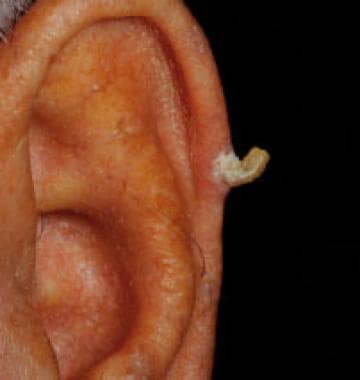
The image below depicts a typical presentation of a cutaneous horn.
See Nonmelanoma Skin Cancers You Need to Know, a Critical Images slideshow, to help correctly identify these lesions.
History
Cutaneous horns usually are asymptomatic. Because of their excessive height, they can be traumatized. This may result in inflammation at the base with resulting pain. Rapid growth is common in presentation.
Diagnostics
Determination of the histology of the underlying lesion is crucial to rule out malignancy and to determine proper follow-up, as no reliable clinical features clearly delineate the etiology of the lesion at the base of the horn. Diagnosis is confirmed with a skin biopsy. An adequate specimen usually can be obtained with a simple shave biopsy. The specimen must be of sufficient depth to ensure that the base of the epithelium is obtained for histologic examination.
Also see Histologic Findings.
Treatment
Treatment recommendation is contingent upon the type of lesion at the base. In order to rule out a malignancy, it is essential to perform a biopsy of the lesion that includes the base of the horn. In the case of benign lesions at the base of the horn, the biopsy is both diagnostic and therapeutic.
Excise malignancies with appropriate margins. Patients discovered to have horns with an underlying squamous cell carcinoma also should be evaluated for metastasis.
Local destruction with cryosurgery is first-line treatment for verruca vulgaris, actinic keratosis, and molluscum contagiosum. Benign lesions do not require any further therapy after the diagnostic biopsy.
In patients with squamous cell carcinoma or basal cell carcinoma, follow-up examinations to screen for a recurrence or a new primary are recommended for the first 3 years after diagnosis. Other diagnoses require follow-up as routinely recommended for that entity.
Pathophysiology
Cutaneous horns usually arise on sun-exposed skin but can occur even in sun-protected areas. The hyperkeratosis that results in horn formation develops over the surface of a hyperproliferative lesion. While the protruding, compact keratin may be the most prominent clinical feature, it is the process at the base of the lesion that is of most importance.
Most often, this is a benign verruca or seborrheic keratosis; however, cutaneous horns complicate a number of conditions, including premalignant actinic keratoses and frank malignancy. More than half of all of the inciting lesions at the base of cutaneous horns are benign, and a further 23-37% are derived from actinic keratoses. A malignancy has been reported at the base of a cutaneous horn in up to 20% of lesions. [3, 4, 5, 6]
The true pathobiology for the formation of cutaneous horns over each underlying base lesion remains unknown.
Benign lesions associated with cutaneous horns include angiokeratoma, angioma, benign lichenoid keratosis, cutaneous leishmaniasis, [7] dermatofibroma, discoid lupus, [8] infundibular cyst, epidermal nevus, [9] epidermolytic acanthoma, fibroma, granular cell tumor, [10] inverted follicular keratosis, [11] keratotic and micaceous pseudoepitheliomatous balanitis, organoid nevus, prurigo nodularis, pyogenic granuloma, [12] sebaceous adenoma, seborrheic keratosis, trichilemmoma, [13] and verruca vulgaris. [14, 15]
Lesions with premalignant or malignant potential that may give rise to cutaneous horns include adenoacanthoma, actinic keratosis, arsenical keratosis, basal cell carcinoma, [16] Bowen disease, Kaposi sarcoma, [17] keratoacanthoma, [18, 19] malignant melanoma, [20, 21] Paget disease, [22] renal cell carcinoma, [23, 24] sebaceous carcinoma, [25, 26] and squamous cell carcinoma. [27]
Etiology
Malignant lesions at the base of the horn usually are squamous cell carcinoma, although basal cell carcinoma has been rarely reported. These are predominately precipitated by ultraviolet radiation. Rare tumors at the base include Paget disease of the breast, sebaceous adenoma, and granular cell tumor. The premalignant lesion, actinic keratosis, is a frequent finding at the base. The human papilloma virus most frequently causes infectious etiology resulting in a verruca vulgaris. [28] Molluscum contagiosum of the poxvirus group occasionally has formed a cutaneous horn. The only other infectious cause has been leishmaniasis.
Benign idiopathic causes are frequent and include seborrheic keratosis, epidermal nevus, trichilemmal cyst, trichilemmoma, prurigo nodule, and intradermal nevus.
Epidemiology
Frequency
While these lesions are almost uniformly referred to as “rare” in the literature, neither the true incidence nor prevalence has been well described. [18, 29]
Race-,sex-, and age-related information
Because of the proportion of cutaneous horns that arise from actinic keratoses and squamous cell carcinomas, races with lighter complexions tend to be preferentially affected. However, several cases of cutaneous horns have been reported in patients of darker complexion, including those of African and Mexican descent. [1, 30, 31]
A sex predilection for cutaneous horn has not been shown consistently. In men, the rate of malignancies at the base of the lesion is increased when compared with age-matched women, and some studies also have shown premalignant lesions to be slightly more common in men. [32]
The peak occurrence of cutaneous horn is in persons aged 60 years to mid 70s. Lesions with malignancy at the base occur more frequently in patients aged 70 years or older.
Prognosis
The prognosis is dependent upon the classification of the underlying proliferative lesion at the base of the horn (eg, actinic keratosis, squamous cell carcinoma)
Cutaneous horns often cause little physical discomfort unless struck or if arising in areas prone to physical irritation. Cosmetically, however, they can cause significant concern for the patient, as they can be socially disconcerting. In fact, historically, prior to modern medicine, people with giant cutaneous horns had been persecuted as having “magical power” and were often displayed as sideshow attractions. [18, 2]
The lesion at the base of the keratin mound is benign in the majority of cases. Malignancy is present in up to 20% of cases, with squamous cell carcinoma being the most common type. The incidence of squamous cell carcinoma increases to 33% when the cutaneous horn is present on the penis. [28, 33] Tenderness at the base of the lesion is often a clue to the presence of a possible underlying squamous cell carcinoma. [4] Bleeding at the base of the lesion, as well as larger size, have been suggested as an indication of underlying malignancy. [2, 32]
-
A typical presentation of a cutaneous horn on the ear.
-
An unusually large cutaneous horn extending from the ear.
-
Medium power view of the mixed hyperparakeratosis and hyperorthokeratotic keratin that makes up the actual horn (hematoxylin and eosin [H&E] stain, original magnification 20x).
-
Low power view of a cutaneous horn overlying an actinic keratosis (hematoxylin and eosin [H&E] stain, original magnification 20x).
