Practice Essentials
Dermatitis herpetiformis is an exquisitely pruritic eruption classically seen on the buttocks and the extensor surfaces of the arms and legs. Severe cases may involve larger surface areas.
Dermatitis herpetiformis is an autoimmune subepidermal blistering disorder that is often associated with a gluten-sensitive enteropathy (GSE). It is considered a cutaneous variant of celiac disease. It was described and named in 1884 by Dr. Louis Duhring at the University of Pennsylvania. [1] and is also known as Duhring disease or Duhring-Brocq disease. [2]
Dermatitis herpetiformis is characterized by grouped excoriations, erythematous, urticarial plaques, and papules with vesicles. The classic location for dermatitis herpetiformis lesions is on the extensor surfaces of the elbows, knees, buttocks, and back. It is extremely pruritic, and the vesicles are often excoriated to erosions by the time of physical examination, as shown in the image below.

Diagnosis requires direct immunofluorescence of a skin biopsy specimen showing deposition of immunoglobulin A (IgA) in a granular pattern in the upper papillary dermis. Although most patients are asymptomatic, greater than 90% have an associated gluten-sensitive enteropathy upon endoscopic examination. [3] Among patients with celiac disease, 15-25% develop dermatitis herpetiformis. The mainstays of treatment are dapsone and a gluten-free diet. A vertically oriented fibrillar staining pattern is noted in a subset of patients, with immune deposits along dermal microfibrils, creating a characteristic “picket fence” on immunofluorescence. This fibrillar pattern is present in a third of Japanese patients, and this group lacks the typical distribution of skin lesions and has a low association with celiac disease. [4] .
There is data to support that dermatitis herpetiformis and celiac disease can be associated with both T-cell and B-cell lymphoma.
Signs and symptoms
Patients typically present with a waxing and waning pruritic eruption on the extensor surfaces of the arms, knees, and buttocks. It may become generalized. Small vesicles may have been noted but have often been excoriated by the time of presentation to the physician. They may have associated worsening of disease with dietary intake of gluten. Many do not report any GI symptoms, even when prompted.
The diagnosis is suspected based on the distribution of the eruption.
Flesh-colored–to–erythematous excoriated papules or plaques with herpetiform (ie, small, clustered) vesicles are symmetrically distributed over extensor surfaces, including the elbows, knees, buttocks, and shoulders.
Dermatitis herpetiformis rarely occurs on the posterior (nuchal) scalp and face. Lesions occur infrequently on the oral mucosa, but males are more likely than females to have involvement of the oral and genital membranes. Palms and soles are spared. Digital purpura resembling vasculitis can occur. Erythematous papules and urticarialike plaques occur less frequently; bullae are rare.
The eruption is intensely pruritic; patients often present with erosions and crusts in the absence of vesicles, which have ruptured due to excoriation.
Typical symptoms include burning, stinging, and intense itching. Rarely, if ever, are patients totally asymptomatic, although the degree of itching varies.
See the image below.
Complications
Complications are related to the gluten-sensitive enteropathy, the risk of developing lymphomas, and the potential adverse effects of medications, particularly dapsone.
Diagnostics
Also see Workup.
The diagnosis of dermatitis herpetiformis (DH) is made on the basis of skin biopsy results. However, other tests can be performed depending on the presence of symptoms of associated syndromes. Serum markers, such as IgA endomysial antibodies, are negative in as many as 10-37% of patients with dermatitis herpetiformis. [5, 6] Arguments have been made in favor of testing for tissue transglutaminase for diagnosis, [7] but tissue transglutaminase enzyme-linked immunosorbent assay positivity can occur in many autoimmune diseases because of impurities and cross-reactivity. [8]
Management
Also see Treatment and Medication.
Treatment of dermatitis herpetiformis (DH) includes avoidance of gluten by consuming a gluten-free diet and pharmacotherapy. [9]
A gluten-free diet is a lifelong commitment, and adherence to a strict diet is difficult to achieve. Improvement of skin disease with a gluten free diet takes several months. Gluten is present in various foods that are consumed on an everyday basis, most importantly wheat, barley, and rye. Concern has surrounded oats containing gluten, but studies have shown that consumption of a moderate amount of oats does not worsen dermatitis herpetiformis or celiac disease. [10, 11] Contamination of gluten-free products with gluten remains a potential problem. [12] Nutritional supplementation with multivitamins and iron may be prudent in patients on a strict gluten-free diet. [13]
Dapsone (diaminodiphenyl sulfone) and sulfapyridine (no longer available in most countries) are the primary medications used to treat dermatitis herpetiformis. The exact mechanism of action is unknown but is thought to be related to inhibition of neutrophil migration and function. Patients report a symptomatic improvement within hours after initiation of dapsone therapy. Patients should be monitored for the adverse effects of dapsone, primarily hemolytic anemia, methemoglobinemia, agranulocytosis, and neuropathy. For patients unable to tolerate dapsone, particularly those who develop hemolysis, sulfapyridine may be substituted. New lesion formation is suppressed for up to 2 days after a dose of dapsone; however, dapsone does not improve GI mucosal pathology.
Pathophysiology
Dermatitis herpetiformis is the result of an immunologic response to chronic stimulation of the gut mucosa by dietary gluten. [14] It is a complex disease of the skin caused by the deposition of IgA in the papillary dermis; this triggers an immunologic cascade resulting in neutrophil recruitment, complement activation, and a variety of other immunologic responses.
An underlying genetic predisposition to the development of dermatitis herpetiformis has been demonstrated. Both dermatitis herpetiformis and celiac disease (CD) are associated with an increased expression of HLA-A1, HLA-B8, HLA-DR3, and HLA-DQ2/DQ8 haplotypes. First-degree relatives of patients with dermatitis herpetiformis and celiac disease have a 15- times higher likelihood of developing either disease [15] . Environmental factors are also important; monozygotic twins may have dermatitis herpetiformis, celiac disease, and/or gluten-sensitive enteropathy with variable symptomatology.
The leading theory for dermatitis herpetiformis is that it is a late manifestation of chronic, subclinical celiac disease. A genetic predisposition for gluten sensitivity, coupled with a diet high in gluten, leads to the formation of IgA antibodies against gluten-tissue transglutaminase (t-TG or TG2), which is found in the gut. These antibodiescirculate and cross-react with epidermal transglutaminase (e-TG or TG3), [16] which is highly homologous with t-TG. Serum from patients with gluten-sensitive enteropathy, with or without skin disease, contains IgA antibodies to both skin and gut types. [17] Deposition of IgA and e-TG complexes in the papillary dermis triggers the immunne response that results in the lesions of dermatitis herpetiformis.
In patients with gluten-sensitive enteropathy, levels of circulating antibodies to t-TG and e-TG have been found to correlate with each other, and both appear to correlate with the extent of enteropathy. [18] Most patients with dermatitis herpetiformis have histologic evidence of enteropathy, even in the absence of symptoms of malabsorption. In one study, all dermatitis herpetiformis patients had increased intestinal permeability (as measured by the lactulose/mannitol ratio) and up-regulation of zonulin, a regulator of tight junctions. [19] Thus, increased expression of zonulin may be involved in the pathogenesis of enteropathy in patients with dermatitis herpetiformis.
Co-localized IgA and eTG deposits have been demonstrated in the papillary dermis in patients with dermatitis herpetiformis and, to lesser extent, in the healthy skin of gluten-sensitive enteropathy patients. [20] eTG has not been demonstrated in normal papillary dermis, suggesting it is part of the circulating complex that is deposited in the papillary dermis, rather than originating from the papillary dermis.
Cutaneous IgA deposits in dermatitis herpetiformis have been shown to function in vitro as a ligand for neutrophil migration and attachment. Although IgA deposition is pivotal for disease, an increased serum IgA is not necessary for pathogenesis; in fact, case reports have described dermatitis herpetiformis in patients with a partial IgA deficiency. [21] When the disease is active, circulating neutrophils express a higher level of CD11b on the cell surface and demonstrate an increased ability to bind IgA. The characteristic histologic finding of dermatitis herpetiformis is neutrophil accumulation at the dermoepidermal junction, frequently localizing to the papillary tips of the basement membrane zone.
Gluten has been shown to induce interleukin-8 (IL-8), a potent activator of neutrophils. [22]
Collagenase and stromelysin 1 may be induced in basal keratinocytes either by cytokines released from neutrophils or by contact with keratin from damaged basement membrane matrix. Stromelysin 1 may contribute to blister formation. Deposits of C3 may also be present in a granular pattern at the dermo-epidermal junction.
One study found levels of E-selectin mRNA expression in normal-appearing skin of patients with dermatitis herpetiformis to be 1271 times greater that that of controls. [23] Additionally, the same study observed increased soluble E-selectin, IgA antitissue transglutaminase antibodies, tumor necrosis factor-alpha, and serum interleukin 8 (IL-8) levels in patients with dermatitis herpetiformis, providing further evidence of endothelial cell activation and a systemic inflammatory response as part of the pathogenic mechanism of the disease. Mild local trauma may also induce the release of cytokines and attract the partially primed or activated neutrophils, which is consistent with the typical location of dermatitis herpetiformis lesions on frequently traumatized areas, such as the knees and elbows.
Deposits of C3 also may be present in a similar pattern at the dermoepidermal junction. The membrane attack complex, C5-C9, also has been identified in perilesional skin, although it may be inactive and not contribute to cell lysis. [24]
A 2009 study showed an increased expression of disintegrin and metalloproteinase (ADAMs) 8, 10, 15, and 17 in lesional skin of patients with dermatitis herpetiformis compared with controls. [25] The high affinity of ADAMs for the basement membrane led the authors to hypothesize a role in blister formation in dermatitis herpetiformis.
Hormonal factors may also play a role in the pathogenesis of dermatitis herpetiformis, and reports describe dermatitis herpetiformis induced by treatment with leuprolide acetate, a gonadotropin-releasing hormone analog. [26] Androgens have a suppressive effect on immune activity, including decreased autoimmunity, and androgen deficient states may be a potential trigger for dermatitis herpetiformis exacerbation. Exacerbation of dermatitis herpetiformis by oral contraceptives has also been reported.
Apoptosis may contribute to the pathogenesis of epidermal changes in dermatitis herpetiformis, and research demonstrates a markedly increased apoptotic rate within the epidermal compartment in dermatitis herpetiformis. [27] In addition, Bax and Bcl-2 proteins are increased in the dermal perivascular compartment and Fas proteins showed epidermal staining in dermatitis herpetiformis lesions.
Keratinocytes express elafin to down-regulate neutrophil-mediated inflammatory responses. Patients with dermatitis herpetiformis have deficient expression of elafin. [28]
Etiology
Dermatitis herpetiformis is generally accepted as a cutaneous manifestation of celiac disease. The standard criterion for the diagnosis of dermatitis herpetiformis is on skin biopsy. Direct immunofluorescence shows granular deposits of IgA in normal-appearing perilesional skin. It is positive in 92.4% of patients.
Gluten is a protein present in grasses of the species Triticeae, which includes barley, rye, and wheat. Gliadin protein in these grains are high-affinity substrates for tissue transglutaminase (TTG). Gliadins can also be found in rice, corn, and oats, but these proteins are poor substrates for tTG and thus these tend to be tolerated. Buckwheat is also tolerated. [29, 30] Strict compliance with a gluten-free diet results in normalization of the small bowel mucosal changes and control of the cutaneous manifestations of dermatitis herpetiformis in most patients and levels of circulating antibodies also tend to normalize. Resumption of gluten in the diet results in return of the characteristic skin disease.
Although cornstarch does not contain gluten, two case reports describe patients with well-controlled dermatitis herpetiformis who had disease flares after ingesting cornstarch. [31] Ingestion of iodide-rich dietary supplements can also cause a flare. [32]
The gluten-sensitive enteropathy does not cause symptoms in most dermatitis herpetiformis patients. Less than 10% exhibit symptoms of bloating, diarrhea, or malabsorption. However, greater than 90% show abnormalities upon endoscopic examination. Two thirds have villous atrophy detected on intestinal biopsy specimens. The other third shows elevated intraepithelial lymphocyte counts, increased T-cell receptor gamma/delta intraepithelial lymphocyte counts, or both.
Mild steatorrhea or other signs of mild malabsorption (eg, altered D-xylose absorption, iron or folate deficiency) can be demonstrated in 20-30% of patients with dermatitis herpetiformis.
Patients with dermatitis herpetiformis and no apparent GI disease can be induced into developing dermatitis herpetiformis by increasing gluten intake, which is often termed latent gluten-sensitive enteropathy.
IgA circulating immune complexes are present in 25-35% of patients with dermatitis herpetiformis, although no association with disease severity has been noted. These immune complexes also have been noted in patients with isolated gluten-sensitive enteropathy and are believed to be related to the presence of the gut disease. [18]
IgA antibodies to gliadin (a portion of wheat protein), reticulum, and smooth muscle endomysium have also been noted in patients with dermatitis herpetiformis and in those with isolated gluten-sensitive enteropathy.
IgA endomysial antibodies are most specific for gluten sensitivity and are found in 80% of patients with dermatitis herpetiformis and greater than 95% of patients with celiac disease. The presence of IgA antiendomysial antibodies correlates with the extent of the gut disease; [18, 33] however, some dermatitis herpetiformis patients do not have detectable IgA antiendomysial antibodies, even during episodes of active skin disease.
Patients with bullous pemphigoid, [34] cicatricial pemphigoid, Henoch-Schönlein purpura, and alcoholic liver disease also may have IgA deposits in normal skin; however, the pattern of IgA deposits is different from that seen in patients with dermatitis herpetiformis.
In patients with dermatitis herpetiformis, 10-15% of their first-degree relatives have dermatitis herpetiformis or celiac disease. HLA studies have conclusively established the presence of a genetic predisposition for dermatitis herpetiformis. Patients with dermatitis herpetiformis have an increased expression of the HLA-A1, HLA-B8, HLA-DR3, and HLA-DQ2 haplotypes. This is identical to the HLA association found in patients with isolated gluten-sensitive enteropathy. Most persons with these HLA types do not have dermatitis herpetiformis or gluten-sensitive enteropathy. Associations of HLA and dermatitis herpetiformis are as follows:
-
For HLA-B8: Association with dermatitis herpetiformis is 58-87%, versus 20-30% for control patients.
-
For HLA-DR3: Association with dermatitis herpetiformis is 90-95%, versus 23% for control patients.
-
For HLA-DQ2: Association with dermatitis herpetiformis is 95-100%, versus 40% for control patients.
Other associations
Associated GI conditions include gluten enteropathy, gastric atrophy, gastric hypochlorhydria, and pernicious anemia.
Associated autoimmune diseases include dermatomyositis, type 1 diabetes mellitus, myasthenia gravis, rheumatoid arthritis, Sjögren syndrome, systemic lupus erythematosus, and thyroid abnormalities. Thyroid abnormalities are present in as many as 50% of dermatitis herpetiformis patients and include hypothyroidism, hyperthyroidism, thyroid nodules, and thyroid cancer. [35]
Neurologic manifestations such as ataxia have been rarely described. [36]
Associated neoplastic conditions include GI lymphomas and non-Hodgkin lymphoma; patients are at increased risk of developing these cancers. [37] A gluten-free diet may reduce the incidence of dermatitis herpetiformis–associated lymphomas.
Celiac disease usually involves more severe and widespread intestinal involvement. Celiac disease has been associated with genetic abnormalities, including Down syndrome, Turner syndrome, and William syndrome. Liver disease, neurologic disorders, and other skin diseases are also increased in celiac disease, possibly due to common HLA regions on chromosome 6 or immune molecule cross-reactivity.
Gastric manipulation (surgery) may induce dermatitis herpetiformis.
Several chemicals have been associated with induction of dermatitis herpetiformis, including potassium iodide and cleaning solutions.
Case reports have described dermatitis herpetiformis induced by medications. Leuprolide acetate, inhibitors of tumor necrosis factor-alpha, anti-influenza medications, and progesterone-containing contraceptives have been reported in association with development of dermatitis herpetiformis. [26, 38] NSAIDs, specifically indomethacin and ibuprofen, have been reported to cause flares. [32]
Epidemiology
Frequency
United States
The only US study showed a dermatitis herpetiformis prevalence of 11.2 cases per 100,000 population.
International
Prevalence of dermatitis herpetiformis has been reported as high as 10 cases per 100,000 population.
Race
Dermatitis herpetiformis occurs more frequently in individuals of Northern European ancestry and is rare in Asians and persons of African descent. Dermatitis herpetiformis is most common in Ireland and Sweden. This can be attributed to the shared HLA associations of dermatitis herpetiformis and celiac disease including DQA1*0501 and B1*-02, which encode HLA-DQ2 heterodimers. It also corresponds to areas with high intake of wheat.
Sex
US studies show a male-to-female ratio of 1.44:1, but international studies have demonstrated a male-to-female ratio up to 2:1. In one study of patients with gluten-sensitive enteropathy, 16% of the men and 9% of the women had dermatitis herpetiformis. [39]
Age
Typically, the onset of dermatitis herpetiformis is in the second to fourth decade; however, persons of any age may be affected. [40] Dermatitis herpetiformis is rare in children.
Prognosis
Dermatitis herpetiformis (DH) is a life-long disease that waxes and wanrs. The prognosis is good for patients who can tolerate dapsone and the few who can maintain a gluten-free diet (which may decrease the risk of lymphoma).
Mortality/morbidity
In an English study, patients with dermatitis herpetiformis (152 total) were followed from the date of diagnosis to the end of 1989 for mortality and from 1971 or the date of diagnosis (if later) to 1986 for cancer incidence. [41] Death occurred in 38 patients younger than 85 years, slightly fewer than expected on the basis of national general population rates. Cancer incidence was significantly increased. Cancer of the small intestine caused 1 death, and lymphoma caused 1 death. Another English study, which compared 846 dermatitis herpetiformis patients with 4225 controls, found that dermatitis herpetiformis conferred no increased risk of lymphoproliferative cancer and no increase in fracture, malignancy, or mortality. [42]
A 30-year population-based study of 1147 celiac disease and dermatitis herpetiformis patients in Finland also revealed an overall good prognosis for patients with dermatitis herpetiformis. [43] The total occurrence of malignancies was equal to that of the general population in both celiac disease and dermatitis herpetiformis patients, but an increased incidence of lymphoma was noted among both celiac disease and dermatitis herpetiformis patients, with standardized incidence ratios of 3.2 and 6.0, respectively. Most were T-cell lymphomas, although B-cell lymphoma has occurred as well. Interestingly, a lower incidence of lymphoma was seen in the first-degree relatives of dermatitis herpetiformis patients. [44] Overall mortality was actually decreased in dermatitis herpetiformis patients compared with that in the general population.
Dermatitis herpetiformis lesions are extremely pruritic. Morbidity results from scarring, discomfort, and insomnia due to itching. Secondary infection may also develop, requiring antibiotic therapy.
Patient Education
Educate patients regarding the use of a gluten-free diet as well as the adverse effects and complications of dapsone. Patients should also be aware of medications, particularly NSAIDs, that can cause flares.
-
Light micrograph shows neutrophils in the dermal papillae, with fibrin deposition, neutrophil fragments, and edema (hematoxylin and eosin stain).
-
Papillary microabscesses form and progress to subepidermal vacuolization and vesicle formation in the lamina lucida, the weakest portion of the dermoepidermal junction (hematoxylin and eosin stain).
-
Classic vesicles of dermatitis herpetiformis.
-
Immunofluorescence showing immunoglobulin A at the dermoepidermal junction (direct immunofluorescence stain).
-
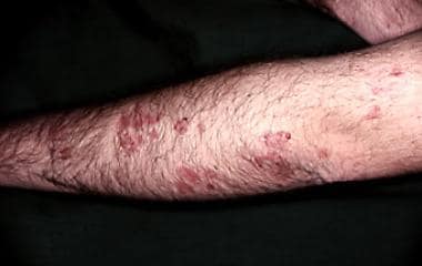
Polymorphic lesions on extensor surface of arm.
