Background
The shoulder is the most frequently dislocated joint. [1] It moves almost without restriction but pays the price of stability. The shoulder's integrity is maintained by the glenohumeral joint capsule, the cartilaginous glenoid labrum (which extends the shallow glenoid fossa), and muscles of the rotator cuff. Most dislocations are anterior, but less frequently, posterior, inferior (luxatio erecta), superior, and intrathoracic dislocations are also possible. [2, 3] Arthroscopic stabilization is performed in nearly 90% of shoulder stabilization surgeries in the United States. [4] Patients with shoulder dislocation generally complain of severe shoulder pain and an associated decreased range of motion of the affected extremity.
Shoulder dislocations constitute up to 50% of all major joint dislocations. Anterior dislocations occur in as many as 97% of cases. Anterior displacement of the humeral head is the most common dislocation seen by emergency physicians and is depicted in the image below.
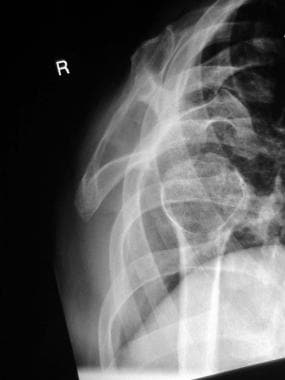 Y-view radiograph of the right shoulder shows anterior dislocation of the humeral head relative to the glenoid fossa.
Posterior displacement is the next most frequently occurring dislocation (2-4%). Inferior (luxatio erecta), superior, and intrathoracic dislocations are rare (< 1%)and are usually associated with complications. [5, 6, 7]
Anterior dislocation is characterized by subcoracoid position of the humeral head in the anteroposterior (AP) view. The dislocation is often more obvious in a scapular view, where the humeral head lies anterior to the "Y." In an axillary view, the "golf ball" (ie, humeral head) is said to have fallen anterior to the "tee" (ie, glenoid). In posterior dislocation, the AP view may show a normal walking stick contour of the humeral head, or it may resemble a light bulb or ice cream cone, depending on the degree of rotation. The scapular "Y" view reveals the humeral head behind the glenoid (the center of the "Y"). Arteriography, angiography, and Doppler flow studies may be used to evaluate suspected vascular injury. Electromyography (EMG) may be used later to evaluate nerve injuries. [8]
Procedural sedation and analgesia (PSA) protocols, intra-articular lidocaine, and ultrasound-guided brachial plexus nerve block assist in making reduction an easier and more comfortable procedure.
Etiology
Anterior shoulder dislocations usually result from abduction, extension, and external rotation, such as when preparing for a volleyball spike. [9] Falls on an outstretched hand are a common cause in older adults. The humeral head is forced out of the glenohumeral joint, rupturing or detaching the anterior capsule from its attachment to the head of the humerus or from its insertion to the edge of the glenoid fossa. This occurs with or without lateral detachment.
Posterior dislocations are caused by severe internal rotation and adduction. This type of dislocation usually occurs during a seizure, a fall on an outstretched arm, or electrocution. Occasionally, a severe direct blow may cause a posterior dislocation. Bilateral posterior dislocation is rare and almost always results from seizure activity. Misinterpretation of the radiograph appearance of a posterior dislocation may result in misdiagnosis as a soft tissue injury in up to 79% of cases.
Rare, but serious, inferior dislocations (luxatio erecta) may be due to axial force applied to an arm raised overhead, such as when a motorcycle collision victim tumbles to the ground. More commonly, the shoulder is dislocated inferiorly by indirect forces hyperabducting the arm. The neck of the humerus is levered against the acromion and the inferior capsule tears as the humeral head is forced out inferiorly. Luxatio erecta almost always has an associated fracture or soft-tissue injury. One series found 80% of patients to have fracture of the greater tuberosity or tear of the rotator cuff. Neurologic compromise was found in 60% of patients, with the axillary nerve the most commonly injured nerve. Inferior dislocations have the highest incidence (3.3%) of vascular compromise.
Epidemiology
United States statistics
In the United States, the incidence of shoulder dislocations is 23.9 per 100,000 person years, and approximately 85-98% of shoulder dislocations are anterior dislocations. Dislocated shoulders tend to occur more often in males than in females. In males, the peak age of incidence is 20-30 years (with a male-to-female ratio of 9:1), and in females it is 61-80 years (with a female-to-male ratio of 3:1). The incidence of proximal humerus fractures increases with age, with a population-adjusted incidence of 101 per 100,000 person years in those older than 65 years. [10]
Shoulder dislocation occurs more frequently in adolescents than in younger children because the weaker epiphyseal growth plates in children tend to fracture before dislocation occurs. In older adults, collagen fibers have fewer cross-links, making the joint capsule and supporting tendons and ligaments weaker and dislocation more likely. Anterior dislocation is most commonly seen in those aged 18-25 years resulting from sporting injury. The second most common age group to sustain anterior dislocation is the elderly, because of their susceptibility to falls. [10]
In an epidemiologic study of patients treated in emergency departments, Becker et al demonstrated that the majority of shoulder dislocations occurred in men (72.1%). More than half (52.5%) of shoulder dislocations were associated with sports activities, and most dislocations were related to participation in basketball (16.4%), American football (15.6%), or cycling (9.0%). [11]
In a study of shoulder dislocation data from the High School Reporting Information Online (RIO) and the National Collegiate Athletic Association (NCAA) Injury Surveillance Program (ISP) databases, high school athletes were found to have an overall shoulder dislocation rate of 2.04 per 100,000 athletic exposures, and college athletes had an overall injury rate of 2.58 per 100,000 athletic exposures. Surgery was performed in 28% of high school shoulder dislocations and 29.6% of college shoulder dislocations. [12]
A subsequent examination of data on shoulder dislocation from the High School RIO and NCAA ISP databases showed an overall incidence rate of 38.19 per 100,000 persons at risk among high school and college athletes. [13]
Congenital dislocation of the shoulder is a very rare condition, and the dislocation of the glenohumeral joint in infants is usually associated with a fracture or a neurologic problem (eg, brachial plexus injury). If there is no history of trauma or a brachial plexus injury, congenital dislocation should be considered as a possible diagnosis. [14]
International statistics
A Greek study examined the demographic data and recurrence rates of shoulder dislocations of 308 patients (170 men and 138 women) and found that the most frequent mechanism of injury was falling, and 92% of reductions were in the ED. The overall recurrence rate in all age groups was 50% but rose to almost 89% in the 14- to 20-year-old age group. [4]
A German study showed that the incidence of shoulder dislocations among professional football players is 9.1 per 1000 matches played. [15]
Prognosis
Age is a major factor in the likelihood of sustaining a recurrent shoulder dislocation. [16] Approximately 80-94% of patients younger than 20 years at the time of the initial dislocation have a recurrence. The major pathology in this age group is thought to be a Bankart lesion with associated inferior glenohumeral ligament injury.
Of patients younger than 40 years, 26-48% develop recurrent dislocation. The major pathology for this age group is thought to be disruption of the labral attachment of the glenohumeral ligaments. Dislocation recurs in only 0-10% of patients older than 40 years. Rotator cuff tear is the major pathology.
Minor trauma that results in a dislocation is associated with an 86% recurrence rate. Many orthopedic surgeons believe that more than one complete anterior dislocation justifies considering surgical repair.
There is general agreement that before being allowed to return to sports after anterior shoulder dislocation, athletes should be pain free and should demonstrate symmetric shoulder and bilateral scapular strength with functional range of motion. Usually, returning to play can occur 2-3 weeks after dislocation; however, athletes with in-season shoulder injury who return to play during the season have demonstrated recurrence rates of 37-90%. [9, 17]
Complications
Complications include fractures, soft tisue injuries, nerve injuries, and vascular injuries. [16, 18, 19, 20, 21, 22, 23]
Fractures and soft-tissue injuries
Hill-Sachs lesions occur when the edge of the glenoid causes an impaction fracture in the posterolateral aspect of the humeral head during anterior dislocation and in the anterolateral aspect in posterior dislocation (referred to as a "reverse Hill-Sachs" lesion).
A Bankart lesion is fracture of the anterior rim of the glenoid labrum associated with joint capsule rupture and inferior glenohumeral ligament injury. Significantly displaced anterior or posterior glenoid rim fractures require operative management. Most initial shoulder dislocations produce a Bankart lesion, particularly in younger patients.
Fracture of the greater tuberosity, acromion, coracoid, clavicle, and humeral neck also occur.
Rotator cuff traction injury is most common in elderly patients and in association with inferior dislocations. This is a commonly missed injury, with an average time of 7 months from injury to diagnosis of rotator cuff rupture in patients older than 40 years.
Patients who experience anterior shoulder dislocation are at increased risk for glenohumeral arthropathy. Average overall T1p values on MRI for humeral head cartilage in dislocated shoulders have been shown to be significantly greater than that in control patients. [23]
Nerve injury
Approximately 3% (and higher in some series) of dislocations involve injury to the axillary nerve. Injury may resolve spontaneously or require surgical exploration and possible nerve grafting.
Patients exhibit numbness in the area of the deltoid muscle and weakness with abduction and external rotation.
Axillary nerve injury does not change initial treatment, but pre-reduction and post-reduction neurologic examinations are important.
Radial nerve injury should also be determined. The axillary and radial nerves both arise from the posterior cord. The thumb, wrist, and elbow will be weak on extension, and the dorsal hand will be numb.
Vascular injury
Axillary artery injuries are rare but have been reported to occur with anterior, inferior, and intra-thoracic dislocations. Especially susceptible are older adults with atherosclerotic axillary arteries. Arterial injury may be associated with decreased radial pulse.
Lateral chest wall ecchymosis with associated axillary hematoma and bruit may be noted on physical examination.
Angiography should be considered with any brachial plexus injury.
-
Y-view radiograph of the right shoulder shows anterior dislocation of the humeral head relative to the glenoid fossa.
