Practice Essentials
Hidradenitis suppurativa (HS) is a disorder of the terminal follicular epithelium in the apocrine gland–bearing skin. This condition is a chronic disabling disorder that relentlessly progresses, frequently causing keloids, contractures, and immobility. (See the image below.)
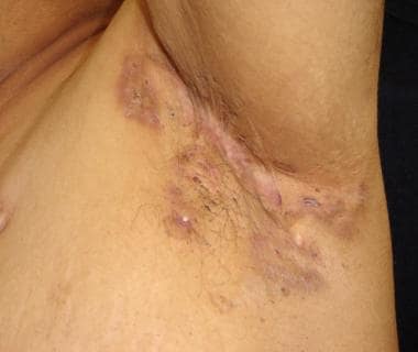 Close-up view of axillary hidradenitis suppurativa in a patient with pyoderma gangrenosum.
Presentation
HS usually occurs in otherwise healthy adolescents and adults. It rarely may begin before puberty. Hidradenitis suppurativa is characterized by comedolike follicular occlusion, chronic relapsing inflammation, mucopurulent discharge, and progressive scarring.
The onset of HS is insidious, with the earliest sign being erythema. Later, the lesions become painful. Arthropathy associated with HS may present with variable clinical features, ranging from asymmetric pauciarticular arthritis to a symmetric polyarthritis and/or polyarthralgia syndrome, as well as spondyloarthropathy. Moreover, epidemiological data also suggest an association of HS with other diseases, including metabolic syndrome. Therefore, it is important to approach HS as a systemic disease with an interprofessional team. [1, 2, 3, 4, 5]
See Clinical Presentation for more detail.
The diagnosis is primarily clinical, no pathognomonic test exists, and biopsy is rarely required, especially in well-developed lesions. [6] The consensus approach indicates that three key elements are required to diagnose HS: typical lesions, characteristic distribution, and recurrence. Arbitrarily, two recurrences over a period of 6 months have been used as a qualifier for a diagnosis.
Pathophysiology
Hidradenitis suppurativa (HS) has traditionally been considered a disorder of the apocrine glands. It was first described as a distinct entity in 1839, when Velpeau reported a patient with superficial abscess formation in the axillary, mammary, and perianal regions. [22] In 1854, Verneuil associated the suppurative process with the sweat glands, and the condition was given its current name. For many years, the condition was described as Verneuil disease, but it subsequently became known as HS. Not having performed any histopathologic studies himself, Verneuil conceded that his conclusion was based purely on the characteristic distribution of the condition. [23]
In 1922, Schiefferdecker classified the sweat glands as eccrine and apocrine, and he subsequently localized HS to the apocrine glands. [24] In 1939, Brunsting provided a detailed description of the histologic features of HS. He observed the primary cellular reaction in the lumen of the apocrine glands and in the neighboring periglandular connective tissue. Detailing the clinical features of HS, Brunsting highlighted its frequent association with acne. Noting that HS, dissecting cellulitis of the scalp and the neck, and acne conglobata commonly occur in the same patient, he thought that the central pathogenetic event in all three was a tendency for follicular hyperkeratinization with secondary bacterial infection. [25]
In 1956, Pillsbury et al combined acne conglobata, HS, and dissecting cellulitis under the term follicular occlusion triad. [26] The only flaw in the concept was the focus on apocrine sweat gland involvement. In 1975, Plewig and Kligman added pilonidal sinus as another component to the ensemble, and they introduced the term acne tetrad. [27] They stated that HS was a misnomer because of the lack of apocrine gland involvement, but they did not present a detailed explanation.
In 1989, Plewig and Steger suggested the term acne inversa as an inclusive and accurate name for what was previously called the follicular occlusion triad (or tetrad). [28] Eventually, HS was accepted as an acneiform disorder that begins with follicular occlusion rather than an infection of the sweat glands. [29, 30]
HS is actually a defect of the follicular epithelium; consequently, there has been a movement toward calling the disease acne inversa instead. The term acne inversa links the pathogenesis to acne and reflects the fact that this conditionis an expression of follicular occlusion in localizations inverse to acne vulgaris. [31] However, HS differs from acne in that it is not associated with any increase in sebaceous secretions.
Etiology
The exact etiology of HS remains obscure. All proposed etiologic factors, such as occlusion and bacterial infection, genetics, host defense defects, hormones, cigarette smoking, and irritants, are likely to be only secondary factors. The primary events in the hair follicles of the affected areas remain unidentified. [32] Also, thickened skin may play a role in the pathogenesis of HS (see Imaging Studies).
The classic view of hidradenitis suppurativa is that it is an occlusive and pyogenic disease of the apocrine glands, a hypothesis that seemed to be confirmed with its experimental reproduction by Shelley and Cahn in 1955. [33] After manually depilating the skin and applying atropine-impregnated tape, they induced initial keratinous obstruction, dilatation, and inflammation of the apocrine duct, which occurred in only 25% of the experimental lesions. No progression to the characteristically chronic condition of hidradenitis suppurativa occurred. [33]
In other studies, hidradenitis suppurativa is identified as a disorder of follicular occlusion rather than apocrine occlusion. [29, 30] Yu and Cook found inflammatory changes involving the apocrine glands in only one third of cases; these occurred only when the inflammation extensively involved the hair follicles and eccrine glands. [29] Attanoos et al reported follicular occlusion in all specimens, when compared with controls, regardless of the disease duration. The inflammation of the apocrine glands did not occur in the absence of an adjacent folliculitis; thus, apocrine gland involvement was incidental or secondary. [30] Therefore, hidradenitis suppurativa is best considered a disorder of the terminal follicular epithelium in the apocrine gland–bearing skin.
The earliest change is plugging, which occurs in follicular hyperkeratosis with infundibulofolliculitis. This obstructs the apocrine gland ducts and perifolliculitis around the ducts. Whether this initial inflammatory change is due to a bacterial infection or factors similar to those involved in acne formation is not known.
In the later stages of hidradenitis suppurativa, bacterial infection seems to be a risk factor for the destructive scarring and extension of hidradenitis suppurativa lesions, and, once the sinuses have formed, the risk of secondary infection is obvious. [34]
Among the most commonly isolated bacteria are coagulase-negative staphylococci and anaerobic bacteria. [3, 34] Sweat ducts can become occluded with periodic acid-Schiff (PAS)–positive extracellular polysaccharide substance, the cause of which has been suggested to be Staphylococcus epidermidis. [35] Such strains induced miliaria under experimental conditions. A similar mechanism may be important in the pathogenesis of hidradenitis suppurativa. [34]
The earliest inflammatory event in hidradenitis suppurativa is the rupture of the follicular epithelium. The cause of the rupture is not known, although friction in intertriginous locations may be a contributing factor. The rupture is followed by the spillage of foreign-body material into the dermis, which initiates an inflammatory response, resulting in foreign-body granuloma formation. Epithelial strands form draining sinuses in this inflammatory tissue. Colonization with bacteria, usually coagulase-negative staphylococci, can aggravate the chronic inflammation. [32, 34]
Regarding the current controversies nonfollicular-based theories on what causes hidradenitis suppurativa, some authors suggest the following [36] :
-
The apocrine glands may play a role in hidradenitis suppurativa since an abnormal secretion (either the excess or absence) could be influencing an effect on the acroinfundibulum, distal from the gland itself.
-
The sinus tract formation is an early feature of hidradenitis suppurativa, arising not from hair follicles but rather from invaginations of epidermis as cysts.
-
Resident bacteria, such as coagulase-negative staphylococci may cause adherence of the epidermis in the closed serrated tissue of intertriginous areas, leading to the formation of cysts and hidradenitis suppurativa lesions.
Although the inciting influences for the follicular occlusion and sinus tract formation have not been fully elucidated, genetic factors may play a role. More than 15 years ago, the existence of a familial form of hidradenitis suppurativa with autosomal dominant inheritance was proposed. The disease frequency among first-degree relatives of patients with familial hidradenitis suppurativa was 34%. [37]
Heterozygous mutations were have been reported in the gamma-secretase genes PSENEN, PSEN1, and NCSTN in some patients with hidradenitis suppurativa. [38] One member of this gene complex, termed nicastrin (NCSTN), lies on chromosome 1 within the previously reported region 1p21.1-1q25.3 on chromosome 1, where the AI (acne inversa) gene (one putative genetic locus in hidradenitis suppurativa), was mapped. [39]
Gamma-secretase is a transmembrane protease composed of four essential protein subunits: one catalytic presenilin (PSEN1) subunit and three cofactor subunits [presenilin enhancer 2 (PSENEN), nicastrin (NCSTN), and anterior pharynx defective 1 (APH1)]. Gamma-secretase appears integral to normal skin function, through effects on notch signaling, such as the biological role in the hair follicle. The pattern of mutations suggests that loss of function of components of the gamma-secretase complex underlies the disease: follicular keratinization, follicular atrophy, the formation of epidermal cysts, absence of sebaceous glands, and epidermal hyperplasia. Most frequently, gamma-secretase mutations corresponded to nicastrin (NCSTN) mutant proteins.
Although these mutations only appear in a minority of cases of hidradenitis suppurativa, their identification delineated the first genetically defined clinical subgroup of patients with hidradenitis suppurativa and primary involvement of the hair follicle instead apocrine gland, suggesting that the primary event is follicular occlusion. [38] Owing to research efforts over the last 3 years, the genetic reasons for the disease are known in approximately 5% of the hidradenitis suppurativa patients. They are different heterozygous mutations in subunits of gamma-secretase. Genetic factors might influence not only the appearance of hidradenitis suppurativa, but also the phenotype of disease. [3]
Host-defense defects
Host-defense defects in patients with hidradenitis suppurativa are suspected but not proven. [40]
Hyperreactive neutrophils have been proposed to be of pathophysiologic importance in many chronic inflammatory diseases involving the destruction of the surrounding tissue by the simultaneous release of reactive oxygen species and active proteases.
The release of oxygen radicals from peripheral neutrophils that are activated in vitro was studied in patients with inactive hidradenitis suppurativa. The generation of free oxygen radicals after the stimulation of peripheral neutrophils with protein kinase C (PKC) activator and phorbolmyristate acetate (PMA) was significantly higher in patients with hidradenitis suppurativa than in healthy control subjects.
The higher sensitivity of PKC to PMA in patients with hidradenitis suppurativa is unlikely to have been induced by the disease and its local lesions because the systemic effects were minor in the quiescent state. Therefore, a defect in the function of the neutrophils might be of pathogenic importance in hidradenitis suppurativa. [40]
A reduction in the percentage of natural killer cells over time and a lower response of monocytes to triggering by bacterial components were found in patients with hidradenitis suppurativa. Further study is needed to elucidate if these changes are related to an autoimmune mechanism in the pathogenesis of hidradenitis suppurativa. [41]
Toll-like receptors play an integral role in the innate immune response to bacteria. A highly increased expression of toll-like receptor 2 (TLR2) by CD68+ macrophages and CD209+ dendritic cells in acne inverse skin lesions was found. [42]
Apart from Toll-like receptor 2 activating microbial products as an important trigger factor in the chronic inflammatory process, the inflammatory reactions leading to hidradenitis suppurativa are only poorly understood, but they show many similarities with other inflammatory reactions such as with psoriasis; proinflammatory cytokines interleukin (IL)‒12 and IL-23 are mediators in autoimmune tissue destruction and are abundantly expressed by macrophages in hidradenitis suppurativa; IL-23 has been shown to be involved in the induction of a T-helper cell subset named Th17; antimicrobial peptides beta-defensin 2, Psoriasin, and cathelicidin are highly up-regulated in lesions of psoriasis and in hidradenitis suppurativa, which may at least in part, explain the clinical finding that hidradenitis suppurativa patients only rarely have skin infections. [3]
Hormonal effects
Apocrine sweat glands are stimulated by androgen and suppressed by estrogen. Evidence for the hormonal effects in hidradenitis suppurativa exists; however, the exact roles of androgens in the pathogenesis of hidradenitis suppurativa remain controversial, and they may prove to be secondary. [14, 43, 44, 45]
Many women describe a worsening of the condition with menses, whereas others report alleviation with pregnancy, followed by postmenstrual flaring. These observations suggest that the low level of estrogen predisposes women to disease activity. Premenstrual flare has been shown to be unrelated to menstrual disturbances. These flares were not predictive of the overall course, and the effect of pregnancy was not constant. [46] The female preponderance may also be explained by other specific factors such as the estrogenic influence on inflammation. [47]
The following evidence supports the association of androgens and hidradenitis suppurativa: the disease is rarely present until after puberty begins, [48] Hidradenitis suppurativa is not present in eunuchs or eunuchoids, and hidradenitis suppurativa may occur as the presenting feature of premature adrenarche. [49] Also, antiandrogen therapy is of some benefit in patients with hidradenitis suppurativa. [50]
The relationship between hidradenitis suppurativa and hyperandrogenism is largely based on the finding that the free androgen index is increased due to a low level of sex hormone–binding globulin (SHBG). SHBG is now believed to be regulated by factors that influence body weight. [46]
Hirsutism and obesity are common findings among women with hidradenitis suppurativa. [51] Obesity can alter sex hormone metabolism, leading to an androgen-excess state. Excessive androgens can enhance keratin production and coarsening of the hair shaft, promoting follicular occlusion. [30] Although hirsutism, obesity, and acne among women with hidradenitis suppurativa are more common than expected, [46] the incidences are not significantly different from those in the general population. [43, 44, 52] However, neither evidence for biochemical hyperandrogenism nor suppression of SHBG has been shown in women with hidradenitis suppurativa when compared with age-, body weight-, and hirsuties-matched controls. [46] In one study, obesity and oral contraceptive use were not common or pronounced among patients; this observation suggested that, while these parameters may influence preexisting disease, they are unlikely to be pathogenically important. [53] In obesity, increased skin-to-skin contact may promote follicular hyperkeratosis. [30]
An abnormal end-organ response to normal circulating levels of androgens is proposed. [50] The normal apocrine gland contains 5-alpha reductase, which converts testosterone to the potent androgen dihydrotestosterone. Finasteride is a competitive inhibitor of the 5-alpha reductase type II isoenzyme. The benefits of finasteride in some patients with recalcitrant and persistent forms of hidradenitis suppurativa, [50] raised the issue of whether 5-alpha reductase type I or type II is expressed in this disease and whether this expression applies to the apocrine gland, sebaceous gland, or both. On the other hand, sebum excretion is not an important factor in the development of hidradenitis suppurativa. [52] Thus, hormonal influence remains controversial.
Other
Cigarette smoking may be among the major triggering factors in hidradenitis suppurativa, and its cessation should be encouraged, although whether cessation improves the course of disease is unknown. [14, 54] It remains unclear precisely what pathogenetic mechanism would be responsible for the effect of smoking in the manifestation of acne inversa, but an altered chemotaxis of polymorphic neutrophils could play a part. [55]
Chemical irritants (eg, deodorants) and mechanical irritation (eg, depilation, shaving) have been considered risk factors. However, in one study, no significant difference was found in patients who were exposed to these factors compared with age-matched control subjects. [56] Factors associated with disease activity, such as heat, sweating, stress, and menstruation in females, are the most commonly cited factors that exacerbate the disease. In one study, 32% of responders observed a deterioration in their disease during the summer. [7]
Hidradenitis suppurativa is rarely a side effect of drug use, but oral contraceptives and lithium have been associated with its development. [47]
Lung and buccal cancer are more common among hidradenitis suppurativa patients than in the general population as would be expected with increased tobacco smoking and chewing. [47]
Hidradenitis suppurativa may rarely be complicated by hidradenocarcinoma. [57]
In conclusion, being overweight and obesity are clearly associated factors in hidradenitis suppurativa; their role as severity factors is highly probable. The relationship between the severity of hidradenitis suppurativa and cigarette smoking has been studied, with conflicting results. Mechanical stress as a trigger for hidradenitis suppurativa has still to be proven. [3]
Epidemiology
United States and international statistics
In the United States, the prevalence of HS appears to be 1-2% in the general population.
Worldwide, the prevalence of hidradenitis suppurativa appears to be 1% of the general population [14, 4] ; it was 4% in a group of young adults who were treated at a clinic for sexually transmitted diseases. [14] A 2008 study showed that the prevalence among persons aged 55 years and older was significantly lower than that in younger age groups (0.5% vs 1.4%). [54] . Studies that provide prevalence or incidence estimations have been performed under different settings (hospital vs population-based) and in different periods. [4]
Age-, sex-, and race-related demographics
In the great majority of cases, the onset of HS comes between the ages of 11 and 50 years (average patient age, 23 y) [7] ; it comes before age 11 years in fewer than 2% of cases. [48] It is extremely rare for HS to occur before puberty, but it has been suggested that a prepubertal onset is not uncommon in severely affected patients [58, 59] or after menopause. [46]
Although HS is widely considered to occur more frequently in females than in males, with a ratio as high as 2-5:1, [7, 53] reports on sex prevalence have been controversial. [7, 60, 61] A review by Wang et al noted that in fact, females were more likely to have a family history of HS, and men tended to have more severe disease and associated severe acne. [61] Active genitofemoral lesions occur significantly more often in females, whereas perianal involvement is more common in males. No sex difference is seen in the axillary lesions. Comedones have been suggested as precursor lesions for HS, and they appear to be equally distributed in both sexes and sites. [47]
Most authors report no specific racial predilection. One report suggested an increased observed incidence in Blacks, possibly because Blacks have a greater density of apocrine glands than Whites do. [16]
Prognosis
In general, HS is a chronic disease, as underscored by the finding that 90% of patients in one large series still had active disease in the last year despite an average disease duration of nearly 19 years. [7] The impression of HS as a relentlessly progressive disorder may be explained by the finding that almost two thirds of patients acknowledge the existence of persistently painful boils that failed to heal. Possibly, new boils develop at an unchanged rate throughout the course of the disease, but some fail to subside in the usual manner and become chronic. [7]
With rare exceptions, surgical intervention is sufficient to stop the disease. [62] Shame, frustration, and despair may cause patients to delay radical surgical procedures. No single treatment has shown overwhelmingly positive outcomes. [63] Spontaneous resolution is rare. [10]
Specific factors appear to influence the prognosis. Larger excisions may offer a better outcome. Better results can be obtained by leaving wounds to secondary healing. Perianal surgery, axillary surgery, and older patient age are associated with lower recurrence rates, irrespective of the preoperative duration. [47]
The recurrence rate in patients treated with radical surgery varies considerably, depending on the site affected; the highest rate is 50% in the submammary region. [16] An overall recurrence rate of 2.5% has been estimated after wide surgical excision (median postoperative follow-up, 36 mo). [10]
The postoperative relapse risk is higher in women after surgery under general anaesthesia in severe HS. [59]
If untreated, HS causes significant morbidity, particularly in women and patients with moderate and severe disease who are more likely to experience a significant diagnostic delay (>2 y). [64]
HS has significant socioeconomic effects as well. In the Danish population, Jemec et al documented an annual average of 2.7 lost workdays due to HS (overall workdays lost, 7.5). [53] The general self-reported level of health, which is well correlated with more objective parameters of morbidity, was also significantly worse among HS patients than among healthy control subjects. The mean Dermatology Life Quality Index (DLQI) score is higher for HS than for previously studied skin diseases, indicating significant morbidity for those affected. [7]
Patient Education
Patients should be educated about the initial treatments, which include the following [19] (see Treatment):
-
Practicing proper hygiene
-
Using soaps and antiseptic and antiperspirant agents
-
Using warm compresses
-
Wearing loose-fitting clothing
-
Smoking cessation
Patients who are obese should be educated about weight loss (see Diet). Additionally, patients should be educated about activities that may provide some relief of their condition [7] (see Activity). These activities include swimming, bathing, and avoiding smoking.
-
Vulvar hidradenitis suppurativa.
-
Vulvar and inguinal indurations.
-
Sinus tract.
-
Draining sinus tract.
-
Axillary hidradenitis suppurativa in a patient with pyoderma gangrenosum.
-
Close-up view of axillary hidradenitis suppurativa in a patient with pyoderma gangrenosum.
-
Submammary hidradenitis suppurativa in a patient with pyoderma gangrenosum.
-
Double-ended-comedones. Hidradenitis suppurativa in a patient with pyoderma gangrenosum.
-
Inguinal hidradenitis suppurativa in a patient with pyoderma gangrenosum.
-
Close-up view of inguinal hidradenitis suppurativa in a patient with pyoderma gangrenosum.
-
Pyoderma gangrenosum in a patient with hidradenitis suppurativa.
-
Close-up view of pyoderma gangrenosum in a patient with hidradenitis suppurativa.
-
Coexisting hidradenitis suppurativa and pyoderma gangrenosum.
-
Coexisting hidradenitis suppurativa and pyoderma gangrenosum.
-
Hidradenitis suppurativa in a patient with pyoderma gangrenosum.
Tables

What would you like to print?
- Overview
- Presentation
- DDx
- Workup
- Treatment
- Guidelines
- Medication
- Tetracyclines
- Antibiotics, Lincosamide
- Macrolides
- Antibiotics, Combos
- Antitubercular Agents
- Antileprosy Agents
- Retinoid-like Agents
- Corticosteroids
- Aldosterone Antagonists, Selective
- Monoclonal Antibodies
- Interleukin Inhibitors
- Estrogens/Progestins
- 5-Alpha-Reductase Inhibitors
- Trace Elements/Metals
- Show All
- Media Gallery
- References