Continually Updated Clinical Reference
 
 
  All Sources     eMedicine     Medscape     Drug Reference     MEDLINE
 
You are in: eMedicine Specialties > Emergency Medicine > Trauma And Orthopedics

Knee Injury, Soft Tissue

Last Updated: May 8, 2006
Email to a Colleague
Synonyms and related keywords: soft tissue knee injury, soft-tissue knee injury, knee injury, knee pain, knee strain, knee ligaments, medial collateral ligament, MCL, lateral collateral ligament, LCL, anterior cruciate ligament, ACL, ACL tear, posterior cruciate ligament, PCL, patella, knee joint, PCL tear, knee sprain, knee dislocations, compartment syndrome, chronic knee instability, degenerative joint disease, chondromalacia patellae, patellar malalignment syndrome, Larsen-Johansson disease of the patella, inferior pole patellar chondropathy, meniscal tear, ACL-deficient knee, Baker cyst, bursitis, tendinitis, knee effusion, cruciate ligament tear, knee dislocation, articular fracture, meniscal injuries, acute ligament rupture, capsular disruption, prepatellar bursitis, meniscal cystic changes, outgrowth of a popliteal cyst, tears in the meniscus, injury of the cruciate ligaments, osteochondral fracture, antalgic gait, Bragard sign, positive patellar apprehension sign, rupture in the quadriceps, Osgood-Schlatter syndrome, valgus stress, varus stress, Lachman maneuver, anterior drawer testing, posterior drawer testing, tibial sag test, Godfrey sign, pivot-shift test, McMurray test, Apley compression, grind test, O'Donahue triad

  AUTHOR INFORMATION Section 1 of 11    Click here to go to the next section in this topic
Author Information Introduction Clinical Differentials Workup Treatment Medication Follow-up Miscellaneous Pictures Bibliography

Author: David Levy, DO, Chairman, Associate Professor of Emergency Medicine, Department of Emergency Medicine, St. Elizabeth Health Center

David Levy, DO, is a member of the following medical societies: American Academy of Emergency Medicine, American College of Emergency Physicians, American Medical Informatics Association, and Society for Academic Emergency Medicine

Editor(s): Eric Kardon, MD, FACEP, Consulting Staff, Department of Emergency Medicine, Athens Regional Medical Center; Francisco Talavera, PharmD, PhD, Senior Pharmacy Editor, eMedicine; Tom Scaletta, MD, Assistant Professor, Department of Emergency Medicine, Rush Medical College; John Halamka, MD, Chief Information Officer, CareGroup Healthcare System, Assistant Professor of Medicine, Department of Emergency Medicine, Beth Israel Deaconess Medical Center; Assistant Professor of Medicine, Harvard Medical School; and Rick Kulkarni, MD, Medical Director, Assistant Professor of Surgery, Section of Emergency Medicine, Yale-New Haven Hospital

Disclosure


  INTRODUCTION Section 2 of 11   Click here to go to the previous section in this topic Click here to go to the top of this page Click here to go to the next section in this topic
Author Information Introduction Clinical Differentials Workup Treatment Medication Follow-up Miscellaneous Pictures Bibliography

Background: Soft tissue injuries of the knee are some of the most common and clinically challenging musculoskeletal disorders in patients presenting to the ED. Therefore, establishing clear-cut diagnostic and therapeutic objectives for these injuries is important.

Knee pain and related symptoms may derive from damage to 1 or more of the soft tissue structures that stabilize and cushion the knee joint (including the ligaments, muscles, tendons, and menisci), from infection to the knee joint or surrounding structures, or from trauma to the bones forming the joint. In addition to the etiology of the patient's presenting complaint, it is important to determine the acuity of the pathologic process as an acute traumatic or infectious event or exacerbation of a chronic overuse or degenerative syndrome.

Accurate and timely diagnosis increases the likelihood of fully restoring normal and pain-free use of the affected knee. For most patients, the severity of the etiology and the injury or pathologic process, acute or chronic, can be determined from a targeted history, focused physical examination, and thoughtful workup including diagnostic imaging (eg, plain radiography).
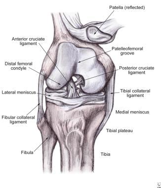
Pathophysiology: To understand the various injury patterns associated with trauma to the knee, it is important to appreciate the anatomy (see Image 1). Knee symptoms arise from an alteration or disruption of the normal anatomic structures that impede normal knee function. In mechanical terms, the knee performs like a rolling cam rather than as a simple hinged (ginglymus) joint. As the knee proceeds from flexion to extension, a complex screw-type of motion takes place, with the femoral condyles locking into the tibial plateau as the femur rotates internally. Full knee extension increases the tautness of the major bracing ligaments, transforming the knee into a mechanically rigid structure. Flexion loosens the knee joint by unlocking and disengaging the bracing structures, including retraction of the menisci, thereby enhancing ligamentous laxity and increasing the range of motion (ROM) of the joint.

Two separate but interdependent joints forming the knee are the tibiofemoral articulation and the patellofemoral coupling. Weight-bearing forces, up to 5 times an individual's body weight, are transmitted through the opposing condyles of the femur and the tibia. Two shock-absorbing cartilaginous menisci interpose between the femur and the tibia, forming the largest synovial joint in the body. The medial meniscus is smaller and more fixed than the lateral meniscus; these features predispose it to injury. A fibrous capsule lined by a synovial membrane also surrounds and bolsters the knee joint but does not contribute to the inherent stability of the joint.

Fitness of the knee joint depends largely on the fortifying ligaments and muscles binding together the femur, tibia, and patella. Two sets of knee ligaments are frequently affected. The first set, lying outside of the knee joint proper, are the extracapsular collateral ligaments. These ligaments consist of the medial collateral ligament (MCL), which opposes extreme abductive and/or valgus forces, and its counterpart, the lateral collateral ligament (LCL), which limits excessive adductive and/or varus pressures. The second set, crisscrossing in the knee joint, are the anterior cruciate ligament (ACL) and the posterior cruciate ligament (PCL), which individually brace against excessive translation in the anteroposterior (AP) plane. The ACL serves as the primary knee stabilizer, preventing forward displacement of the tibia on the femur.

Formed primarily by the quadriceps muscles, the extensor apparatus envelops and stabilizes the patella. At its distal aspect, the quadriceps muscle consolidates into the patellar ligament, ultimately inserting onto the tibial tubercle. Several bursae envelop the knee, including the prepatellar, superficial and deep infrapatellar, and pes anserine bursae, which permit friction-free movement between the various structures. Acute trauma or repetitive occupational stress may incite inflammation, with infection and metabolic disorders (eg, gout) being less common etiologies. Inflammation of the bursa then leads to localized tenderness, erythema, and increased warmth. Extensive bursae in this area alleviate potentially damaging frictional forces between the susceptible structures. Fixed in the back of the knee joint, in the popliteal fossa, are vital neurovascular structures, including the popliteal artery.

Definitions and grades of sprains and strains

Sprains to the knee are characterized by the stretching or tearing of noncontractile structures, such as the investing ligaments or of the joint capsule itself, whereas a strain refers to stretching or severing along the course of muscles or tendons. Both collateral- and cruciate-ligament sprains, as well as muscular strains, are relatively common. Ligamentous (sprain) and muscular (strain) injuries may be classified according to the degree of impairment.

  • Grade I sprain - Stretching but no tearing of the ligament, local tenderness, minimal edema, no gross instability with stress testing, firm end point

  • Grade II sprain - Partial tears of the ligaments, moderate local tenderness, mild instability with stress testing (but firm end point), moderately incapacitating

  • Grade III sprain - Complete tear, discomfort with manipulation but less than expected for degree of injury, variable amount of edema (ranging from negligible to grossly conspicuous), clear instability with stress testing (expressing a mushy end point), severe disability

Specific injuries

  • ACL injury: Rupture of the ACL is among the most serious of the common knee injuries and results from a variety of mechanisms. Most patients with ACL damage complain of immediate and profound pain, exacerbated with motion, and inability to ambulate. Disruption of the ACL may occur alone or with other knee injuries, especially a meniscal injury or tear of the MCL.

  • PCL injury: Patients typically report falling on a flexed knee or sustaining a direct blow to the anterior aspect of the knee (eg, when the knee strikes the dashboard in a motor vehicle accident). PCL harm signifies a major injury and rarely occurs as an isolated injury.

Frequency:

  • In the US: Knee pain is a complaint in up to 20% of the general adult population, accounting for almost 3 million outpatient and emergency department visits per year. Trauma to the knee is the second most common occupational accident. The MCL is the most frequently injured ligament in the knee. ACL damage causes the highest incidence of pathologic joint instability.

    In National Collegiate Athletic Association (NCAA) football, 1 major knee injury occurs per team every year. Sports-related activity accounts for approximately 60% of knee injuries producing ligamentous laxity.

  • Internationally: As in the United States, the incidence of knee injuries is increased in countries where sporting activities, such as skiing, soccer, and basketball, are popular.

Mortality/Morbidity:

  • Patients receiving inappropriate or ill-timed care of knee dislocations may have undue morbidity (eg, amputation) due to vascular complications in the distal leg.
  • Oversight of the magnitude of soft tissue injuries of the knee may result in a failure to expeditiously consider compartment syndrome and its resultant complications, including loss of a limb.
  • Misdiagnosis or mismanagement of damage to supporting structures of the knee may lead to chronic knee instability with subsequent development of degenerative joint disease and/or loss of knee function, including but not limited to an inability to bear weight or ambulate.

Sex:

  • Disorders of the patella and lateral meniscus are generally more common in girls and women than in boys and men. Recent studies also suggest that females are more prone to ACL injuries.
  • Chondromalacia patellae or patellar malalignment syndrome (ie, premature erosion and degeneration of patellar cartilage) predominates in young women.
  • Larsen-Johansson disease of the patella, also known as inferior pole patellar chondropathy, is 9 times more prevalent in boys and men than in girls and women, especially boys aged 10-14 years old.

Age: In general, knee dislocations arise from high-energy trauma, such as motor vehicle accidents. The epidemiology of injury follows that of car accidents; therefore, patients tend to be young men.

  • Ligamentous and meniscal injuries are most likely in young to middle-aged adults, whereas children and adolescents are most susceptible to osseous damage. Most patients with a meniscal tear are aged 20-30 years, but a second peak occurs in patients older than 60 years. Meniscal injuries are rare in children younger than 10 years with morphologically normal menisci.
  • Elderly patients may have fractures after minimal disturbances that typically produce only soft tissue injuries in younger patients.
  • The region of the extensor mechanism susceptible to disruption is correlated with the patient's age. The older the patient, the more proximal the area of rupture. Disruption of the quadriceps tendon most often occurs in elderly patients, whereas more distal severance of the patellar tendon and avulsion of the tibial tubercle occurs in younger patients.


  CLINICAL Section 3 of 11   Click here to go to the previous section in this topic Click here to go to the top of this page Click here to go to the next section in this topic
Author Information Introduction Clinical Differentials Workup Treatment Medication Follow-up Miscellaneous Pictures Bibliography

History: Confirm that an acute traumatic event occurred. Document mechanism of injury, type and location of pain (ie, ask the patient to point to area of pain), associated symptoms, amount of immediate dysfunction, presence and onset of joint edema, and history of past knee problems. Verify that the cause of the knee problem is mechanical by establishing that pain is exacerbated by movement (eg, walking, climbing, jumping).

  • Mechanism of injury
    • Knowledge of the mechanism can help in predicting which structures may be injured.

    • Determine the circumstances, such as the position of the extremity, whether the foot was anchored to the ground, if the forces were direct or indirect, and the specific details of the injury (eg, direction, magnitude, torque of impact).

    • Direct blows and valgus or varus contact may provoke injury to the contralateral collateral ligaments, fractures to the epiphyseal plates in children with open growth plates, and patellar dislocation.
    • Pure valgus forces, such as those occurring when a football player is struck on the lateral aspect of the knee, are more common than varus-directed contact.

    • The MCL is more prone to injury than the LCL.

    • A combination of valgus or varus stress, whether direct or indirect, delivered to a rotated leg accounts for a wide array of injuries.

    • Vulnerable structures include the collateral and cruciate ligaments, the menisci, and the joint capsule.

      • Consider the intricate damage to the knee of a skier who catches the inside edge of a ski, diverting the tip outward as the body continues to advance forward. This action produces torque, forcing the knee into extreme valgus and external tibial rotation.

      • Depending on the magnitude of the force, this mechanism may tear the MCL, the posteromedial capsule, the lateral meniscus, and the ACL.
    • Rupture of the ACL is among the most serious of the common knee injuries and results from a variety of mechanisms. ACL tears are more common than PCL tears by a ratio of at least 9:1.

      • Most patients with ACL damage complain of immediate and profound pain that is exacerbated with motion and an inability to ambulate.

      • Most patients report a snapping or popping sensation or sound at the time of injury.

      • An acute knee injury heralded by a pop or snap, followed by a rapidly evolving effusion, almost always affirms a rupture of the ACL.

      • Disruption of the ACL may occur alone or with other knee injuries, especially a lateral meniscal injury or tear of the MCL.

      • ACL tears are associated with anterior blows that hyperextend the knee, excessive noncontact hyperextension of the knee, and extreme deceleration forces to the knee.
    • Patients with PCL tears typically report falling on a flexed knee or sustaining a severe direct blow to the anterior aspect of the knee (eg, when the knee strikes the dashboard in a motor vehicle accident).

      • This injury patter displaces the tibia backward and pulls apart the PCL.

      • Patellar injury with disruption of normal articulation or fracture may also result.

      • Look for abrasions, contusions, or lacerations over the knee region.

      • Onset of edema and pain tends to occur within the first 3 hours after injury.

      • PCL harm signifies a major injury and rarely occurs as an isolated injury.
    • Rotational movements may cause a meniscal tear. The most common types of injuries are bucket-handle or flap tear where only one portion has torn free. Given that the medial meniscus is more firmly fixated and attached to the MCL capsule, it is more vulnerable to injury. Menisci are without pain fibers; the tearing and bleeding into peripheral attachments, as well as the traction on the joint capsule, causes discomfort.
  • Instability
    • Decide if the patient is describing true instability, characterized by an aberrant displacement of the osseous components of the knee. (The patella dislocates laterally on the femur, or the tibia slides excessively forward in an ACL-deficient knee.)
    • Such terms as giving way or slippage usually denote instability. Buckling tends to be associated with a different phenomenon, often resulting from pain or muscle weakness of the quadriceps.
  • Pain
    • Determine if the pain is acute or chronic in nature.

    • Abrupt onset of anterior knee pain with inability to bear weight indicates damage to the extensor mechanism.

    • Acute pain confined to the proximity of medial or lateral regions of the knee joint tends to result from ligamentous and/or meniscal damage.
    • Recent onset of pain at the posteromedial corner of the knee indicates a tear of the medial meniscus or an expanding or ruptured Baker cyst. Menisci are without pain fibers; it is the tearing and bleeding into peripheral attachments as well as the traction on the joint capsule that causes discomfort.

    • Tumors are characterized by chronic pain that is worse at night.

    • Discomfort from bursitis and/or tendinitis is likely to be chronic and bilateral, it is typically worse with rising or walking after sitting than at other times, and it is provoked by prolonged exercise or use.
  • Knee effusion
    • The rapidity of onset of knee effusion may corroborate a particular diagnosis. Effusion onset within 6 hours supports the assumption of a cruciate ligament tear, articular fracture, or knee dislocation, whereas delayed edema occurs with meniscal injuries.

    • Nearly one half of patients with an acute ligament rupture have localized edema at the site of injury.

    • Complete ligamentous or capsular disruption can give rise to less-than-anticipated swelling if fluid exudes through the tear. Recurrent knee effusion after activity is consistent with a meniscal injury.

    • Localized distension may originate from bursitis (prepatellar bursitis), meniscal cystic changes, outgrowth of a popliteal cyst (Baker cyst), or dilation of an artery (popliteal-artery aneurysm).
  • Locking
    • True locking manifests as a knee joint that is unable to move, usually at 45° of flexion; patients cannot bend further without assistance. It may arise from tears in the meniscus, detached tissue lodging in the knee joint, injury of the cruciate ligament(s), or an osteochondral fracture.

    • Pseudolocking may result from pain and muscle spasm secondary to increasing edema.
    • On occasion, locking of the knee is confused with giving way of the knee. Giving way of the knee may accompany ACL injuries, quadriceps weakness, or patellar disorders.
    • Acute traumatic weakness of the knee often follows a derangement of the extensor mechanism of the knee. Clinically significant atrophy of the quadriceps may appear as early as within 1 week of disuse.
  • Degree of immediate dysfunction: Injuries, including meniscal tears, patellar subluxation, or ligament strains or ruptures, may not entirely preclude a patient from bearing weight.
  • History of previous knee problems

Physical: Developing a standard routine to avoid omitting important aspects of the knee joint examination is important.

  • When performed by properly trained physicians, the physical examination is reasonably sensitive (74-81%) for detecting meniscal, ACL, PCL, and collateral ligament injuries. However, accurate examination becomes difficult with greater elapsed time because of swelling secondary to bleeding and inflammation.

  • Ensure adequate exposure of both lower extremities, from the groin to the toes, and examine the patient in a supine position. It is important to compare the symptomatic knee to the contralateral, normal knee joint.

  • Attempt to alleviate the patient's fears, and convince the patient to relax as much as possible during palpation and during stress testing of the injured knee joint.

  • Examine the uninjured knee first to approximate baseline values and to allow the patient to understand what will be done to the injured knee.

  • Focus the initial examination on inspection, palpation, and neurovascular evaluation. In the event of a suspected knee dislocation, any discrepancy in distal pulses compared to the unaffected leg, or an ankle-brachial index less than 0.8, an arteriogram is needed to rule out a vascular injury.

  • Begin distal to the injury. If possible, observe the patient standing and/or walking.
    • A grossly antalgic gait indicates a clinically significant problem with 1 or both lower limbs.

    • When an uninjured patient stands with feet together, the medial aspects of both knees and ankles are normally in contact.
  • Inspect the knee for edema, ecchymosis, effusion, masses, patella location and size, muscle mass, erythema, and evidence of local trauma, such as abrasions, contusions, or lacerations. Carefully check for a communication of a skin defect with the underlying knee joint (eg, open intra-articular injury).

  • The knee appears ordinarily hollow on either side of the patella and slightly indented just above the patella.
  • Confirm the position of the patella.

    • A superiorly displaced patella (patella alta) may accompany a disruption of the patellar ligament, while a breach of the quadriceps tendon might cause an inferior position of the patella (patella baja).

    • If time permits and it is relevant to the patient's complaint, measure the Q angle which is calculated by drawing a line from the tibial tubercle to the center of the patella and then from the center of the patella to the anterior superior iliac spine. Any angle exceeding 15° may make the patella vulnerable to subluxation or dislocation.

  • Conspicuous atrophy of the quadriceps muscles indicates the presence of a long-standing or preexisting disorder. Atrophy of the vastus medialis muscle commonly follows surgical repair of the knee.

  • Inspect and palpate the popliteal fossa; this evaluation is best performed with the patient prone.
    • The only palpable structure in this space is ordinarily the popliteal artery.

    • Abnormal bulges may derive from popliteal-artery aneurysms, popliteal thrombophlebitis, or Baker cysts. A Baker cyst originates from a herniation of the synovial membrane through the posterior aspect of the joint capsule, and tends to be associated with intra-articular disease. Rupture of a Baker cyst with subsequent dissection of the synovial fluid results in a clinical picture often indistinguishable from that of thrombophlebitis of the calf.
  • Palpation
    • Palpate the knee in slight flexion. This position is facilitated by placing a pillow under the popliteal fossa. Direct palpation at specific areas.

    • Tenderness strictly confined to the joint line suggests a meniscal tear.

    • The Bragard sign indicates a medial meniscus injury. It refers to point sensitivity along the medial aspect of the joint line. The tenderness intensifies with internal rotation and extension of the tibia.

    • Additional clinical clues suggestive of meniscal disease are the first and second Steinmann signs and the Payr sign.

      • With the patient supine and the hip and knee flexed to 90° the examiner vigorously and quickly rotates the tibia internally and externally. Pain in the lateral compartment with forced internal rotation indicates a lateral meniscus lesion. Medial-compartment pain during forced external rotation indicates a lesion of the medial meniscus. Test for a second Steinmann sign when the point of tenderness is most pronounced along the anterior joint line. When the examiner moves the knee from extension into flexion, the meniscus is displaced posteriorly, and the point of tenderness is displaced from the anterior joint line back towards the collateral ligaments. This maneuver helps in differentiating meniscal injuries from ligamentous problems, as ligament pain does not shift with flexion.

      • Test for Payr sign with the patient sitting cross-legged and exerting downward pressure along the medial aspect of the knee. Medial knee pain indicates a posterior horn lesion of the medial meniscus.

    • Damage to the MCL may reveal tenderness along the entire course of the ligament.

      • The tenderness can be present from the origin on the medial femoral condyle to its tibial insertion.

      • Tenderness of the MCL limited to the insertion or origin can occur with avulsion-type fracture.

    • A damaged LCL may be tender from its attachment on the lateral femoral epicondyle to its insertion on the fibular head.

    • Placing the knee in slight flexion with the patient supine optimizes palpation of the MCL and improves LCL evaluation by positioning the affected knee in a figure 4 arrangement.

    • A positive patellar apprehension sign implies a preceding patellar dislocation.

      • With the leg held and supported in 30° of flexion, apply a firm, laterally directed force toward the medial aspect of the patella. A positive test manifests as trepidation on the patient's part, such as grabbing for the examiner's arm when pressure is applied.

      • Tenderness or edema about the medial retinaculum is associated with spontaneously reduced patellar dislocation.

    • Quadriceps rupture

      • Palpate the anterior aspects of both thighs, particularly noting any muscle wasting.

      • A rupture in the quadriceps commonly emerges just proximal to the tibia, revealing a transverse tract more pliable than the surrounding musculature. The extent and completeness of the tear correlates with the size of the defect. This palpable defect above the patella is known as the gap test or sulcus sign.

    • Bursal injuries

      • Palpate the clinically significant bursae accordingly.

      • Prepatellar bursitis (ie, housemaid's knee) presents with tenderness, erythema, warmth, and swelling superficial to the patella, and does not usually restrict ROM of the knee.

      • With superficial infrapatellar bursitis, tenderness is present over the tibial tubercle and can be confused with Osgood-Schlatter disease, as tenderness and edema at the site where the infrapatellar bursa inserts into the tibial tubercle accompanies this disease.

      • The deep infrapatellar bursa is located under the patellar tendon (separating it from the underlying fat pad and tibia). When inflamed, it causes pain during active ROM, but no pain during passive flexion-extension.

      • Located between the pes anserine tendon and MCL, inflammation of this bursa (anserine bursitis) causes marked tenderness approximately 5 cm below the medial joint line. Pain is often times worst at night and particularly pronounced when the patient walks up stairs or rises from a sitting position. Anserine bursitis may be mistaken for an injury to the MCL.

      • Bursitis can be distinguished from an intra-articular injury by both location of the pain and the fact that movement of the joint itself should not cause discomfort (up to the point of skin tension).

    • In children, be sure to palpate the epiphyses, as growth plate injuries may be confused with soft-tissue injuries.

    • Patellar tendonitis or jumper's knee is an overuse syndrome caused by repetitive actions presenting with localized tenderness along the superior or inferior aspects of the patella. Pain increases with active resistance against extension.

    • Traumatic prepatellar neuralgia presents with persistent, intense point tenderness over the middle or outer borders of the patella. The pain may be exacerbated even with the slight pressure of clothing.

    • Discrete tenderness over the lateral femoral epicondyle (approximately 3 cm above the lateral joint line) is seen with iliotibial band tendonitis. Soft tissue swelling and crepitus may also be present. Pain is most pronounced with weight bearing on a flexed knee and occurs most commonly in long-distance runners.
  • ROM testing
    • Evaluate the knee for active flexion and extension. Inability to extend the knee to any extent implies damage to the extensor mechanism.

    • Clinically significant effusions may hinder complete extension of the knee joint and may be incorrectly identified as locking of the knee.
  • Stress testing
    • Assess knee joint stability by applying various stresses to the joint.

    • Clarify the examination by employing a standard routine and directing gentle firm pressure, rather than applying sudden forceful manipulation, causing reflexive contraction of adjacent muscles.

    • Excessive joint motion (laxity) indicates an injury.

    • The appearance of a soft or mushy end point versus the healthy hard (ie, abrupt increase in joint stiffness) end point implies possible ligament damage.

    • Assess the quantity and the quality of translation, comparing the injured knee to the unaffected side. Side-to-side differences in knee translation hold greater significance than absolute measurements of motion.

    • Characterize knee joint unsteadiness by the direction of tibial displacement. For example, medial knee instability means that the tibia moves abnormally away from the femur on the medial side.

    • Assess the collateral stability in 30° flexion and in full extension. Position the patient supine with the thigh resting on the edge of the examination table and support the foot. The MCL is assessed by applying valgus stress to the knee. The LCL is assessed by applying varus stress to the knee.

      • Provide valgus stress by pressing with 1 hand on the lateral aspect of the knee; with the other hand, direct the ankle laterally, attempting to open the knee joint on the medial side.

      • Inducing a substantial gap in the medial aspect of the knee joint implies impairment of the MCL.

      • Increased laxity of the medial side of the knee with the knee in extension indicates additional damage to posterior structures, such as the posterior joint capsule and the PCL.

      • Test for lateral knee joint stability by reversing the hand positions.

      • Push medially upon the knee and laterally against the ankle (varus stress), trying to open the knee joint on the lateral side.

      • Gapping of the knee joint may be visible and palpable.

      • A difference in the degree of lateral knee tautness when comparing the affected knee to the uninjured side indicates disruption of the LCL.

      • Lateral instability with the knee extended suggests injury to the LCL and the lateral capsule, as well as likely damage to the ACL or the PCL.

      • Relieving stress on the injured knee when following testing for collateral ligament impairment may provoke a clunking sensation.

    • The Lachman maneuver confirms ACL integrity.

      • This maneuver is the most sensitive bedside test, about 87% for ACL tear (see Image 3).

      • Place the patient supine and flex the knee 20-30°.

      • With 1 hand, grasp and stabilize the patient's thigh just above the patella. With the opposite hand, try to dislodge the proximal tibia forward.

      • In patients with large thighs, the position may need to be modified. Position the large thigh of the patient over the knee of the examiner. Push downward on the patient's femur with one hand, while the other hand grasps the proximal tibia and attempts to move it anteriorly.

      • Damage to the ACL manifests as excessive forward motion of the tibia without a firm end point.

      • If viewed from the lateral aspect, the natural concave silhouette of the knee, extending from the tibial tubercle to the superior aspect of the patella, obliterates when ACL damage occurs.
    • Anterior drawer testing also evaluates the soundness of the ACL (see Image 4).

      • Place the patient in the supine position, flex the hip to 45°, and bend the knee to 90° with the patient's foot planted firmly on the examination table.

      • Sitting on the dorsum of the foot, place both hands behind the knee.

      • Once the hamstrings appear relaxed, discreetly try to displace the proximal leg anteriorly.

      • The anterior drawer test is less sensitive for ACL damage than the Lachman maneuver.

    • Posterior drawer testing is done in a similar manner to anterior drawer testing, except that the pressure is directed backward on the proximal tibia.

      • Posterior instability arising from PCL injury manifests as an abnormal increase in posterior tibial translation.

      • Confusion may occur when trying to distinguish whether abnormal translation of the tibia on the femur originates from excessive ACL or PCL laxity.

      • Unless the examiner knows prior to testing that the resting position of the tibia is shifted posteriorly, anterior drawer testing may yield a false perception of instability.

    • To distinguish disorders of the ACL from those of the PCL, employ the tibial sag test.

      • Flex the patient's hips and knees to 90° while supporting the patient's heels.

      • In this position, the PCL impaired knee will clearly sag backward from the effects of gravity (also referred to as the Godfrey sign).
    • The pivot-shift test helps substantiate capsular tears and injury to the ACL (see Image 5).

      • Elicit by lifting the tibia of the affected side. If the ACL is impaired, the tibia subluxates anteriorly during knee extension.

      • In addition, apply a moderate valgus stress while flexing the knee.

      • As the knee joint approaches 20-40° of flexion, an abrupt jerking movement occurs in the ACL-impaired knee, indicating a reduction in prior anterior subluxation.
  • McMurray testing substantiates meniscal disorders (see Image 6).
    • With the patient supine and the knee in maximum flexion, palpate the posteromedial margin of the affected knee joint with one hand and support the foot with the opposite hand.

    • Externally rotate the lower leg as far as possible, affix varus pressure, and cautiously extend the knee joint.

    • If a tear is present in the medial meniscus, an audible, palpable, and painful clunk transpires, as the femur passes over the damaged portion of the meniscus.

    • To check the lateral meniscus, repeat the above technique, but place one hand over the posterolateral aspect of the knee joint and internally rotate the lower leg to its maximum extent.

    • Slowly extend the leg again, listening and feeling for a click or pop, and observe the patient for distress.

    • Clicks unassociated with pain or joint-line tenderness, especially during lateral meniscus testing, may represent a normal variant and should not be interpreted as evidence of a meniscal tear.

    • Unfortunately, the McMurray test is neither sensitive nor specific for meniscal damage.
  • Another method for establishing the soundness of the menisci, especially the posterior horn of the menisci, is the Apley compression or grind test (see Image 7).
    • Place the patient in the prone position and flex the knee to 90°.
    • Stabilize the patient's thigh against the examination table with the knee of the examiner, and apply a downward-directed force onto the patient's foot and leg.
    • Rotate the leg, while mildly flexing and extending the knee joint. Any pain in the joint should be noted, as well as any resonance or irritation emanating from the knee.

Causes: The intrinsic structural framework of the knee and its exposure to the environment account for particular injuries.

  • Harm may arise from direct impact, such as in contact or collision sports or blows to the knee connected to motor vehicle accidents.
  • Most soft-tissue injuries sustained by the knee do not, however, involve direct trauma but arise from actions producing excessive torque on the knee joint, especially those activities involving twisting, rapid deceleration, or landing from a jump.
  • If tensile forces placed on the knee exceed the intrinsic tone of the ligaments, injury to the ligaments results.
  • Low-intensity forces may provoke a reversible injury, with only transient deformation of the elastic ligament; however, profound loads applied to the knee joint produce irreversible rupture of the ligament fibers.
  • Valgus-directed blows sustained by the externally rotated knee occur commonly.

  • High-intensity impact to the lateral side of the knee tends to be serious and befalls skiers when catching a ski tip or football players following a blind-side clipping type of injury.
  • Valgus contact injuries may cause a series of injuries to the knee, including a tear of the MCL, followed by damage to the posterior medial capsule, and, finally, damage to the ACL. This combination of injuries is referred to as O'Donahue or unhappy triad.
  • An intense varus stress to the knee joint gives rise to a sequence of injuries, depending on the position of the knee.
  • Impact to the medial side of the knee in a neutral position causes disruption of the LCL, the iliotibial band, and/or the biceps femoris.
  • Profound varus strain to the extended and internally rotated knee harms not only the LCL and the ACL, but also may mar the PCL and the lateral posterior capsule.
  • Undue varus stress to the flexed and internally rotated knee brings about LCL injury, proceeding to damage of the lateral posterior capsule and/or lateral meniscus and, if extreme, impairment of the PCL.
  • Extensor-mechanism injuries result from direct or indirect forces.

    • Direct injuries result from a blow to the anterior aspect of the knee, often causing the patella to fracture.

    • Indirect injury occurs when forced flexion of the knee occurs against a contracted quadriceps.

    • Depending on the patient's age and the type of force applied, disruption of the extensor mechanism can occur by means of quadriceps tendon tear, patellar fracture, rupture of the patellar tendon, or avulsion of the tibial tuberosity.

    • Many conditions have been reported to be associated with rupture of the quadriceps tendon including hyperparathyroidism, chronic renal failure, gout, obesity, leukemia, rheumatoid arthritis, diabetes, systemic lupus erythematosus, steroid use (both local injection and systemic use), and fluoroquinolone use.

    • Bilateral simultaneous ruptures of the quadriceps tendon are rare, but they have been reported numerous times in the literature. In 1 series of bilateral quadriceps rupture, 41 (65%) were attributed to falls or missteps, and 22 (35%) occurred spontaneously while the patient was walking.
  • Patients with meniscal injuries frequently report pain after twisting (rotational force) their knee while bearing weight on the affected side.
  • Imposing extreme hyperextension force may disrupt the cruciate ligaments, conceivably anywhere along their span.
  • ACL tears
    • Rupture of the ACL is among the most serious of the common knee injuries and results from a variety of mechanisms.
    • ACL tears are associated with anterior blows that hyperextend the knee, excessive noncontact hyperextension of the knee, and extreme deceleration forces to the knee.
    • Disruption of the ACL may occur alone or with other knee injuries, especially a meniscal or MCL tear.
  • PCL tears
    • Patients typically report falling on a flexed knee or sustaining a direct blow to the anterior aspect of the knee (eg, when the knee strikes the dashboard in an motor vehicle accident).
    • PCL harm signifies a major injury and rarely occurs as an isolated injury.
  DIFFERENTIALS Section 4 of 11   Click here to go to the previous section in this topic Click here to go to the top of this page Click here to go to the next section in this topic
Author Information Introduction Clinical Differentials Workup Treatment Medication Follow-up Miscellaneous Pictures Bibliography

Arthritis, Rheumatoid


Other Problems to be Considered:

ACL rupture
Baker cyst
Bursitis
Collateral ligament tear, medial or lateral
Knee dislocation
Meniscal tear
Osteochondral fracture (osteochondritis dissecans and osteonecrosis)
Patellar dislocation
Patellar fracture
Patellar tendon rupture
PCL rupture
Segond fracture

Quick Find
Author Information
Introduction
Clinical
Differentials
Workup
Treatment
Medication
Follow-up
Miscellaneous
Pictures
Bibliography

Click for related images.

Related Articles
Arthritis, Rheumatoid


Patient Education


  WORKUP Section 5 of 11   Click here to go to the previous section in this topic Click here to go to the top of this page Click here to go to the next section in this topic
Author Information Introduction Clinical Differentials Workup Treatment Medication Follow-up Miscellaneous Pictures Bibliography

Lab Studies:

  • For patients with significant injuries or individuals who may require surgery, appropriate pre-operative laboratory investigations might include blood typing and screening and determination of the complete blood count (CBC) and electrolyte, serum glucose, blood urea nitrogen (BUN), and creatinine levels.
  • If warranted by the initial history and physical findings, laboratory analysis of joint aspiration may assist in confirming a diagnosis.
    • Aspirated synovial fluid should be analyzed for white blood cell (WBC) count and differential and for glucose and protein levels. Gram staining, cultures, and sensitivity testing should be performed.
    • A CBC, erythrocyte sedimentation rate (ESR), and serum glucose and uric acid levels should also be considered at the time of arthrocentesis if infectious or gouty arthritis is a consideration.

Imaging Studies:

  • Plain images of the knee generally include AP and lateral views at minimum.
  • Some centers add 2 oblique projections that are helpful in detecting tibial-plateau fractures.
  • Special views include tunnel view to aid in evaluating the tibial and femoral articular surfaces and a sunrise view to assist in assessing the articular surface and the body of the patella.

  • In most patients with severe ligamentous or meniscal damage, plain radiographic findings are of limited value, and images often appear normal.
  • Fewer than 15% of knee radiographs reveal clinically significant findings.

  • Various clinical prediction rules exist (Ottawa, Pittsburgh, Weber, Fagan-Davies), with the Ottawa rules being the most validated.

  • Plain images are recommended for the following scenarios:
    • Patients older than 55 years (because of an increased risk of pathologic fracture associated with osteoporosis)

    • Patients with tenderness over the fibular head
    • Patients with discomfort confined to the patella upon palpation

    • Patients unable to flex the knee to 90°
    • Patients incapable of bearing weight, immediately and in the ED, for at least 4 steps
  • Although plain radiography tends to be unproductive in diagnosing soft-tissue injuries, certain radiographic findings are strongly suggestive of ligamentous, meniscal, or tendon damage.
    • Particularly review the lateral radiograph for fluid within the suprapatellar pouch. The extensor tendon mechanism is normally well outlined.

    • As an effusion accumulates in the suprapatellar pouch, the posterior margin of the quadriceps is apt to be obliterated, and, with additional fluid collecting, the space between the prefemoral and the anterior superior suprapatellar fat pads widens.
  • A cross-table lateral radiograph may expose a fat-fluid level, also known as a lipohemarthrosis, a pathognomonic sign of an intra-articular fracture.

  • Avulsion fractures of the tibial spine or the femoral condyles imply ligamentous rupture.
  • Occult fractures that are commonly missed on plain radiographs include those of the patellar, tibial plateau, of fibular head, as well as Segond fractures (small vertical avulsion crack of the proximal lateral tibia, also called the lateral capsular sign).
  • Three radiographic findings associated with ACL injuries are avulsion of the intercondylar tubercle, anterior displacement of the tibia with respect to the femur (labeled the radiographic drawer sign), and a Segond fracture.

  • Avulsion of the fibular head aligns with LCL or biceps femoris injury.
  • The most common radiographic finding with a PCL injury is avulsion at the site of the ligamentous origin on the posterior tibia.
  • Unilateral widening of the joint space may denote ligamentous instability.
  • Chronic MCL damage, usually lasting longer than 6 weeks, may heal with calcification, presenting as Pellegrini-Stieda syndrome.
  • Patellar abnormalities can be misleading for an acute fracture.
    • A bipartite or multipartite patella is a normal variant that may be difficult to distinguish from a patellar fracture.

    • An unfused secondary ossification center forming the bipartite patella typically appears in the upper lateral quadrant, tends to occur bilaterally, and reveals well-defined margin lines.
  • CT scans prove effective for corroborating areas questionable for fracture in the knee region, particularly tibial-plateau fractures in elderly patients.
  • Ultrasound imaging has been employed to diagnose tendon ruptures.
  • The relative sensitivity and specificity of clinical examination and MRI findings were comparable (96.5% sensitivity of clinical examination versus 98% of MRI; 87% specificity of clinical examination versus 85.5% of MRI).

Other Tests:

  • MRI has supplanted the arthrography as the procedure of choice for evaluating soft-tissue injuries of the knee. Although MRI plays an important role in future surgical management of acute knee disorders, it is rarely an essential part of the ED workup.
  • Ultrasonography simplifies differentiation of a Baker cyst, popliteal-artery aneurysm, and thrombophlebitis, and it has also been used to diagnose tendon ruptures.
  • An arteriogram is required when a knee dislocation is strongly suspected, even in the absence of reliable signs or symptoms of vascular impairment. Palpation of a regular pulse or Doppler confirmation of pedal pulses does not exclude vascular injury, as intimal tears may be undetectable. The incidence of a concomitant popliteal artery injury is 20-50%. Do not delay emergency surgery for arteriography; imaging can be performed intraoperatively.
  • Although not 100% sensitive, an injection of methylene blue into the affected knee until the joint is fully distended and observing for extravasations can be done to test for open joint injury.

Procedures:

  • Knee joint aspiration
    • Because the knee is the largest synovial joint in the body and given its relatively accessible location, the knee joint provides one of the easiest sites in which to perform arthrocentesis.
    • Indications for knee joint aspiration include confirmation of a diagnosis (The knee is a common site for septic and inflammatory arthritis.) and pain arising from a tense effusion.
    • Place the patient supine with the knee joint extended, trying to ease any contraction of the quadriceps muscles.
    • After properly cleansing the skin, infiltrate the skin and underlying dermis with local anesthetic at the point where aspiration will take place.
    • Approach the joint for aspiration from the medial aspect of the knee, with the site of puncture being 1 cm medial to the anteromedial border of the patella.

    • Insert an 18-gauge needle or catheter through the same tract used to inject local anesthetic, at the midpoint or superior position of the patella, aiming for a point between the posterior surface of the patella and the femoral intercondylar notch.
    • Approach the anterolateral border in a similar manner, mirroring the access sites.

    • Occasionally, the examiner perceives a slight give as the needle perforates the joint capsule.

    • Aspirate as much fluid as possible, recalling that the knee may contain 50 mL or more of fluid.
    • When the fluid stops flowing freely, compress the suprapatellar pouch and attempt to push additional fluid into the adjacent pouch.

    • If the bore appears obstructed during the arthrocentesis, try rotating the needle or injecting some aspirate, attempting to clear the needle. If this fails, try reinserting the needle one fourth of an inch deeper.
    • Aspiration of blood indicates a ligamentous tear (eg, ACL, PCL), osteochondral fracture, peripheral meniscus tear, capsular tear, or patellar dislocation.

    • Presence of fat globules in the aspirant is pathognomonic for an intra-articular fracture.
    • Hypertrophied synovial tissue or clot formation may hinder aspiration.
    • Instilling 5-10 mL of 1% lidocaine into the joint and then reexamining the knee may facilitate knee testing.
    • After withdrawing the needle, dress the iatrogenic puncture wound with an antibiotic ointment and appropriate adhesive sterile dressing.
    • In the presence of a hot and swollen knee joint, consider the possibility of septic or acute inflammatory arthritis. Joint aspirate should be analyzed for a CBC with differential, glucose levels, and protein measurements. Also order polarized light microscopy for crystals, and culture and sensitivity testing of the joint fluid.
    • Although rare, infection and hemarthrosis may complicate arthrocentesis.
  TREATMENT Section 6 of 11   Click here to go to the previous section in this topic Click here to go to the top of this page Click here to go to the next section in this topic
Author Information Introduction Clinical Differentials Workup Treatment Medication Follow-up Miscellaneous Pictures Bibliography

Prehospital Care:

  • Basic responsibilities in the prehospital setting, if knee trauma is the primary injury, include stabilizing the lower extremity and monitoring the neurovascular status of the limb.
  • The deformed knee should be realigned only if associated neurovascular structures are compromised.
    • Always recheck and document pulses after splinting or manipulation of the limb.

    • If initial efforts meet with resistance, prehospital personnel should not force realignment.
  • Cover open wounds with saline-soaked sterile gauze.
  • Frequently, the joint reduces spontaneously or is reduced at the scene by trained emergency medical services (EMS) personnel; in such cases, the ED physician must obtain information about the time and mechanism of injury and the original position of the limb.

Emergency Department Care: Adhere to the conventional dictums of emergency and trauma care by first corroborating the absence of life-threatening (primary survey) or limb-threatening (secondary survey) injuries before focusing on soft-tissue damage sustained by the knee. A cardinal error occurs when earliest attention is diverted to an obvious extremity injury, such as a knee dislocation, while neglecting possibly lethal trauma.

  • Always determine the mechanism of injury and verify hemodynamic stability.

  • Assess vascular perfusion and control any bleeding.

    • Hard signs of vascular injury include absent or diminished pulses, active hemorrhage, and expanding or pulsatile hematoma.

    • Signs of distal ischemia include pain out of proportion to the injury, pallor, paralysis, and paresthesias.

    • For knee dislocations or grossly malaligned fractures with potential vascular compromise, attempt immediate reduction or realignment of the knee if an orthopedic specialist is not immediately available.

    • Popliteal-artery injuries must be repaired within 6-8 hours to avoid amputation because of limb ischemia.

  • Splint all obvious fractures and unstable knee injuries, stabilizing the femur above and the tibia below.
  • Damage to essential nerves and vessels may be subtle on presentation.

  • Observe for signs and symptoms of compartment syndrome. Measurement of compartment pressures is often needed to exclude the possibility of compartment syndrome.

  • Remove any constricting clothes and bandages.

  • Make sure patients ingest nothing by mouth (NPO) until the need for emergency or urgent surgery is ascertained.

  • General treatment principles
    • Aside from the particular injury, treatment plans depend on the patient's age and activity level and the presence of additional injuries.

    • Obtain orthopedic consultation when appropriate.

    • Initial nonpharmaceutical treatment includes rest, ice, compression, and elevation (RICE).

    • For the first 1-3 days, use therapeutic measures that minimize incipient damage and reduce pain and inflammation.

    • Consider splinting the injured knee to provide support and to prevent further injury.

    • Serviceable devices include commercially available immobilizers and handcrafted compressive dressings, such as the Robert Jones dressing, which incorporates coaptation plaster.

    • Detrimental effects of immobilization include joint stiffness, degenerative changes in articular cartilage, muscle atrophy and weakness, and decreased vascularity.
  • Therapy for specific injuries
    • For first-degree sprains, provide symptomatic treatment, essentially the RICE regimen. Normal function usually returns quickly.

    • Second-degree sprains require protection by using a cast, cast brace, or a restrictive movement brace. Arrange for timely follow-up care.

    • Treatment of third-degree sprains depends on the severity and type of instability; some third-degree sprains of ligaments necessitate surgical repair. Factoring into the deliberation for surgery is the patient's age, relative health, associated injuries, activity demands, and individual desires.

    • Treatment for ACL injuries is individualized. Various conditions influence decisions on the optimum management of ACL tears, such as the presence or absence of comorbid pathology, age of the patient, baseline activity level, degree of instability, and associated ligamentous injuries miring the knee.

    • Presence of a meniscal tear does not automatically lead to surgical intervention. If the knee is not locked or unstable, conventional treatment (ie, RICE therapy) ordinarily suffices; however, the meniscus cartilage of the knee generally supports a precarious blood supply, and tears are prone to inadequate mending. Therefore, timely follow-up care is critical.
    • Most knee dislocations reduce spontaneously before the patient's arrival in the ED. However, when examination findings suggest neurovascular compromise, an attempt to restore circulation with traction and/or reduction and emergency orthopedic consultation are needed.

    • Classify dislocations with respect to the relationship of the tibia on the femur.

    • Anterior dislocations occur most commonly.

    • In the ideal case, perform reductions in the operating room under general anesthesia. However, if circumstances preclude this scenario, an attempt in the ED is warranted.

    • Barring contraindications, administer conscious sedation.

    • With an assistant providing stabilization and countertraction of the thigh, a second person applies longitudinal traction to the leg. This maneuver usually suffices for reduction.

    • Reduction of an anterior knee dislocation may be aided by trying to transpose the femur anteriorly.

    • Avoid affixing pressure over the popliteal space as it may exacerbate arterial damage.

    • For posterior dislocations (where the tibia lies posterior to the femur), attempt to reinstate the tibia anteriorly by gently lifting the tibia forward.

    • After relocation of the knee, confirm neurovascular status and immobilize the knee in 15° of flexion.

    • Order postreduction images and consultation with the orthopedic surgeon, and obtain an emergency arteriogram.
  • Reduction of patellar dislocation
    • Patellar dislocations typically occur in predisposed individuals and tend to recur.

    • Patellar dislocations are identified with respect to the patella's position on the knee joint, with lateral dislocations being most common.

    • If the joint has not already reduced spontaneously, verify the dislocation radiographically.

    • After administering necessary analgesia, place the hip in a mild amount of flexion, and gently press anteriorly and medially on the patella while extending the knee joint.

    • Postreduction films should include a sunrise view, as osteochondral fractures may result.

    • Other types of patellar dislocations tend to be resistant to closed reduction.

    • Aspiration of a Baker cyst may render temporizing relief. This procedure is best performed electively by a qualified physician.
  • Injection therapy

    • Ensure that the joint is not infected.

    • Pain secondary to aseptic inflammation of the prepatellar, suprapatellar, and pes anserine bursae may be relieved by instilling 2-4 mL of lidocaine mixed with 15-20 mg of a prednisolone suspension into the affected bursae.

    • Recent data have suggested that an intra-articular installation of morphine 1-5 mg confirms a local analgesic effect and possibly prolongs pain relief compared with systemic opioids.

  • Reducing the locked knee

    • Avoid undue manipulation because it may aggravate internal derangement.

    • A simple measure to reduce the locked knee involves positioning the patient with the knee dangling over the edge of the examination table in 90° of flexion.

    • Gravity usually expedites distraction of the tibia from the femur, thereby promoting unlocking.

    • If this technique fails after a reasonable time period, tempered rotation of the knee along with caution traction on the leg usually frees the knee joint.

Consultations: Depending on the degree of knee instability, affected ligaments, and the patient's age and baseline activity level, early surgical intervention may be the best option.

  • Indications for emergency or urgent orthopedic consultation include the following:
    • Gross knee dislocation or unstable knee
    • All knee injuries with associated neurologic or vascular injury
    • Complete quadriceps tendon rupture or a complete patellar tendon rupture
    • First-time patellar dislocations and patellar displacements accompanied by an osteochondral fracture

    • Open joint injuries of the knee

  MEDICATION Section 7 of 11   Click here to go to the previous section in this topic Click here to go to the top of this page Click here to go to the next section in this topic
Author Information Introduction Clinical Differentials Workup Treatment Medication Follow-up Miscellaneous Pictures Bibliography

In addition to resolving the etiology of the pain, the ED physician must make every effort to relieve suffering as completely and expeditiously as possible. Distinguish acute injury from chronic distress. Consider the intensity of the pain. The perception of pain is subjective, and therapy should be individualized.

Objective parameters, such as tachycardia, are unreliable. Minor trauma to the knee that involves the ligaments, muscles, and joints usually causes a mild to moderately severe, self-limiting discomfort. An enormous choice of analgesics is available for use by the ED physician, but pharmacologic agents tend to fall into 2 general categories: nonnarcotic and narcotic analgesics. Furthermore, consider the best route for delivering the drug.

Drug Category: Nonnarcotic analgesics -- Patients with pain accompanying minor acute soft-tissue injuries of the knee generally benefit from a short course of nonnarcotic analgesics, with acetaminophen and nonsteroidal anti-inflammatory drugs (NSAIDs) being the most frequently prescribed agents. If inflammation is a component of the injury and if no contraindication is present, prescribe NSAIDs, as acetaminophen lacks anti-inflammatory properties. An increasing number of NSAIDs are available for use. NSAIDs share a common mechanism of action, which involves inhibiting the production of pain-mediating prostaglandins. In general, NSAIDs provide a comparable degree of pain and inflammatory relief, but they differ in dosing schedules.

Choosing an NSAID to prescribe can be perplexing; data comparing these agents are meager, and individual responses are inconsistent. Because no individual NSAID is clearly superior, base decisions on personal experience, safety profiles, cost, and convenience.
Drug Name
Acetaminophen (Tylenol, Feverall, Aspirin Free Anacin) -- Most commonly ingested pain reliever; marketed in combination with other drugs to provide analgesia. Advantages include availability, cost, and relatively good safety profile. Onset of relief usually in 20-30 min. Extended-release preparations do not appear to offer major benefits (other than dosing convenience) and may increase incidence of toxicity. For children, available as drops (80 mg/0.8 mL), elixir (160 mg/5 mL), tablets (80, 160, or 325 mg), and suppositories (125 or 325 mg).
Adult Dose650-1000 mg PO q4h; not to exceed 4000 mg/d PO
Pediatric Dose10-15 mg/kg/dose PO q4-6h
ContraindicationsDocumented hypersensitivity; known G-6-P deficiency
Interactions Rifampin can reduce analgesic effects; barbiturates, carbamazepine, alcohol, hydantoins, zidovudine, and isoniazid may increase hepatotoxicity
Pregnancy B - Usually safe but benefits must outweigh the risks.
PrecautionsHepatotoxicity possible in chronic alcoholism following various dose levels; severe or recurrent pain or high or continued fever may indicate serious illness; many over-the-counter (OTC) products contain acetaminophen, and combined use may result in cumulative doses exceeding recommended maximum
Drug Name
Acetylsalicylic acid (Bayer Aspirin, Bufferin, Anacin, Ecotrin) -- Prototype NSAID; used in combination with many other drugs; available OTC. Offers anti-inflammatory benefits, unlike acetaminophen. Effective in PO, suppository, and topical preparations. Therapeutic anti-inflammatory serum level 15-30 mg/dL.
Adult Dose650 mg PO q4-6h
Pediatric Dose10-15 mg/kg/dose PO q 4-6h
ContraindicationsDocumented hypersensitivity; liver damage; hypoprothrombinemia; vitamin K deficiency; bleeding disorders; asthma; because of association with Reye syndrome, do not use in children (<16 y) with flu
InteractionsEffects may decrease with antacids and urinary alkalinizers; corticosteroids decrease serum levels; additive hypoprothrombinemic effects and increased bleeding time may occur with anticoagulants; may antagonize uricosuric effects of probenecid and increase toxicity of phenytoin and valproic acid; doses >2 g/d may potentiate glucose-lowering effect of sulfonylurea drugs
Pregnancy D - Unsafe in pregnancy
PrecautionsMay cause transient decrease in renal function and aggravate chronic kidney disease; avoid use in patients with severe anemia, with history of blood coagulation defects, or taking anticoagulants
Drug Name
Ketorolac (Toradol) -- Choice of parenteral pain medications dispensed in ED. Frequently overlooked fact is that this medication is an NSAID, with all of the group's attendant risks and that it costs almost 20 times more than morphine (and 140 times more than ibuprofen). Data supporting superiority over other analgesics scarce.
Adult Dose10 mg PO q6h prn
15-30 mg IV/IM q6h prn; administer IV dose over 15-30 sec; not to exceed 5 d of treatment
Pediatric Dose<16 years: Not established
>16 years: Administer as in adults
ContraindicationsDocumented hypersensitivity; peptic ulcer disease; recent GI bleeding or perforation; renal insufficiency; high risk of bleeding; do not administer into CNS
InteractionsAspirin increases risk of inducing serious NSAID-related adverse effects; probenecid may increase concentrations and possibly toxicity; may decrease effects of hydralazine, captopril, and beta-blockers; may decrease diuretic effects of furosemide and thiazides; may increase prothrombin time (PT) in patients taking anticoagulants; monitor PT closely, and instruct patients to watch for signs of bleeding; may increase risk of methotrexate toxicity; may increase phenytoin levels
Pregnancy C - Safety for use during pregnancy has not been established.
PrecautionsCategory D in third trimester of pregnancy; acute renal insufficiency, hyperkalemia, hyponatremia, interstitial nephritis, and renal papillary necrosis may occur; increases risk of acute renal failure in patients with preexisting renal disease or compromised renal perfusion; low WBC counts (rare) usually return to normal during ongoing therapy; discontinue therapy if persistent leukopenia, granulocytopenia, or thrombocytopenia occur
Drug Name
Ibuprofen (Motrin, Advil, Nuprin) -- Widely used NSAID, also available OTC, derivative of propionic class of NSAIDs and considered safest of NSAIDs. Available as tablets 200, 400, 600, and 800 mg; pediatric dosage forms available as tab and PO suspension (20 mg/mL). Advise taking with food or milk if possible; caution in children with flulike illnesses.
Adult Dose400-600 mg PO q6h or to 800 mg PO q8h
Pediatric Dose30-50 mg/kg/d PO divided qid; not to exceed 2400 mg/d
ContraindicationsDocumented hypersensitivity; peptic ulcer disease; recent GI bleeding or perforation; renal insufficiency; high risk of bleeding
InteractionsAspirin increases risk of inducing serious NSAID-related adverse effects; probenecid may increase concentrations and possibly toxicity; may decrease effects of hydralazine, captopril, and beta-blockers; may decrease diuretic effects of furosemide and thiazides; may increase PT in patients taking anticoagulants; monitor PT closely, and instruct patients to watch for signs of bleeding; may increase risk of methotrexate toxicity; may increase phenytoin levels
Pregnancy C - Safety for use during pregnancy has not been established.
PrecautionsCategory D in third trimester of pregnancy; caution in congestive heart failure, hypertension, and decreased renal and hepatic function; caution in coagulation abnormalities or during anticoagulant therapy
Drug Category: Narcotic analgesics -- Patients reporting inadequate pain relief from NSAIDs may benefit from short-term supplementation with an opioid compound. A wide array of products is available. PO hydrocodone (Lortab, Lorcet, Vicodin, Anexsia), schedule III narcotic, and oxycodone (Roxicet, Percodan, Tylox), schedule II substance, usually provide additional pain relief. Codeine-containing products (schedule III) are not as reliable for alleviating pain. Although relative potencies of oxycodone and hydrocodone are approximately 0.33 of those with parenteral morphine, PO codeine is 0.05. Mixed agonist-antagonist PO agents (eg, butorphanol, nalbuphine, pentazocine), offer no real advantages to opioid agents yet increase incidence of adverse effects. Common adverse effects include constipation, nausea, respiratory depression, sedation, and urinary retention.

General approved dosage of hydrocodone is 5-10 mg combined with acetaminophen 500-750 mg administered PO q6h prn. Oxycodone analgesic preparations typically combine 2.5-5 mg of oxycodone with 325 mg of acetaminophen, dosed as 1-2 tabs PO q4h prn for moderate to severe pain. Acetaminophen with codeine (Tylenol #3) contains 30 mg of codeine with 325 mg of acetaminophen. Usually, 1-2 pills q4h prn is recommended.

Elixirs containing hydrocodone (Hycodan) are convenient for children older than 6 years with moderately severe to severe pain and who are unable to swallow pills. One teaspoon (5 mL) of Hycodan contains 5 mg of hydrocodone, with 1.25-2.5 mg administered q4h, depending on the child's size and severity of pain. The elixir of Tylenol with codeine for children contains 120 mg of acetaminophen and 12 mg/5 mL of codeine in an alcohol base (7%).

Orally administered drugs generally impart a slower onset of action. For patients in severe pain or for those patients who must be kept NPO, parenteral agents may be necessary. Although the IM route may be more convenient for the staff, the IV route offers a number of advantages. Narcotics administered IV provide a rapid and predictable onset of action and are easier to titrate. Morphine and meperidine are the most commonly used parenteral narcotic agents.
Drug Name
Hydrocodone and acetaminophen (Vicodin, Lorcet, Lortab, Anexsia) -- Drug combination indicated for relief of moderately severe to severe pain.
Adult Dose1-2 tab or cap PO q4-6h prn
Pediatric Dose<12 years: 10-15 mg/kg/dose of acetaminophen PO q4-6h prn; not to exceed 2.6 g/d of acetaminophen
>12 years: 750 mg of acetaminophen PO q4h
Single dose not to exceed 10 mg of hydrocodone bitartrate; do not exceed 5 doses in 24 h
ContraindicationsDocumented hypersensitivity; elevated intracranial pressure
InteractionsPhenothiazines may decrease analgesic effects; CNS depressants or tricyclic antidepressants increase toxicity
Pregnancy B - Usually safe but benefits must outweigh the risks.
PrecautionsTablets contain metabisulfite, which may cause hypersensitivity; caution in patients dependent on opiates because substitution may result in acute opiate-withdrawal symptoms; caution in severe renal or hepatic dysfunction
Drug Name
Oxycodone and acetaminophen (Percocet, Tylox, Roxicet) -- Drug combination indicated for relief of moderately severe to severe pain.
Adult Dose1-2 tab or cap PO q4-6h prn
Pediatric Dose0.05-0.15 mg/kg/dose oxycodone PO q4-6h prn; not to exceed 5 mg/dose of oxycodone
ContraindicationsDocumented hypersensitivity
InteractionsPhenothiazines may decrease analgesic effects; CNS depressants or tricyclic antidepressants increase toxicity
Pregnancy C - Safety for use during pregnancy has not been established.
PrecautionsDuration of action may increase in the elderly; be aware of total daily dose of acetaminophen patient is receiving; do not exceed 4000 mg/24h of acetaminophen; higher doses may cause liver toxicity
Drug Name
Acetaminophen and codeine (Tylenol #3) -- Drug combination indicated for treatment of mild to moderately severe pain.
Adult Dose30-60 mg/dose (based on codeine content) PO q4-6h or 1-2 tab q4h; not to exceed 12 tabs/d
Pediatric Dose0.5-1 mg/kg/dose (based on codeine content) PO; 10-15 mg/kg/dose q4h (based on acetaminophen content); not to exceed 2.6 g/d
ContraindicationsDocumented hypersensitivity
InteractionsCNS depressants or tricyclic antidepressants increase toxicity
Pregnancy C - Safety for use during pregnancy has not been established.
PrecautionsCaution in patients dependent on opiates (substitution may result in acute opiate-withdrawal symptoms); caution in severe renal or hepatic dysfunction
Drug Name
Morphine sulfate (Oramorph, MS Contin, Duramorph) -- Criterion standard for relief of acute severe pain; may be administered in a number of ways; commonly titrated until desired effect obtained. IV morphine demonstrates half-life of 2-3 h; however, half-life may be 50% longer in the elderly.
Adult DoseLoading dose: 0.1 mg/kg IV/IM/SC
Maintenance dose: 5-20 mg/70 kg IV/IM/SC q4h
Relatively hypovolemic patients: Start with 2 mg IV/IM/SC and reassess hemodynamic effects of dose
Pediatric Dose0.11-0.2 mg/kg/dose IV; then titrate carefully to adequate pain relief
ContraindicationsDocumented hypersensitivity; hypotension; potentially compromised airway in which establishing rapid airway control would be difficult
InteractionsPhenothiazines may antagonize analgesic effects; tricyclic antidepressants, MAOIs, and other CNS depressants may potentiate adverse effects
Pregnancy C - Safety for use during pregnancy has not been established.
PrecautionsAvoid in hypotension, respiratory depression, nausea, emesis, constipation, and urinary retention; caution in atrial flutter and other supraventricular tachycardias; has vagolytic action and may increase ventricular response rate
Drug Name
Meperidine (Demerol) -- Narcotic analgesic with multiple actions similar to morphine; however, may cause less constipation, smooth muscle spasm, and depression of cough reflex than similar analgesic doses of morphine. Serum half-life 2 h. Metabolism occurs by hepatic demethylation into normeperidine. Patients with moderately severe to severe liver disease may develop excessive levels of metabolites, precipitating CNS adverse effects, including tremors and seizures.
Adult Dose50 mg PO/IV/IM/SC initially; then, titrate up to 150 mg
Pediatric Dose0.5-1 mg/kg/dose PO/IV/IM/SC; not to exceed 150 mg
ContraindicationsDocumented hypersensitivity; concurrent MAOIs; upper airway obstruction or significant respiratory depression; during labor when delivery of premature infant anticipated
InteractionsMonitor for increased respiratory and CNS depression with coadministration of cimetidine; hydantoins may decrease effects; avoid with protease inhibitors
Pregnancy C - Safety for use during pregnancy has not been established.
PrecautionsCaution in patients with head injuries since meperidine may increase respiratory depression and CSF pressure (use only if absolutely necessary); caution when using postoperatively and with history of pulmonary disease (suppresses cough reflex)
Substantially increased dose levels given because of tolerance may aggravate or cause seizures even if no history of convulsive disorders; monitor closely for meperidine-induced seizure activity if prior seizure history
  FOLLOW-UP Section 8 of 11   Click here to go to the previous section in this topic Click here to go to the top of this page Click here to go to the next section in this topic
Author Information Introduction Clinical Differentials Workup Treatment Medication Follow-up Miscellaneous Pictures Bibliography

Further Inpatient Care:

Further Outpatient Care:

Transfer:

Deterrence/Prevention: