Practice Essentials
Lumbar disc disease accounts for a large amount of lost productivity in the workforce. Accurate diagnosis can be difficult and often requires interpretation. Treatment is controversial. Treatment failures are not uncommon, are often related to posttraumatic or work-related injuries, and may result in litigation. As a consequence, this disease can generate distrust of physicians on the part of patients and vice versa. Surgical treatment was not widespread until the 1950s. Today, lumbar discectomy is one of the most commonly performed elective operations in the United States. [1, 2, 3, 4, 5, 6]
Lumbar disc disease is a rather encompassing term. For example, some physicians include back pain alone as a symptom of disc disease. Others make the diagnosis without evidence of disc disease on MRI. The discussion of this article is limited to well-defined lumbar disc herniation. The pathophysiology, clinical presentation, radiographic diagnosis, treatment, and outcome are discussed.
A disc herniation (lumbar disc disease) most frequently irritates the displaced nerve root. One of the more difficult concepts for beginning medical students to grasp is the anatomic relationship of the fifth lumbar (L5) nerve root to the L4-5 disc herniation. Equally important to understand is the concept of the far lateral or foraminal disc herniation in which the root above the disc herniation is irritated.
With very large herniations, the entire cauda equina can be compressed and functionally compromised. [7] This causes saddle anesthesia and can cause urinary retention and incontinence.
A herniated disk fragment comes from the nucleus pulposus of the disc (a remnant of the embryonic notochord). In the normal condition, this nucleus is in the disk center securely contained by the annulus fibrosus. When a fragment of nucleus herniates, it irritates and/or compresses the adjacent nerve root. This can cause the pain syndrome known as sciatica and, in severe cases, dysfunction of the nerve. Although most people experience back pain during their lifetime, only a fraction experience lumbar radiculopathy or sciatica as a consequence of root compression or irritation. Almost 5% of males and 2.5% of females experience sciatica at some time in their lifetime. [8]
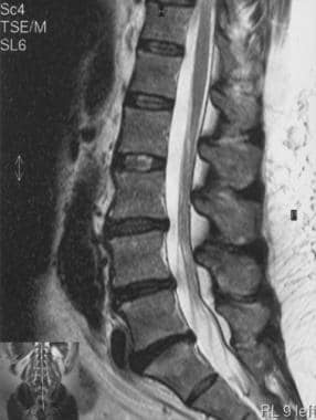
MRI is by far the most commonly ordered test to evaluate patients with sciatica. Often, MRI is performed prior to plain radiographs. MRI is very sensitive in delineating lumbar disc herniations. Far lateral discs are best evaluated with this test. In reoperations, MRI can delineate the full extent of scar tissue and, with moderate reliability, differentiate it from recurrent disc herniation. [2, 3, 9, 10, 11, 6]
Lumbar discectomy is the most common operation performed in the United States for lumbar-related symptoms. Almost all patients with sciatica and disc herniations deserve a trial of medical therapy. The one obvious exception is a patient presenting with cauda equina syndrome or profound motor deficits. a large multicenter trial found that surgical and nonsurgical outcomes at 2 years were similar, but that the surgical group experienced faster pain relief. [12, 13] The limitations of this study are outlined in an editorial. [14]
See the images below of herniated disc disease.
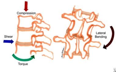 Degenerative lumbar disc disease. The various forces placed upon the discs of the lumbar spine that can result in degenerative changes.
Epidemiology
The lifetime prevalence of low back pain is 80%, with disk disorders being the most common cause of adult low back pain. The most consistent risk factor for degeneration is increasing age. [15]
Correlations have been found with the following [15] :
-
Body mass index, mechanical loading, and genetic predisposition.
-
Genes coding for collagen, aggrecan, vitamin D receptors, matrix metalloproteinase, cartilage intermediate layer protein, and interleukins.
-
Smoking and increased rates of disk degeneration, with animal models showing increased proinflammatory markers, alterations to annular structure, vasoconstriction, and altered nutrient distribution to the disc.
The prevalence of a symptomatic herniated lumbar disc is about 1 to 3% in Finland and Italy, depending on age and sex. The highest prevalence is in persons 30 to 50 years of age, with a male-to-female ratio of 2:1. In persons 25 to 55 years of age, about 95% cases of herniated disc occur at the lower lumbar spine (L4–5 level); disc herniation above this level is more common in persons older than 55 years. [16]
Almost 5% of males and 2.5% of females experience sciatica at some time in their lifetime. [8]
Presentation
Most lumbar disc herniations (lumbar disc diseases) are preceded by bouts of varying degrees and duration of back pain. In many cases, an inciting event cannot be identified. Pain eventually may radiate into the leg. It may be characterized as less achy, burning, or similar to an electrical shock and is often described as a shooting or stabbing pain. The distribution of the leg pain is somewhat dependent on the level of nerve root irritation. Higher herniations (third or fourth lumbar levels) can radiate into the groin or anterior thigh. Lower radiculopathies (first sacral level) cause pain in the calf and bottom of the foot.
Fifth lumbar radiculopathy, which occurs most commonly, causes lateral and anterior thigh and leg pain. Often, accompanying numbness or tingling occurs with a distribution similar to the pain. Accompanying muscle weakness may be unrecognized if the pain is incapacitating. The pain usually improves when the patient is in the supine position with the legs slightly elevated. Patients are more comfortable when changing positions. Short walks can bring relief. Long walks or extended sitting (especially driving) can aggravate the pain.
On examination, patients may be neurologically normal, may have a profound radiculopathy, or may even demonstrate a cauda equina syndrome. A positive straight-leg raising sign is almost always present. However, a crossed straight-leg raising sign may be even more predictive of a lumbar disc herniation (lumbar disc disease). The back may appear scoliotic. Gait is often abnormal. Muscle weakness may be revealed particularly when testing walking on heels and toes.
Indications
The indications for surgical treatment of symptomatic lumbar disc disease are not clearly delineated. Nevertheless, situations exist in which most spine surgeons would probably agree on operative intervention. These situations include the following:
-
A patient with cauda equina syndrome
-
A patient demonstrating progressive neurologic deficit during a period of observation
-
A patient with persistent bothersome sciatic pain, despite conservative management, for a period of 6-12 weeks (a time period that varies from surgeon to surgeon)
Notably missing from this list is a patient presenting with a profound motor deficit of varying duration. In the absence of pain, whether such patients benefit from surgery is unclear. No consensus has been reached concerning how urgent surgery is for a patient who presents with a clinical picture of painful disc herniation. Unfortunately, the decision to operate emergently is often based on fear of legal repercussions rather than on scientific evidence of actual patient benefit.
Contraindications
Any claim of absolute contraindications for lumbar disc disease would invariably be challenged. Most spine surgeons adhere to some guidelines, including the following:
-
A patient with unrelenting back pain: Patients who have back pain after a bout of sciatica has resolved are not good candidates for operative treatment. Often, these patients are the most insistent and difficult to manage. Occasionally, these are patients whose back pain improved after discectomy for a large central disc herniation.
-
A patient with an incomplete workup: When diagnosis is uncertain, postpone surgery. Disc herniations are so ubiquitous that being cavalier in diagnosis is easy. Ensure the completeness of the workup prior to proceeding with the operation. All surgeons can recall several cases in which a diabetic plexopathy or an epidural metastasis was missed.
-
A patient not provided adequate conservative treatment: Spine surgeons rarely commit a patient with a short period of sciatica and without bedrest and a steroid trial to an operation that will permanently alter the patient's back mechanics and strength.
-
Degenerative lumbar disc disease. The various forces placed upon the discs of the lumbar spine that can result in degenerative changes.
-
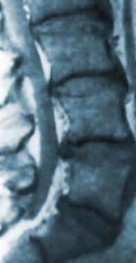
Degenerated lumbar disc disease.
-
Magnetic resonance image of a herniated nucleus pulposus of the lumbar spine.
