Background
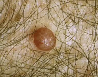
Molluscum contagiosum virus causes a benign viral infection that is largely (if not exclusively) a disease of humans. Molluscum contagiosum virus causes characteristic skin lesions consisting of single or, more often, multiple, rounded, dome-shaped, pink, waxy papules that are 2-5 mm (rarely up to 1.5 cm in the case of a giant molluscum) in diameter. The papules, or bumps, are umbilicated and contain a caseous plug. Although treatment is not required, it may help to reduce autoinoculation or transmission to close contacts and improve clinical appearance. Intervention may also be indicated if lesions persist. Therapeutic modalities include topical application of various medications, radiation therapy, and/or surgery.
See the images below for examples. (See Presentation and Workup.)
 Molluscum contagiosum. Approximately 10% of patients develop eczema around lesions. Eczema associated with molluscum lesions spontaneously subsides following removal.
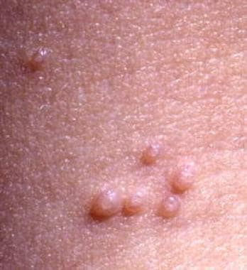
 Molluscum contagiosum. Larger lesions may have several clumps of molluscum bodies rather than the more common single central umbilication. This may make them difficult to recognize as molluscum contagiosum.
See 15 Rashes You Need to Know: Common Dermatologic Diagnoses and 20 Signs of Sexually Transmitted Infections, Critical Images slideshows, to help identify and treat various rashes.
Molluscum contagiosum virus is an unclassified member of the Poxviridae family. It cannot be grown in tissue culture or eggs; it has been grown in human foreskin grafted to athymic mice but has not been transmitted to other laboratory animals (see Etiology).
Through restrictive endonuclease analysis of the genomes of isolates, molluscum contagiosum virus types I-IV have been identified. In a study of 147 patients, molluscum contagiosum virus I caused 96.6% of infections, and molluscum contagiosum virus II caused 3.4%; however, no relationship was observed between virus type and lesional morphology or anatomical distribution. Molluscum contagiosum viruses III and IV are rare. In patients with human immunodeficiency virus (HIV) infection, molluscum contagiosum virus II causes most infections (60%).
Bateman first described the disease in 1817, and Paterson demonstrated its infectious nature in 1841. In 1905, Juliusburg proved its viral nature. Infection follows contact with infected persons or contaminated objects, but the extent of epidermal injury necessary is unknown. Lesions may spread by autoinoculation.
Etiology
Transmission of molluscum contagiosum
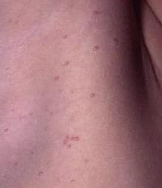
The molluscum contagiosum virus may be inoculated along a line of minor skin trauma (eg, from shaving), resulting in lesions arranged in a linear pattern (see the image below). This process, termed autoinoculation, can also result from manipulation of lesions by the patient. Autoinoculation is different from the Koebner phenomenon, which is also called an isomorphic response. In the Koebner phenomenon, new lesions develop along a line of trauma and the etiology of the underlying condition is unknown. Psoriasis and lichen planus are examples of skin conditions that commonly koebnerize.
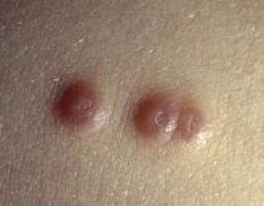 In a patient who had preexisting molluscum contagiosum, the virus was inoculated along a line of minor skin trauma, resulting in the development of the 3 new lesions.
Molluscum contagiosum virus transmission through direct skin contact between children sharing a bath and between athletes sharing gymnasium equipment and benches has been reported. An association between school swimming pool use and molluscum contagiosum infection has also been reported. [1]
Three distinct disease patterns are observed in 3 different patient populations: children, adults who are immunocompetent, and patients who are immunocompromised (children or adults). The prognosis and therapy are different for each of these groups.
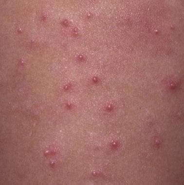
Molluscum contagiosum is most common in children who become infected through direct skin-to-skin contact or indirect skin contact with fomites, such as bath towels, sponges, and gymnasium equipment. Lesions typically occur on the chest, arms, trunk, legs, and face. Hundreds of lesions may develop in intertriginous areas, such as the axillae and intercrural region (see the image below). Lesions may rarely occur on the mucous membranes of the lip, tongue, and buccal mucosa. The palms are spared. Patients with atopic dermatitis may develop large numbers of lesions.
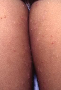 Molluscum lesions may become quite numerous in intertriginous areas. This child has autoinoculated lesions to both inner thighs.
In adults, molluscum contagiosum most commonly is a sexually transmitted disease (STD). Healthy adults tend to have few lesions, which are limited to the perineum, genitalia, lower abdomen, or buttocks. Molluscum contagiosum in healthy children and adults is usually a self-limited disease.
Widespread, persistent, and atypical molluscum contagiosum may occur in patients who are significantly immunocompromised or have acquired immunodeficiency syndrome (AIDS) with low CD4 T-lymphocyte counts (see the images below). Molluscum contagiosum may be the presenting complaint in patients with AIDS. Molluscum contagiosum virus infection in immunocompromised patients may be particularly resistant to therapy. Other opportunistic infections in these patients may closely resemble molluscum contagiosum.
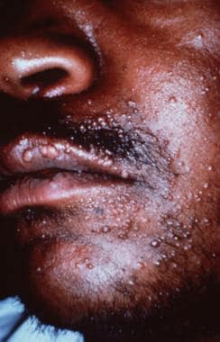
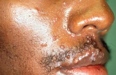 Molluscum contagiosum rarely occurs on the face in an adult unless the patient is infected with HIV. When molluscum contagiosum occurs in individuals infected with HIV, facial lesions are common and frequently numerous.
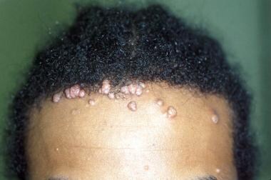 Molluscum contagiosum lesions in individuals infected with HIV may number in the hundreds. In addition, they may become quite large and prominent.
Case reports have detailed molluscum contagiosum eruptions in areas that were treated with tacrolimus 0.1% (Protopic). [2, 3, 4]
Infection with molluscum contagiosum
The molluscum contagiosum virus replicates in the cytoplasm of epithelial cells, producing cytoplasmic inclusions and enlargement of infected cells. This virus infects only the epidermis. Infection follows contact with infected persons or contaminated objects, but the extent of necessary epidermal injury is unknown. The initial infection seems to occur in the basal layer, and the incubation period is usually 2-7 weeks. This is suggested by the fact that, although viral particles are noted in the basal layer, viral deoxyribonucleic acid (DNA) replication and the formation of new viral particles do not occur until the spindle and granular layers of the epidermis are involved. Infection may be accompanied by a latent period of as long as 6 months.
Following infection, cellular proliferation produces lobulated epidermal growths that compress epidermal papillae, while fibrous septa between the lobules produce pear-shaped clumps with the apex upwards. The basal layer remains intact.
Cells at the core of the lesion show the greatest distortion and are ultimately destroyed, resulting in large hyaline bodies (ie, molluscum bodies, Henderson-Paterson bodies) containing cytoplasmic masses of virus material. These bodies are present in large numbers and appear as a white depression at the center of fully developed lesions. Occasionally, the lesions can progress beyond local cellular proliferation and become inflamed with attendant edema, increased vascularity, and infiltration by neutrophils, lymphocytes, and monocytes. Viral-derived proteins inhibit mitochondrial-mediated apoptosis. [5]
As with other poxviruses, molluscum contagiosum virus does not appear to develop latency but evades the immune system through the production of virus-specific proteins. Cell-mediated immunity is most important in modulating and controlling the infection. Children and patients with HIV infection generally have more widespread lesions. Prevalence of molluscum contagiosum virus in patients with HIV may be as high as 5-18%, and the severity of infection is inversely related to the CD4 T-lymphocyte count. More extensive and resistant infections also are noted in patients receiving prednisone and methotrexate.
The virus is not strongly immunogenic, as it infrequently induces antibody formation. Specific antibodies have been found in approximately 80% of patients and in about 15% of control subjects. A role for humoral immunity in regression of lesions is not established. Reinfection is common.
Viral characteristics of molluscum contagiosum
Molluscum contagiosum is a viral disease caused by a DNA poxvirus and is largely, if not exclusively, a disease of humans. It is an unclassified member of the Poxviridae family (ie, poxviruses).
The poxviruses are a large group of viruses with a high molecular weight. They are the largest animal viruses, only slightly smaller than the smallest bacteria, and are just visible using light microscopy. They are complex DNA viruses that replicate in the cytoplasm and are especially adapted to epidermal cells. They cannot be grown in tissue culture or eggs. Molluscum contagiosum virus has been grown in human foreskin grafted to athymic mice but not in other laboratory animals.
Humans are the host for the following 3 types of molluscum contagiosum virus:
-
Orthopoxvirus - This resembles variola (smallpox) and vaccinia, which are ovoid (300 x 250 nm)
-
Parapoxvirus - These are orf and milker’s nodule viruses, which are cylindrical (260 x 160 nm)
-
Unclassified (with features that are intermediate between those of the orthopox and parapox groups) - These are intermediate in structure (275 X 200 nm); they include molluscum contagiosum virus and tanapox
The primary structure and coding capacity of molluscum contagiosum virus was determined by Senkevich et al. [6] Analysis of the molluscum contagiosum virus genome has revealed that it encodes approximately 182 proteins, 105 of which have direct counterparts in orthopoxviruses.
Restriction endonuclease analysis of the genomes has identified 4 types. Molluscum contagiosum virus I and molluscum contagiosum virus II have genomes of 185 kilobases (kb) and 195 kb, respectively. Molluscum contagiosum virus III and IV are very rare.
No relationship between virus type and lesional morphology or anatomical distribution is known. Molluscum contagiosum virus encodes an antioxidant protein (MC066L), selenoprotein, which functions as a scavenger of reactive oxygen metabolites and protects cells from damage from ultraviolet (UV) light and peroxide. The particular role of this protein is not known.
In one study, type I caused 96.6% and type II caused 3.4% of infections in 147 patients, but no relationship was observed between virus type and lesional morphology or anatomic distribution.
Epidemiology
Occurrence in the United States
Molluscum contagiosum is a common infection throughout the United States and accounts for approximately 1% of all skin disorders diagnosed. Data reported from 1969-1983 by the National Disease and Therapeutic Index Survey show an increasing number of patient visits. The prevalence rate in patients with HIV is reported to be 5-18%, and, if the CD4 cell counts are less than 100 cells/μL, the prevalence of molluscum contagiosum is reported to be as high as 33%.
International occurrence
The molluscum contagiosum virus occurs throughout the world, and its incidence in most areas is not reliably known. It is more prevalent in tropical areas. In Mali, molluscum contagiosum is among the most frequent dermatoses in children, with an incidence of 3.6%. [7] In Australia, an overall seropositivity rate of 23% is reported. [8] The lowest antibody prevalence was in children aged 6 months to 2 years (3%), and seropositivity increased with age to reach 39% in persons aged 50 years or older.
Childhood molluscum contagiosum is common in Papua New Guinea, Fiji, and certain parts of Africa. During a regional outbreak in East Africa, it was estimated that 17% of the village population and as many as 52% of children older than age 2 years developed lesions. Epidemiologic studies suggest that transmission may be related to poor hygiene and climatic factors such as warmth and humidity.
Race- and sex-related demographics
During a US longitudinal study performed from 1977-1981, 2-4 times as many cases were found in whites than in persons of other races. [9] Whether the noted difference was secondary to differences in access to medical care, other socioeconomic factors, or genetic predisposition is unclear. [10]
Several studies have shown that males are affected by molluscum contagiosum more commonly than are females. Data from STD clinics in England and Wales revealed that more than twice as many men as women were diagnosed with the infection.
Age-related demographics
Molluscum contagiosum is rare in children younger than age 1 year, perhaps because of maternally transmitted immunity and a long incubation period; otherwise, incidence seems to reflect exposure to others. The greatest incidence is in children younger than age 5 years and in young adults. The peak among the pediatric age group correlates with casual contact, whereas the peak in young adults correlates with sexual contact. [11, 12]
Spread of the virus among households is common in warm climate countries where children are lightly dressed and in close contact with one another and where personal hygiene may be poor. The age of peak incidence is reported to be 2-3 years in Fiji and 1-4 years in the Congo (formerly Zaire). In New Guinea, the annual infection rate for children younger than age 10 years was found to be 6%.
In cooler climates, spread within households is less common, and infection is more common at a later age. Use of school swimming pools is correlated with childhood infections, with a peak incidence in children aged 10-12 years in Scotland and 8 years in Japan. Prevalence appears to be increasing in all age groups.
Prognosis for Molluscum Contagiosum
The prognosis in molluscum contagiosum is generally excellent because the disease is usually benign and self-limited. Spontaneous resolution generally occurs by 18 months in immunocompetent individuals; however, lesions have been reported to persist for as long as 5 years. In healthy patients, treatments are usually effective, although lesions can be disfiguring and may produce anxiety in the patient, family, and daycare facility or school.
Recurrences occur in as many as 35% of patients after initial clearing. The significance of these recurrences is unknown. They may represent reinfection, exacerbation of ongoing disease, or new lesions arising after a prolonged latent period.
The disease often becomes generalized in patients who are infected with HIV or are otherwise immunocompromised. A direct correlation has been found between increasing severity of the disease and lower CD4 counts. The duration of infection is uncertain in populations with HIV infection and in populations that are otherwise immunocompromised (eg, patients who have undergone renal transplant), because molluscum contagiosum may not be self-limiting in these cases.
Morbidity and mortality
Molluscum contagiosum is generally a benign and self-limited infection. For the most part, morbidity is caused by temporary adverse cosmetic results. Morbidity is higher in immunocompromised patients because they tend to have more lesions and more widespread infection. Most lesions resolve with no permanent residual skin defect; however, occasional lesions may produce a slightly depressed scar. This may represent deeper skin damage in lesions that were particularly inflammatory or secondarily infected. Involvement of the margin of the eyelids may produce keratoconjunctivitis. No mortality has been associated directly with the molluscum contagiosum virus.
Patient Education
Before attempting any therapy, educate the patient or parents in-depth about the diagnosis, prognosis, risk of autoinoculation or infection of others, therapeutic options, and risks of therapy. [13, 14] More than 1 treatment session is frequently required. Providing this information at the first clinical visit is particularly important when treating benign lesions, such as those of molluscum contagiosum and common warts. A few extra minutes of explanation at this stage can prevent or mitigate numerous problems and questions during later visits. [15]
When lesions fail to respond to initial therapy, a temptation to be overzealous in treatment may occur. Patients and families are more understanding and less likely to demand aggressive therapy when reasonable goals and limitations of therapy are thoroughly discussed.
The US Centers for Disease Control and Prevention (CDC) has advised on at-home therapies patients or guardians (for pediatric patients) may wish to consider. [16] The CDC suggests that oral therapy with cimetidine can be administered at home and is a reasonable option for widespread lesions or in children, who may be averse to physical removal techniques. They suggest home use of cimetidine is effective, safe, and well tolerated, but that it is less effective for facial lesions versus lesions elsewhere on the body. The CDC also suggests that topical therapies are effective for home use, but the agents involved must be prescribed by a healthcare professional.
Stress the benign nature of this ubiquitous disease to the patient and his or her parents. Limiting physical contact with infected areas of skin and good handwashing may reduce transmission. Instruct the patient to avoid scratching, which may result in autoinoculation.
Keeping children out of school is not necessary; however, discourage physical contact and sharing of clothes and towels. In smaller children in whom physical contact is more difficult to prevent, keeping infected areas covered with clothing is reasonable. Cover exposed lesions with tape or an adhesive bandage. Infection of other children cannot be completely prevented. Because the disease is extremely common and of very little clinical significance, the decision to limit infected children from daycare centers must be approached on a case-by-case basis.
In adolescent and adult patient populations, this disease is usually sexually transmitted. Encourage safe sex and abstinence; however, whether condoms and other barrier methods provide adequate protection against transmission is unclear.
Emphasize that not all STDs are as benign as molluscum contagiosum virus (eg, herpes simplex, gonorrhea, chlamydia, HIV). Stress adherence to abstinence until lesions resolve. In the patient with multiple sexual partners or other risk factors, HIV testing is strongly recommended. Note that not all cases in adults are sexually transmitted. This diagnosis can cause significant relationship stress.
-
Note the central umbilication in these classic lesions of molluscum contagiosum.
-
Molluscum contagiosum. Approximately 10% of patients develop eczema around lesions. Eczema associated with molluscum lesions spontaneously subsides following removal.
-
Molluscum contagiosum on the shaft of the penis. Molluscum contagiosum in the genital region of adults is most commonly acquired as a sexually transmitted disease.
-
Molluscum contagiosum. Larger lesions may have several clumps of molluscum bodies rather than the more common single central umbilication. This may make them difficult to recognize as molluscum contagiosum.
-
Molluscum lesions may become quite numerous in intertriginous areas. This child has autoinoculated lesions to both inner thighs.
-
After trauma, or spontaneously after several months, inflammatory changes result in suppuration, crusting and eventual resolution of the lesion. This inflammatory stage does not usually represent secondary infection and seldom requires antibiotic therapy.
-
Lesions of molluscum contagiosum have a characteristic histopathology. Lobules containing hyalinized molluscum bodies, also known as Henderson-Paterson bodies, are diagnostic.
-
This lesion of cutaneous coccidioidomycosis could be included among the differential diagnoses of molluscum contagiosum.
-
This keratoacanthoma could be included among the differential diagnoses of molluscum contagiosum.
-
Molluscum contagiosum. Lesions on the upper eyelid of a 3-year-old child.
-
In a patient who had preexisting molluscum contagiosum, the virus was inoculated along a line of minor skin trauma, resulting in the development of the 3 new lesions.
-
Molluscum contagiosum on the right axilla.
-
Presented here are the classic umbilicated papules of molluscum contagiosum lesions on the cheek of a child. Facial lesions occur frequently in children, although lesions generally are few.
-
Molluscum contagiosum rarely occurs on the face in an adult unless the patient is infected with HIV. When molluscum contagiosum occurs in individuals infected with HIV, facial lesions are common and frequently numerous.
-
Molluscum contagiosum lesions in individuals infected with HIV may number in the hundreds. In addition, they may become quite large and prominent.
-
This low-power view of a molluscum contagiosum lesion shows the classic cup-shaped invagination of the epidermis into dermis. The Henderson-Paterson bodies are identified readily and stained purple to red in this image.
-
This is a medium-power view of a molluscum contagiosum lesion. Magnification allows better demonstration of the intracytoplasmic molluscum bodies (staining purple-pink) within the keratinocytes.
-
This molluscum contagiosum body is an intracytoplasmic inclusion body. Notice in the image that the keratinocyte nuclei are displaced to the periphery of the cell and that the intracytoplasmic inclusions have a granular quality.
-
Multiple papules on the face of a man with HIV.
-
Cytoplasmic viral inclusions become progressively larger toward the epidermal surface (hematoxylin and eosin, 200X)
-
Low-power histopathologic examination reveals an overall cup-shaped appearance.
-
Viral particles have a dumbbell-shaped appearance. Courtesy of Alvin Zelickson, MD.
Tables

What would you like to print?
- FDA Warns Of Potential Risk From Hologic's Devices Implanted in Soft Tissue
- Posttransplant Skin Disease: Consider Skin Cancer, Infection Risks
- Presurgery Skin Antisepsis Affects Patient Infection Risk
-
 In Lupus, How to Spot Hidden Heart Risk
-
Is Microwave Ablation Better for Treating Thyroid Nodules?
-
Is Scarless Thyroidectomy an Option for Your Patient?


