Background
Orbital cellulitis and preseptal cellulitis are the major infections of the ocular adnexal and orbital tissues. Orbital cellulitis is an infection of the soft tissues of the orbit posterior to the orbital septum. Preseptal cellulitis is an infection of the soft tissue of the eyelids and periocular region anterior to the orbital septum. (See Presentation.) Orbital cellulitis and preseptal cellulitis can sometimes be a continuum.
Orbital cellulitis has various causes and may be associated with serious complications. As many as 11% of cases of orbital cellulitis result in visual loss. Prompt diagnosis and proper management are essential for curing the patient with orbital cellulitis (see the images below). (See Etiology, Prognosis, Presentation, Workup, Treatment, and Medication.)
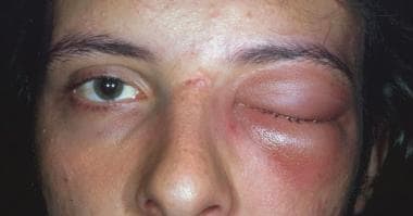 A male patient with orbital cellulitis with proptosis, ophthalmoplegia, and edema and erythema of the eyelids. The patient also exhibited pain on eye movement, fever, headache, and malaise.
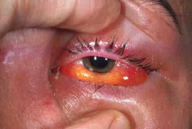 A male patient with orbital cellulitis who demonstrated proptosis, ophthalmoplegia, and edema and erythema of the eyelids. The patient also exhibited chemosis and resistance to retropulsion of the globe.
Anatomy
The orbital septum is a layer of fascia extending vertically from the periosteum of the orbital rim to the levator aponeurosis in the upper eyelid and to the inferior border of the tarsal plate in the lower eyelid.
Patient education
For patient education information, see the Diabetes Center, as well as Cellulitis.
Etiology
Orbital cellulitis occurs in the following 3 situations [1] :
-
Extension of an infection from the paranasal sinuses or other periorbital structures such as the face, globe, or lacrimal sac
-
Direct inoculation of the orbit from trauma or surgery
-
Hematogenous spread from bacteremia
Extension of infection
Orbital cellulitis is commonly associated with sinus infection and can be caused by direct extension of infection from the globe, eyelids, ocular adnexa, and other periocular tissues. Orbital cellulitis may follow dacryocystitis, osteomyelitis of the orbital bones, phlebitis of the facial veins, and dental infections.
Orbital cellulitis is caused most commonly in all age groups by ethmoid sinusitis, accounting for more than 90% of all cases; aerobic, non-spore–forming bacteria are the organisms that most frequently are responsible. The process involves edema of the sinus mucosa, which leads to narrowing of the ostia and subsequent reduction or cessation of normal sinus drainage. Microflora indigenous to the sinuses and upper respiratory tract proliferate and invade the edematous mucosa, resulting in suppuration. It is enhanced by the reduced oxygen tension within the obstructed sinus cavity.
The organisms gain access to the orbit through thin bones of the orbital walls, venous channels, foramina, and dehiscences. Then, subperiosteal and intraorbital abscesses may occur. The resulting elevation of intraorbital pressure results in the typical signs of proptosis, ophthalmoplegia, and chemosis.
Orbital cellulitis resulting from infection of the maxillary sinus secondary to dental infections can be caused by microorganisms indigenous to the mouth, including anaerobes, commonly Bacteroides species.
Those cases stemming from dacryocystitis most commonly are caused by S aureus, S pneumoniae, Streptococcus pyogenes, and nontypeable H influenzae. Infections spreading from the soft tissues of the eyelids and face are due most commonly to staphylococci and S pyogenes. The initial antibiotic regimen can be modified if the response is inadequate or if the cultures dictate otherwise.
Traumatic causes
Infectious material may be introduced into the orbit directly through accidental (eg, orbital fracture) or surgical trauma. Indeed, orbital cellulitis may be caused by any injury perforating the orbital septum. Orbital inflammation [2] may be noted within 48-72 hours after injury, or, in the case of a retained orbital foreign body, it may be delayed for several months.
Surgical procedures, including orbital decompression, dacryocystorhinostomy, eyelid surgery, [3] strabismus surgery, retinal surgery, and intraocular surgery, have been reported as the precipitating cause of orbital cellulitis. Postoperative endophthalmitis can extend to the orbital soft tissues.
Bacterial causes
Streptococcus species, Staphylococcus aureus, and Haemophilus influenzae type B are the most common bacterial causes of orbital cellulitis. Pseudomonas, Klebsiella, Eikenella, and Enterococcus are less common culprits. Polymicrobial infections with aerobic and anaerobic bacteria are more common in patients aged 16 years or older.
Fungal causes
Fungal causes of orbital cellulitis are most commonly Mucor and Aspergillus species. Fungi can enter the orbit. Orbital cellulitis due to fungal infections carries a high mortality rate in patients who are immunosuppressed.
Zygomycosis (also known as mucormycosis or phycomycosis) [4, 5, 6] has a wide distribution, whereas aspergillosis more commonly is seen in warm, humid climates. Mucormycosis can cause rapid-onset thrombosing vasculitis (1-7 days), whereas some forms of aspergillosis can be chronic and indolent (months to years).
Aspergillosis initially results in chronic proptosis and decreased vision, while mucormycosis gives rise to the orbital apex syndrome (involving cranial nerves II, III, IV, V-1, and VI, and orbital sympathetics). More commonly, mucormycosis presents with pain, lid edema, proptosis, and visual loss. While aspergillosis and mucormycosis each may result in nasal and palatal necrosis, mucormycosis also may lead to thrombosing arteritis and ischemic necrosis, whereas aspergillosis gives rise to chronic fibrosis and a nonnecrotizing granulomatous process.
Path of infection
The medial orbital wall is thin and is perforated by numerous valveless blood vessels and nerves, as well as by numerous defects (lamina papyracea/Zuckerkandl dehiscences). This combination of thin bone, foramina for neurovascular passages, and naturally occurring defects in the bone allows for easy communication of infectious material between the ethmoidal air cells and the subperiorbital space in the medial aspect of the orbit. The most common location of a subperiorbital abscess is along the medial orbital wall. The periorbita is adherent relatively loosely to the bone of the medial orbital wall, which allows abscess material to easily move laterally, superiorly, and inferiorly within the subperiosteal space.
In addition, the lateral extensions of the sheaths of the extraocular muscles, the intermuscular septa, extend from one rectus muscle to the next and from the insertions of the muscles to their origins at the annulus of Zinn, posteriorly. Posteriorly in the orbit, the fascia between the rectus muscles is thin and often incomplete, allowing easy extension between the extraconal and intraconal orbital spaces.
Venous drainage from the middle third of the face, including the paranasal sinuses, mainly is via the orbital veins, which are without valves, allowing the passage of infection anterograde and retrograde.
Epidemiology
An increased incidence of orbital cellulitis occurs in the winter nationally and internationally, because of the increased incidence of sinusitis in cold weather.
In the United States, an increase has been noted in the frequency of orbital cellulitis owing to community-acquired methicillin-resistant S aureus infections. [7, 8, 9, 10, 11, 12, 13]
Sex- and age-related demographics
In children, orbital cellulitis has been reported as twice as common in males as in females. In adults, however, no difference in the frequency of orbital cellulitis exists between the sexes, except for cases caused by methicillin-resistant Saureus, which are more common in females than in males by a ratio of 4:1.
Orbital cellulitis, in general, is more common in children than in adults. [14] The median age range of children hospitalized with orbital cellulitis is 7-12 years.
Prognosis
Prior to the availability of antibiotics, patients with orbital cellulitis had a mortality rate of 17%, and 20% of survivors were blind in the affected eye. As a result of prompt diagnosis and the appropriate use of antibiotics, however, this rate has been reduced significantly, although blindness still occurs in up to 11% of cases. Orbital cellulitis caused by methicillin-resistant S aureus can lead to blindness despite antibiotic treatment.
Morbidity and mortality
Orbital cellulitis can result in orbital and intracranial complications. Subperiosteal or orbital abscess formation may occur (7-9%), whereas permanent vision loss may result from corneal damage secondary to exposure or neurotrophic keratitis, destruction of intraocular tissues, secondary glaucoma, optic neuritis, or central retinal artery occlusion. Blindness also may occur secondary to elevated intraorbital pressure or the direct extension of infection to the optic nerve from the sphenoid sinus.
Direct involvement of the ocular motor nerves or the extraocular muscles may lead to decreased ocular motility.
Intracranial complications include meningitis (2%), cavernous sinus thrombosis (1%), and intracranial, epidural, or subdural abscess formation. Cavernous sinus thrombosis has a mortality rate of 50% or higher, but it has become relatively rare in industrialized countries with proper treatment. Cavernous sinus thrombosis should be considered in any patient with orbital cellulitis and should be suspected in the presence of rapid progression of the clinical signs (eg, increasing proptosis, mydriasis, dilation of retinal veins, decreasing visual acuity, development of an afferent pupillary defect).
Intracranial abscess formation is suggested by altered consciousness, signs of central nervous system disturbance, persistent fever despite adequate antibiotic therapy, and resolution of the sinusitis and orbital cellulitis components of the disease.
-
A male patient with orbital cellulitis with proptosis, ophthalmoplegia, and edema and erythema of the eyelids. The patient also exhibited pain on eye movement, fever, headache, and malaise.
-
A male patient with orbital cellulitis who demonstrated proptosis, ophthalmoplegia, and edema and erythema of the eyelids. The patient also exhibited chemosis and resistance to retropulsion of the globe.
Tables

What would you like to print?
- Triamcinolone Improves Eyelid Retraction in Thyroid Eye Disease
- Remote Infections Increase Risk for Surgical Site Infections in Neurosurgery
- Infections Down in Acute Care Hospitals
-
 Cases of Meat Allergy Linked to Tick Bites on the Rise: CDC
-
Recalled Eye Drops Lead to More Deaths, Removals of Eyes
- Surgical Tips for Aesthetic Lower Lid Blepharoplasty: Prevention of Round Eye


 Common Eye Conditions: Slideshow