Continually Updated Clinical Reference
 
 
  All Sources     eMedicine     Medscape     Drug Reference     MEDLINE
 
eMedicine - Pediatrics, Foreign Body Ingestion : Article by

Quick Find
Authors & Editors
Introduction
Clinical
Differentials
Workup
Treatment
Medication
Follow-up
Miscellaneous
Multimedia
References

Related Articles
Appendicitis, Acute

Disk Battery Ingestion

Esophagitis

Foreign Bodies, Trachea

Gastritis and Peptic Ulcer Disease

Gastroenteritis

Munchausen Syndrome

Obstruction, Large Bowel

Obstruction, Small Bowel

Pediatrics, Appendicitis

Pediatrics, Gastroenteritis

Pediatrics, Gastrointestinal Bleeding

Pediatrics, Intussusception

Pediatrics, Pyloric Stenosis

Pediatrics, Reactive Airway Disease

Pharyngitis

Pneumonia, Aspiration

Pneumothorax, Iatrogenic, Spontaneous and Pneumomediastinum


Patient Education
Esophagus, Stomach, and Intestine Center

Battery Ingestion Overview

Battery Ingestion Causes

Battery Ingestion Symptoms

Battery Ingestion Treatment


Author: Gregory P Conners, MD, MBA, MPH, Chief of Pediatric Emergency Medicine, Vice Chair of Emergency Medicine, Professor of Emergency Medicine and Pediatrics, Departments of Emergency Medicine and Pediatrics, University of Rochester School of Medicine and Dentistry

Gregory P Conners is a member of the following medical societies: Ambulatory Pediatric Association, American Academy of Pediatrics, American College of Emergency Physicians, and Society for Academic Emergency Medicine

Editors: Jerry Balentine, DO, Professor of Emergency Medicine, New York College of Osteopathic Medicine; Medical Director, Saint Barnabas Hospital; Mary L Windle, PharmD, Adjunct Assistant Professor, University of Nebraska Medical Center College of Pharmacy, Pharmacy Editor, eMedicine.com, Inc; Wayne Wolfram, MD, MPH, Clinical Associate Professor, Departments of Pediatrics, Children's Hospital and University of Cincinnati; John Halamka, MD, Chief Information Officer, CareGroup Healthcare System, Assistant Professor of Medicine, Department of Emergency Medicine, Beth Israel Deaconess Medical Center; Assistant Professor of Medicine, Harvard Medical School; Richard G Bachur, MD, Assistant Professor of Pediatrics, Harvard Medical School; Associate Chief and Fellowship Director, Associate Clinical Director, Department of Emergency Medicine, Children's Hospital of Boston

Author and Editor Disclosure

Synonyms and related keywords: foreign body ingestion in children, swallowed foreign bodies, coins, pins, screws, button batteries, disk batteries, toy parts, esophageal impaction, aortoenteric fistula, esophageal foreign body, stomach foreign body, lower GI foreign body, mucosal abrasion, intestinal obstruction, esophageal obstruction, retropharyngeal abscess, esophageal perforation


Background

As children explore the world, they will inevitably put foreign bodies into their mouths and swallow some of them.

Most swallowed foreign bodies pass harmlessly through the gastrointestinal (GI) tract. Foreign bodies that damage the GI tract, become lodged, or have associated toxicity must be identified and removed. Children with preexisting GI abnormalities (eg, tracheoesophageal fistula, stenosing lesions, previous GI surgery) are at an increased risk for complications.

Although adults most often present to the ED after ingestion of radiolucent foreign bodies (typically food), children usually swallow radiopaque objects, such as coins, pins, screws, button batteries, or toy parts. Although children commonly aspirate food items, it is less common for small children to present because of foreign body complications due to food ingestion.

Pathophysiology

Esophagus

Most complications of pediatric foreign body ingestion are due to esophageal impaction, usually at 1 of 3 typical locations. The most common site of esophageal impaction is at the thoracic inlet. Defined as the area between the clavicles on chest radiograph, this is the site of anatomical change from the skeletal muscle to the smooth muscle of the esophagus. The cricopharyngeus sling at C6 is also at this level and may "catch" a foreign body. About 70% of blunt foreign bodies that lodge in the esophagus do so at this location. Another 15% become lodged at the mid esophagus, in the region where the aortic arch and carina overlap the esophagus on chest radiograph. The remaining 15% become lodged at the lower esophageal sphincter (LES) at the gastroesophageal junction.

Children with preexisting esophageal abnormalities (eg, repair of a tracheoesophageal fistula) are likely to have foreign body impaction at the site of the abnormality. If a child with no known esophageal pathology has a blunt foreign body lodged at a location other than the 3 typical locations described above, the possibility of a previously unknown esophageal abnormality should be considered.

Pointed objects, such as thumbtacks, may become impaled and, therefore, lodged anywhere in the esophagus. Small objects, such as pills, may adhere to the slightly moist esophageal mucosa at any point. 

Stomach/lower gastrointestinal tract

Once a swallowed foreign body reaches the stomach of a child with a normal GI tract, it is much less likely to lead to complications. Foreign bodies occasionally become lodged at the ileocecal valve. Other exceptions include pointed or toxic foreign bodies or objects too long (ie, >6 cm) or too wide (ie, >2 cm) to pass through the pyloric sphincter. Another important exception is the child who has swallowed more than one magnet; reports exist of swallowed toy magnets attracting and adhering tightly to each other through the GI tract, leading to necrosis of intervening tissues, sometimes with severe sequelae.1

Children with known GI tract abnormalities are more likely to encounter complications. Previous surgery may cause abnormalities of peristalsis, increasing the likelihood of foreign body impaction. For example, children who have had surgery to correct pyloric stenosis are more likely to retain a foreign body in the stomach.

Previously unsuspected lower GI tract abnormalities may present as a complication of foreign body ingestion. For example, a small foreign body may become lodged in a Meckel diverticulum.

Impacted foreign bodies

A foreign body lodged in the GI tract may have little or no effect; cause local inflammation leading to pain, bleeding, scarring, and obstruction; or erode through the GI tract. Migration from the esophagus most often leads to mediastinitis but may involve the lower respiratory tract or aorta and create an aortoenteric fistula. Migration through the lower GI tract may cause peritonitis.

Frequency

United States

Although exact figures are unavailable, foreign body ingestion is clearly common among children. Nearly 111,000 ingestions of foreign bodies by people aged 19 years and younger were reported to American Poison Control Centers in 2005.2 In a recent cross-sectional survey of parents of more than 1500 children, 4% of the children had swallowed a coin (the most commonly swallowed foreign body in many studies).3

International

International data are scant, but pediatric foreign body ingestion is a worldwide problem. Impaction of swallowed fish bones is more commonly observed in countries where fish is a major dietary staple.

Mortality/Morbidity

  • Most foreign bodies pass harmlessly through the GI tract and are eliminated in the stool.

  • Systemic reactions, such as from nickel allergy, are unusual but have been reported, typically in massive ingestions or occupational exposures.
  • Retained foreign bodies may cause GI mucosal erosion, abrasion, local scarring, or perforation.

    • Foreign body migration may lead to peritonitis, mediastinitis, pneumothorax, pneumomediastinum, pneumonia, or other respiratory disease.

    • Migration into the aorta may produce an aortoenteric fistula, a horrific complication with a high mortality rate.
  • Complications of removal procedures may lead to iatrogenic morbidity or mortality from the procedure or from accompanying sedation/anesthesia.

Sex

  • The male-to-female ratio in young children is 1:1.
  • In older children and adolescents, males are more commonly affected than females.

Age

Children of all ages ingest foreign bodies. However, incidence is greatest in children aged 6 months to 4 years. This reflects the tendency of small children to use their mouths in the exploration of their world. Younger children may be "fed" foreign bodies by older children or be intentionally given foreign bodies by abusive adults. In the teenaged years, concomitant psychiatric problems, mental disturbances, and risk-taking behaviors may lead to foreign body ingestion.


History

  • Children commonly come to medical attention after a caregiver witnesses the ingestion of a foreign body or after a child reports an ingestion to a caregiver.
  • Alternatively, the child may present because of signs or symptoms of a complication of ingestion.
  • Occasionally, the caregiver discovers a foreign body that has passed in the stool and brings the child in for evaluation.
  • Children with significant complications of foreign body ingestion may be initially asymptomatic.
  • Children may have vague symptoms that do not immediately suggest foreign body ingestion.
  • When caring for children, always keep the possibility of foreign body ingestion in mind.
  • Esophageal foreign body symptoms
    • Dysphagia

    • Food refusal, weight loss

    • Drooling

    • Emesis/hematemesis

    • Foreign body sensation

    • Chest pain, sore throat

    • Stridor, cough

    • Unexplained fever

    • Altered mental status

  • Stomach/lower GI tract foreign bodies
    • Abdominal distention/pain, vomiting

    • Hematochezia

    • Unexplained fever

Physical

  • Specific physical examination findings are unusual.
  • Physical findings may suggest complications of foreign body migration, such as peritoneal irritation or rales.
  • Abrasions, streaks of blood, or edema in the hypopharynx may be evidence of proximal swallowing-related trauma. Inspection of the oropharynx may occasionally reveal an impacted foreign body.
  • Drooling or pooling of secretions suggests an esophageal foreign body but may be due to an esophageal abrasion as a result of a swallowed foreign body.

Causes

  • Most cases occur as children discover and place small objects in their mouths.
  • Repeated cases may suggest a chaotic home environment and neglect.
  • Children with known GI tract abnormalities or previous complications of foreign body ingestion are more likely to have complications.
  • Older children may be seeking attention or be manifesting psychological abnormalities.
  • Ingestion of unusual foreign bodies may suggest an underlying abnormality. For example, a well-established association exists between toothbrush ingestions and bulimia in teenaged girls.4


Appendicitis, Acute
Disk Battery Ingestion
Esophagitis
Foreign Bodies, Trachea
Gastritis and Peptic Ulcer Disease
Gastroenteritis
Munchausen Syndrome
Obstruction, Large Bowel
Obstruction, Small Bowel
Pediatrics, Appendicitis
Pediatrics, Gastroenteritis
Pediatrics, Gastrointestinal Bleeding
Pediatrics, Intussusception
Pediatrics, Pyloric Stenosis
Pediatrics, Reactive Airway Disease
Pharyngitis
Pneumonia, Aspiration
Pneumothorax, Iatrogenic, Spontaneous and Pneumomediastinum

Other Problems to be Considered

Foreign body aspiration
Gastrointestinal obstruction
Esophageal stricture
Failure to thrive
Meckel diverticulum
Psychiatric diseases - Autism, bulimia, mental retardation, personality disorders


Lab Studies

  • Children with foreign body ingestion typically do not require laboratory testing.
  • Laboratory studies may be indicated for workup of specific complications, such as potential infection.

Imaging Studies

  • Chest/abdominal radiography

    • Most foreign bodies ingested by children are radiopaque (in contrast to inhalation, in which most are radiolucent).
    • If the swallowed object may be radiopaque, a single frontal radiograph that includes the neck, chest, and entire abdomen is usually sufficient to locate the object.
    • If the object is below the diaphragm, further radiographs are generally unnecessary (in the absence of previous GI disorders, such as repaired pyloric stenosis).
    • If the object is in the esophagus, frontal and lateral chest radiographs are necessary to precisely locate and better identify the object and to be sure that the foreign body is not, in fact, two adherent objects. Lateral views of button (disk) batteries reveal a distinctive 2-step border, as opposed to the smooth borders of most coins. Frontal views may suggest a corresponding ring just inside the outermost ring of the battery.
    • Coins and similarly shaped objects may be localized to the esophagus or the airway by their position on a frontal radiograph.
    • With rare exceptions, coins in the esophagus appear in the coronal orientation (ie, coin seen as a disk on frontal view), while coins in the trachea appear in the sagittal orientation (ie, coin seen from the side on frontal view).
    • If the ingested object is radiolucent, the object's location may be inferred from effects (eg, airway compression) seen on plain radiographs. However, such findings are not reliable. 
    • Radiolucent objects in the esophagus may be better visualized by repeating the study after having the child drink a small amount of dilute contrast. This should not be done if endoscopy is planned.
    • Special care must be taken if the esophagus could possibly be obstructed or perforated.
    • When a foreign body is strongly suspected on clinical grounds, visualization by endoscopy, which has the added advantage of allowing removal of the object, may be the most efficient method of management.
  • CT scan or MRI is rarely indicated but may enhance the detection of foreign bodies or complications (eg, perforations) in special cases.

Other Tests

  • Metal detectors
    • The use of handheld metal detectors to identify the location of ingested metallic objects (especially coins) has proven sensitive and specific. In the case of aluminum (eg, flip top of a soda can), a metal detector may be more sensitive since aluminum is often radiolucent. The operator should have experience with this modality before using it for patient care.

    • Patients with coins localized to the abdomen may be safely observed. However, patients with coins localized in the esophagus probably should have the exact locations confirmed by plain radiography.

Procedures

  • Endoscopy
    • Endoscopy (esophagoscopy) may be diagnostic and therapeutic.

    • Children who require extensive radiologic investigation may be best served by referral to a pediatric gastroenterologist or surgeon for endoscopy, which is safe and highly effective.


Prehospital Care

  • Most children who have swallowed a foreign body do not require specialized care.
  • Patients with drooling may require suction.
  • Children benefit by being allowed to remain with their parents and being allowed to assume a position of comfort.
  • Although a theoretical risk of spontaneously vomiting and then aspiration of a foreign body exists, this is unusual. Children should not routinely be intubated to protect their airways.
  • Similarly, do not attempt to dislodge a foreign body from a spontaneously breathing patient by giving abdominal thrusts or syrup of ipecac.

Emergency Department Care

The usual goal of ED management is to localize the position of the ingested foreign body. Patients with drooling, marked emesis, or altered mental status (from excess vagal stimulation) may require supportive measures to protect the airway.

Most patients should undergo radiographic imaging as described above. Metal detectors may be used to locate metallic foreign bodies. Even radiopaque foreign bodies may be difficult to localize. Referral for endoscopy should be considered.

Remember that children with no symptoms may have impacted foreign bodies and that children with foreign body sensation or pain may not. Radiographs of about 15% of children presenting to the ED after witnessed coin ingestions do not show a coin. This suggests that not all foreign bodies whose ingestions were witnessed were really ingested.

  • Esophageal foreign bodies

    • Objects found within the esophagus should generally be considered impacted. Because impacted esophageal foreign bodies may lead to significant morbidity (and even mortality), removal of impacted esophageal foreign bodies is mandatory. An important exception is blunt esophageal foreign bodies (except button [disk] batteries) that are well tolerated and are known to have been in place for less than 24 hours (see Spontaneous passage below).
    • Endoscopy (esophagoscopy) is by far the most commonly used means of removal and is usually the procedure of choice. Most children with esophageal foreign bodies are stable. Endoscopy usually can be delayed until the child's stomach is emptied and a surgical team is assembled. However, pointed objects should be removed as rapidly as possible to avoid further injury to the esophageal mucosa. Impacted button (disk) batteries are notorious for rapidly causing local necrosis and should be removed from the esophagus without delay.
    • Because endoscopy is relatively invasive and expensive, 2 other methods of esophageal foreign body removal have been investigated and are probably more cost-effective when used appropriately. Both have been performed most commonly on children with esophageal coins.
      • Foley catheter method: Blunt foreign bodies may be removed by use of a Foley catheter. The patient is restrained in a head-down position on a fluoroscopy table, and an uninflated catheter is inserted distal to the object. The catheter is then inflated and gently withdrawn, drawing the foreign body with it. Progress is typically monitored fluoroscopically. This procedure is performed without radiographic monitoring at some centers with extensive experience. Only experienced personnel should perform this procedure, and it should be reserved for healthy children whose ingestion of a blunt object was witnessed less than 24 hours prior to the procedure.
      • Bougienage method: Blunt esophageal foreign bodies may be advanced into the stomach with a bougie. While the child is sitting upright, the lubricated instrument is gently passed down the esophagus, dislodging the object. The object is then expected to pass through the rest of the GI tract; thus, this procedure should not be performed on children with known lower GI tract abnormalities. A brief observation period and a repeat radiograph should follow any removal procedure to rule out retained foreign bodies and other complications (eg, pneumomediastinum). Because any esophageal foreign body may pass spontaneously, chest radiography should be performed immediately prior to any removal procedure. Again, only experienced personnel should perform this procedure, and it should be reserved for healthy children whose ingestion of a blunt object was witnessed less than 24 hours prior to the procedure.
      • More recently, use of Magill forceps for removal of foreign bodies high in the esophagus has been described.
    • Spontaneous passage: Blunt foreign bodies located at the LES often spontaneously pass within several hours of ingestion. This has been best studied in coin ingestions. Previously healthy children may be given food and drink and have repeat radiographs 24 hours following ingestion. Often, the coin passes through the LES, and a removal procedure can be avoided. Although blunt foreign bodies located in other areas of the esophagus are less likely to spontaneously pass, this strategy may be an appropriate alternative for stable children with normal esophageal anatomy and a foreign body in the thoracic inlet or the mid esophagus. This may be most successful in asymptomatic children.
    • Complications: Children with significant complications, such as airway involvement, peritonitis, or hematemesis (possibly heralding exsanguination from an aortoenteric fistula), should be referred to an appropriate surgeon without delay.
  • Stomach/lower GI tract

    • Most swallowed foreign bodies harmlessly pass through the GI tract once they have reached the stomach. Treatment of children with known abnormalities of the GI tract or previous problems with foreign bodies should be discussed with a specialist, preferably one familiar with the child.
    • Unusual foreign bodies: Very sharp or pointed objects may perforate the GI tract (sewing needles are notorious). Therefore, such objects should be endoscopically removed from the stomach. If such an object has passed into the intestines, early consultation with a surgeon is recommended. Objects that are too long (eg, >6 cm) or too wide (eg, >2 cm) to pass through the pyloric sphincter should be removed from the stomach.
    • Button (disk) batteries in the stomach or intestines do not need to be removed immediately, as they generally pass through the lower GI tract without difficulty. Button batteries retained in the stomach or at a fixed spot in the intestines should be removed. One strategy is to instruct families to observe the stool for the battery and to return for a repeat radiograph if it is not passed in 2-3 days. If a battery is still in the stomach at that time, it should be endoscopically removed. If it is in the intestines, its progress should be intermittently monitored via radiographs, to be sure it is progressing.
    • Body packers (ie, patients who have ingested wrapped packages of drugs to avoid detection during transport) are at risk of death if the packets rupture. Such patients should be hospitalized and whole-bowel irrigation considered. Consultation with a poison control center is suggested.

Consultations

  • The treatment of children with known GI tract disorders should be discussed with a physician familiar with the child whenever possible.
  • Experienced personnel, such as a pediatric surgeon, otolaryngologist, or gastroenterologist, should perform endoscopy.
  • Psychiatric consultation is indicated for those with a suspected or confirmed associated psychiatric problem.


Although drugs such as glucagon, benzodiazepines, and nifedipine have been successfully used to relax the lower esophageal sphincter in adult patients with esophageal foreign bodies, these measures are generally unsuccessful in children.

The use of meat tenderizer (papain) to attempt to digest meat impacted in the esophagus is no longer recommended. Such usage may severely injure the esophagus.


Further Inpatient Care

  • Children who require endoscopic foreign body removal are usually taken directly to the operating room or endoscopy suite or are admitted preoperatively. These patients should be given nothing by mouth (NPO) and be given glucose-containing intravenous fluids until the procedure.
  • Preprocedure radiographs to verify the location of the foreign body are recommended, as some foreign bodies may pass into the stomach while awaiting endoscopy.
  • General anesthesia often is used for endoscopic foreign body removal. However, sedation performed by experienced personnel may be successful in selected cases.

Further Outpatient Care

  • After an esophageal foreign body is removed, children with uncomplicated courses do not need to undergo further evaluation.
  • A healthy child with repeated foreign body impaction or impaction at an unusual site should be evaluated for an underlying esophageal disorder.
  • Most children with foreign bodies in the stomach or lower GI tract have no complications.
  • Patients with known abnormalities of the GI tract, previous problems with foreign bodies, or unusual foreign bodies may require special treatment.
  • Caregivers of discharged children should be alerted to return if signs or symptoms of the occasional complication (eg, abdominal pain or distention, hematochezia, unexplained fevers, constipation, vomiting) develop.
  • In general, straining of the stool for the foreign body is unnecessary.
  • Except in special instances, serial radiographs to document progress are unnecessary. The continued presence of a metallic foreign body may be documented by serial metal detector scans.

Transfer

  • Most children do not require a removal procedure, and they may be treated at any facility capable of obtaining radiographs of children.
  • Children who require foreign body removal procedures should be referred to a facility with experienced personnel.
  • Familiarity with pediatric airway emergencies is essential.

Deterrence/Prevention

  • Parents and other caregivers of children should be cautioned about leaving small objects where young children may find them and place them into their mouths. This is especially common at times of unusual activity, such as parties, holidays, when visitors are present in the home, or during travel.

Complications

  • Esophageal foreign bodies
    • Mucosal abrasion

    • Esophageal stricture/obstruction

    • Retropharyngeal abscess

    • Failure to thrive

    • Esophageal perforation may lead to mediastinitis, pneumothorax, pneumomediastinum, aortoesophageal fistula formation (and resulting hemorrhage), and tracheal compression.

  • Stomach/lower GI tract foreign bodies
    • Mucosal abrasion

    • Intestinal obstruction

  • Intestinal perforation may lead to peritonitis and sepsis.

Patient Education


Medical/Legal Pitfalls

  • Children without symptoms still may have an esophageal foreign body that requires removal. When in doubt, obtain a radiograph.
  • The Foley catheter and bougienage methods of esophageal foreign body removal can be safe and effective if performed by experienced personnel on appropriate patients. Appropriate personnel and equipment to handle emergency airway complications should always be present in these cases.
  • Button (disk) batteries in the esophagus should be removed immediately.
  • Radiographs to determine the position of foreign bodies should include the entire neck to avoid missing foreign bodies lodged in the pharynx or upper esophagus.
  • Repeat the radiograph before performing a removal procedure to make sure the foreign body has not moved, making the procedure unnecessary.
  • Children with preexisting GI disorders or with problems from previous foreign body ingestions require special attention.
  • Esophageal foreign bodies, at a location other than 1 of the 3 common anatomic locations, may be a clue to an undiagnosed esophageal disorder.
  • Although previously healthy children with foreign bodies in the lower GI tract usually do well, do not forget to warn caregivers about the signs or symptoms of complications.

Special Concerns

  • Children with nickel allergies may have complications of nickel coin ingestions, and patients and their parents should be counseled to watch for rashes, shortness of breath, or other allergic complications.


Media file 1:  
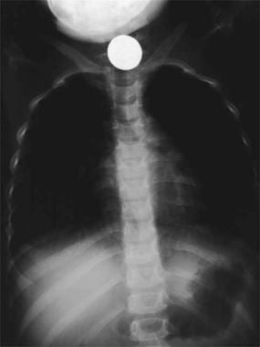
A swallowed coin lodged at the thoracic inlet. Image courtesy of Gregory Conners, MD, MPH.
Click to see larger pictureClick to see detailView Full Size Image
Media type:  X-RAY

Media file 2:  A swallowed radiolucent object (plastic guitar pick) is made visible in the upper esophagus after ingestion of barium. Image courtesy of Raymond K. Tan, MD, and Gregory Conners, MD, MPH.
Click to see larger pictureClick to see detailView Full Size Image
Media type:  X-RAY


  1. Vijaysadan V, Perez M, Kuo D. Revisiting swallowed troubles: intestinal complications caused by two magnets--a case report, review and proposed revision to the algorithm for the management of foreign body ingestion. J Am Board Fam Med. Sep-Oct 2006;19(5):511-6. [Medline].
  2. Lai MW, Klein-Schwartz W, Rodgers GC, Abrams JY, Haber DA, Bronstein AC. 2005 Annual Report of the American Association of Poison Control Centers' national poisoning and exposure database. Clin Toxicol (Phila). 2006;44(6-7):803-932. [Medline].
  3. Conners GP, Chamberlain JM, Weiner PR. Pediatric coin ingestion: a home-based survey. Am J Emerg Med. Nov 1995;13(6):638-40. [Medline].
  4. Riddlesberger MM Jr, Cohen HL, Glick PL. The swallowed toothbrush: a radiographic clue of bulimia. Pediatr Radiol. 1991;21(4):262-4. [Medline].
  5. Bassett KE, Schunk JE, Logan L. Localizing ingested coins with a metal detector. Am J Emerg Med. Jul 1999;17(4):338-41. [Medline].
  6. Bonadio WA, Emslander H, Milner D, Johnson L. Esophageal mucosal changes in children with an acutely ingested coin lodged in the esophagus. Pediatr Emerg Care. Dec 1994;10(6):333-4. [Medline].
  7. Brayer AF, Conners GP, Ochsenschlager DW. Spontaneous passage of coins lodged in the upper esophagus. Int J Pediatr Otorhinolaryngol. Jun 1 1998;44(1):59-61. [Medline].
  8. Brayer AF, Sciera M, Conners GP. Pediatric coin ingestion: an unusual presentation. Int J Pediatr Otorhinolaryngol. Oct 16 2000;55(3):211-3. [Medline].
  9. Buckler JM, Stool SE. Failure to thrive. An exogenous cause. Am J Dis Child. Dec 1967;114(6):652-3. [Medline].
  10. Byard RW, Moore L, Bourne AJ. Sudden and unexpected death--a late effect of occult intraesophageal foreign body. Pediatr Pathol. 1990;10(5):837-41. [Medline].
  11. Calkins CM, Christians KK, Sell LL. Cost analysis in the management of esophageal coins: endoscopy versus bougienage. J Pediatr Surg. Mar 1999;34(3):412-4. [Medline].
  12. Conners GP. A literature-based comparison of three methods of pediatric esophageal coin removal. Pediatr Emerg Care. Apr 1997;13(2):154-7. [Medline].
  13. Conners GP. Diagnostic uses of metal detectors: a review. Int J Clin Pract. Aug 2005;59(8):946-9. [Medline].
  14. Conners GP. Finding aluminum foreign bodies. Pediatr Rev. May 2000;21(5):172. [Medline].
  15. Conners GP. Management of asymptomatic coin ingestion. Pediatrics. Sep 2005;116(3):752-3. [Medline].
  16. Conners GP, Chamberlain JM, Ochsenschlager DW. Conservative management of pediatric distal esophageal coins. J Emerg Med. Nov-Dec 1996;14(6):723-6. [Medline].
  17. Conners GP, Chamberlain JM, Ochsenschlager DW. Symptoms and spontaneous passage of esophageal coins. Arch Pediatr Adolesc Med. Jan 1995;149(1):36-9. [Medline].
  18. Conners GP, Cobaugh DJ, Feinberg R, Lucanie R, Caraccio T, Stork CM. Home observation for asymptomatic coin ingestion: acceptance and outcomes. The New York State Poison Control Center Coin Ingestion Study Group. Acad Emerg Med. Mar 1999;6(3):213-7. [Medline].
  19. Conners GP, Hadley JA. Esophageal coin with an unusual radiographic appearance. Pediatr Emerg Care. Oct 2005;21(10):667-9. [Medline].
  20. Dahiya M, Denton JS. Esophagoaortic perforation by foreign body (coin) causing sudden death in a 3-year-old child. Am J Forensic Med Pathol. Jun 1999;20(2):184-8. [Medline].
  21. Darrow DH, Holinger LD. Aerodigestive tract foreign bodies in the older child and adolescent. Ann Otol Rhinol Laryngol. Apr 1996;105(4):267-71. [Medline].
  22. Ewing S, Miller R. Generalized nickel dermatitis in a 6-year-old boy as a result of swallowing a Canadian nickel. J Am Acad Dermatol. Nov 1991;25(5 Pt 1):855-6. [Medline].
  23. Fleisher AG, Holgersen LO, Stanley-Brown EG, Mones R. Prolonged gastric retention of a swallowed coin following pyloromyotomy. J Pediatr Gastroenterol Nutr. Sep-Oct 1986;5(5):811-3. [Medline].
  24. Gilchrist BF, Valerie EP, Nguyen M, Coren C, Klotz D, Ramenofsky ML. Pearls and perils in the management of prolonged, peculiar, penetrating esophageal foreign bodies in children. J Pediatr Surg. Oct 1997;32(10):1429-31. [Medline].
  25. Karaman A, Cavusoglu YH, Karaman I, Erdogan D, Aslan MK, Cakmak O. Magill forceps technique for removal of safety pins in upper esophagus: a preliminary report. Int J Pediatr Otorhinolaryngol. Sep 2004;68(9):1189-91. [Medline].
  26. Macpherson RI, Hill JG, Othersen HB, Tagge EP, Smith CD. Esophageal foreign bodies in children: diagnosis, treatment, and complications. AJR Am J Roentgenol. Apr 1996;166(4):919-24. [Medline].
  27. Mahafza TM. Extracting coins from the upper end of the esophagus using a Magill forceps technique. Int J Pediatr Otorhinolaryngol. Jan 11 2002;62(1):37-9. [Medline].
  28. Mohiuddin S, Siddiqui MS, Mayhew JF. Esophageal foreign body aspiration presenting as asthma in the pediatric patient. South Med J. Jan 2004;97(1):93-5. [Medline].
  29. Nolte KB. Esophageal foreign bodies as child abuse. Potential fatal mechanisms. Am J Forensic Med Pathol. Dec 1993;14(4):323-6. [Medline].
  30. Paul RI, Christoffel KK, Binns HJ, Jaffe DM. Foreign body ingestions in children: risk of complication varies with site of initial health care contact. Pediatric Practice Research Group. Pediatrics. Jan 1993;91(1):121-7. [Medline].
  31. Paul RI, Jaffe DM. Sharp object ingestions in children: illustrative cases and literature review. Pediatr Emerg Care. Dec 1988;4(4):245-8. [Medline].
  32. Pellerin D, Fortier-Beaulieu M, Gueguen J. The fate of swallowed foreign bodies. Experience of 1250 instances of subdiaphragmatic foreign bodies in children. Prog Pediatr Radiol. 1969;2:286-302.
  33. Sacchetti A, Carraccio C, Lichenstein R. Hand-held metal detector identification of ingested foreign bodies. Pediatr Emerg Care. Aug 1994;10(4):204-7. [Medline].
  34. Seikel K, Primm PA, Elizondo BJ, Remley KL. Handheld metal detector localization of ingested metallic foreign bodies: accurate in any hands?. Arch Pediatr Adolesc Med. Aug 1999;153(8):853-7. [Medline].
  35. Sheikh A. Button battery ingestions in children. Pediatr Emerg Care. Aug 1993;9(4):224-9. [Medline].
  36. Smith SA, Conners GP. Unexpected second foreign bodies in pediatric esophageal coin ingestions. Pediatr Emerg Care. Aug 1998;14(4):261-2. [Medline].
  37. Soprano JV, Fleisher GR, Mandl KD. The spontaneous passage of esophageal coins in children. Arch Pediatr Adolesc Med. Oct 1999;153(10):1073-6. [Medline].
  38. Vargas EJ, Mody AP, Kim TY, Denmark K, Moynihan JA, Barcega BB, et al. The removal of coins from the upper esophageal tract of children by emergency physicians: a pilot study. CJEM. Nov 2004;6(6):434-40. [Medline].
  39. Waltzman ML, Baskin M, Wypij D, Mooney D, Jones D, Fleisher G. A randomized clinical trial of the management of esophageal coins in children. Pediatrics. Sep 2005;116(3):614-9. [Medline].

Pediatrics, Foreign Body Ingestion excerpt

Article Last Updated: Jun 25, 2007