Practice Essentials
Pinworm infection, caused by the nematode Enterobius vermicularis, is prevalent in temperate regions worldwide. It primarily affects children, with transmission often occurring to parents via their children. In the US, general prevalence in children ranges from 0.2-20%, whereas in institutional settings, it can be as high as 50-100%. [1, 2, 3]
-
Diagnosis: Cellophane tape examination to detect eggs in the perianal area (rather than stool examination)
-
Prevention : Handwashing before eating, Cleaning of bedding, clothing, and towels to prevent transmission. [1]
Etiology
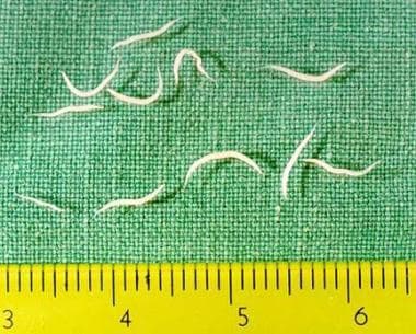
Pinworm infection, also called enterobiasis, is caused by Enterobius vermicularis, a white slender nematode with a pointed tail. In humans, they reside in the cecum, appendix, and ascending colon. Female pinworms are 8-13 mm long, and males are 2-5 mm long. [1, 2]
 Adult female worms of Enterobius vermicularis collected from a 2-year-old girl in a Korean orphanage after treatment with pyrantel pamoate 10 mg/kg.
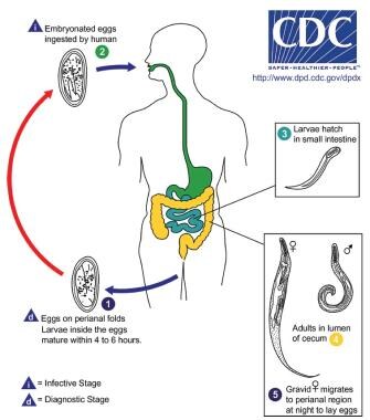
Life cycle of pinworms [5]
-
Pinworms reside in the cecum, appendix, and ascending colon. Unlike other parasites, they do not lay eggs within the intestines. Instead, female worms accumulate around 10,000 eggs in their uterus.
-
At night, gravid female worms migrate to the anus, lay eggs on the perianal skin, and then die. Within 4-6 hours, the larvae develop inside the eggs, becoming infectious. The movement of the female and the ova cause intense local itching. Ova may survive for up to 3 weeks before hatching.
-
Eggs can be transmitted via contaminated surfaces or hands, and once ingested, the larvae hatch in the small intestine, migrate to the cecum, and mature. The entire cycle, from infection to egg release, takes about 3-4 weeks.
-
Retroinfection: There also is a possibility of retroinfection, where larvae hatch around the anus and migrate back into the colon. However, the frequency of this occurrence is not well known.
 Life cycle of Enterobius vermicularis. Image courtesy of DPDx, Centers for Disease Control and Prevention (https://www.cdc.gov/dpdx)
Transmission
Unlike other intestinal nematodes such as roundworm, hookworm, and whipworm, which often involve a life cycle requiring feces-soil-oral transmission, pinworms are transmitted via direct contact with contaminated surfaces (furniture, bedding, clothing, etc.), autoinfection (scratching the anus and ingesting eggs), or through sexual contact.
Embryonated eggs may be released into the air or onto fomites (eg, bedding, clothing, toys, paper money) or onto hands and then placed directly into the mouth and swallowed (autoinfection), after which they settle in the small intestines. [1]
Epidemiology
E vermicularis is the most common helminthic infestation in the United States. General prevalence in children is reported to be 0.2-20%. Pinworm infection is most common in persons who live in crowded living conditions and in individuals who are institutionalized. [1] Prevalence in institutionalized persons is reported to be 50-100%. A similar prevalence of pinworm infestation has been reported in European countries. [6]
The general prevalence of pinworm infection in some regions may be as high as 12%. Pinworm infection is most common in cosmopolitan areas in cool and temperate regions. Egg carrier rates vary by country, from 0.1-98.4%.
Of all age groups, school-aged children are most at risk for pinworm infections. In adults, pinworm infection is most common in parents aged 30-39 years, typically owing to transmission from their children aged 5-9 years.
Overall, males are affected twice as often as females, except in people aged 5-14 years, when infection is predominantly in females. [1]
Risk factors for pinworms include living with a person who is egg-positive, eating before washing hands, and poor personal or group hygiene. [1]
Prognosis
Pinworm infection does not cause severe morbidity unless ectopic infection occurs. This rare complication occurs in individuals with conditions that compromise the integrity of the bowel wall (eg, inflammatory bowel disease). Parasites migrate through the bowel wall and are found in extracolonic sites.
Ectopic enterobiases have been described in various locations, including the vagina, salpinx, inguinal area, genital area, pelvic peritoneum, omentum, liver, salivary glands, male genital tract, and even the lungs. They also have been associated with acute appendicitis, eosinophilic colitis, and eosinophilic gastroenteritis. [1, 7]
Pinworm infestation very rarely is fatal; death and morbidity are from secondary infection. A 28% to 68% increased risk for appendicitis is associated with pinworm infestation. [8]
Eradicating pinworm in groups of institutionalized persons is difficult. Continuous follow-up examination is necessary.
Therapy is much more effective if the child's family and classmates are treated at the same time.
Patient Education
Focus on handwashing, especially before eating. Strict handwashing should be completed after using the toilet or changing a diaper of an affected baby. [1]
Washing sheets, clothes, and towels in a washing machine using regular laundry soap can eliminate pinworm eggs. All bedding and toys should be cleaned every 3-7 days for 3 weeks. Underwear and pajamas should be washed daily for 2 weeks. [1]
-
Adult female worms of Enterobius vermicularis collected from a 2-year-old girl in a Korean orphanage after treatment with pyrantel pamoate 10 mg/kg.
-
Microscopic view of Enterobius vermiculariseggs attached to cellophane tape after a perianal swab from a child in kindergarten in Seoul, Korea. Egg size was 50-60 μm X 20-30 μm. The eggs are elongated and ovoid, distinctly compressed laterally, and flattened on one side.
-
Pinworms in a young patient.
-
Life cycle of Enterobius vermicularis. Image courtesy of DPDx, Centers for Disease Control and Prevention (https://www.cdc.gov/dpdx)

