| Patient Education |
|
Click here for patient education.
|
|
You are in: eMedicine Specialties >
Emergency Medicine > OPHTHALMOLOGY
Retinal Artery Occlusion
Article Last Updated: Jan 14, 2008
AUTHOR AND EDITOR INFORMATION
Section 1 of 12
Author: Kilbourn Gordon III, MD, FACEP, Urgent Care Physician, Primary Medical, Huntington Walk-In and Greenwich Convenient Medical Center
Kilbourn Gordon III is a member of the following medical societies: American Academy of Ophthalmology, American College of Emergency Physicians, and Wilderness Medical Society
Editors: Assaad J Sayah, MD, Chief, Department of Emergency Medicine, Cambridge Health Alliance; Francisco Talavera, PharmD, PhD, Senior Pharmacy Editor, eMedicine; Douglas Lavenburg, MD, Clinical Professor, Department of Emergency Medicine, Christiana Care Health Systems; John D Halamka, MD, MS, Associate Professor of Medicine, Harvard Medical School, Beth Israel Deaconess Medical Center; Chief Information Officer, CareGroup Healthcare System and Harvard Medical School; Attending Physician, Division of Emergency Medicine, Beth Israel Deaconess Medical Center; Robert E O'Connor, MD, MPH, Professor and Chair, Department of Emergency Medicine, University of Virginia Health System
Author and Editor Disclosure
Synonyms and related keywords:
RAO, branch retinal artery occlusion, BRAO, central retinal artery occlusion, CRAO, ocular stroke, embolism of the retinal artery, retinal artery emboli, loss of vision
Background
Painless loss of monocular vision is the usual presenting symptom of retinal artery occlusion (RAO). Ocular stroke commonly is caused by embolism of the retinal artery, although emboli may travel to distal branches of the retinal artery, causing loss of only a section of the visual field. RAO represents an ophthalmologic emergency, and delay in treatment may result in permanent loss of vision.
Immediate intervention improves chances of visual recovery, but, even then, prognosis is poor, with only 21-35% of eyes retaining useful vision. Although restoration of vision is of immediate concern, RAO is a harbinger for other systemic diseases that must be evaluated immediately.
Pathophysiology
Blood supply to the retina originates from the ophthalmic artery, the first intracranial branch of the internal carotid artery that supplies the eye via the central retinal and the ciliary arteries. The central retinal artery supplies the retina as it branches into smaller segments upon leaving the optic disc. The ciliary arteries supply the choroid and the anterior portion of the globe via the rectus muscles (each rectus muscle has 2 ciliary arteries except the lateral rectus, which has 1). Anatomical variants include cilioretinal branches from the short posterior ciliary artery, giving additional supply to part of the macular retina. A cilioretinal artery occurs in approximately 14% of the population. Typical funduscopic findings of a pale retina with a cherry red macula (ie, the cherry red spot) result from obstruction of blood flow to the retina from the retinal artery, causing pallor, and continued supply of blood to the choroid from the ciliary artery, resulting in a bright red coloration at the thinnest part of the retina (ie, macula). These findings do not develop until an hour or more after embolism, and they resolve within days of the acute event. By this time, visual loss is permanent and primary optic atrophy has developed. In those with a cilioretinal artery supplying the macula, a cherry red spot is not observed. An embolism, atherosclerotic changes, inflammatory endarteritis, angiospasm, or hydrostatic arterial occlusion may occlude the retinal artery. The mechanism of obstruction may be obvious from comorbid systemic disease or physical findings. Atrial fibrillation and ipsilateral carotid stenosis are more commonly associated with prolonged visual disturbances. Animal studies have shown that a retina with completely occluded circulation has irreversible ischemic damage at 105 minutes but may recover at 97 minutes. Complete occlusion of retinal artery circulation in humans is rare with retinal artery disease; thus, retinal recovery is possible even after days of ischemia. Branch retinal artery occlusion (BRAO) occurs when the embolus lodges in a more distal branch of the retinal artery. BRAO typically involves the temporal retinal vessels and usually does not require ocular therapeutics unless perifoveolar vessels are threatened. The central retinal artery is affected in 57% of occlusions, the branch retinal artery is involved in 38% of occlusions, and cilioretinal artery obstructions occur in 5% of occlusions.
Frequency
United States
Recent estimates put the incidence of RAO at 0.85 per 100,000 per year, with a 10-year cumulative incidence of 1.5%.
Mortality/Morbidity
Sex
Men are affected slightly more frequently than women.
Age
- The mean age of presentation is early in the seventh decade of life, although a few cases have been reported in patients younger than 30 years.
- The etiology of occlusion changes, depending on the age at presentation.
History
- The most common presenting complaint is an acute persistent painless loss of vision. In central artery occlusions, visual loss is central and dense. In branch artery occlusions, visual loss may go unnoticed if only a section of the peripheral visual field space is affected.
- A complete visual field defect suggests CRAO.
- A sectional visual field defect suggests BRAO and may be an altitudinal defect affecting the upper or lower hemifield but never respecting a vertical axis.
- A history of hypertension or diabetes mellitus is elicited in 67% and 25% of patients with CRAO, respectively.
- Query about any medical problems that could predispose patients to embolus formation (eg, atrial fibrillation, endocarditis, coagulopathies, atherosclerotic disease).
- Prolonged direct pressure to the globe during drug-induced stupor or improper positioning during surgery also may lead to CRAO.
Physical
- Determine the degree of vision loss (eg, no light perception, hand movement, counting fingers); the prognosis for recovery is directly related to initial visual loss.
- Funduscopic examination
- An afferent pupillary defect (ie, paradoxical dilatation of the pupil when a light is shined from the unaffected eye to the affected eye) may be observed within seconds of the occlusive event.
- The cherry red spot and a ground-glass retina are the classic findings but may take hours to develop.
- The funduscopic findings typically resolve within days to weeks of the acute event, sometimes leaving a pale optic disc as the only physical finding.
- Emboli can be observed in approximately 20% of patients with CRAO.
- A dilated funduscopic examination is required to see the pathological signs of RAO.
- BRAO presents with whitening of the retina along the distribution of the occluded vessel.
- Boxcar segmentation of the blood column is observed most often in BRAO and is a sign of severe occlusion and slowing of circulation.
- Direct the physical examination to evaluate for murmurs, carotid bruits, or other signs of cardiovascular disease.
Causes
Causes of CRAO vary, depending on the age of the patient. A detailed analysis of comorbid disease is necessary to elucidate the cause of the acute visual loss.
- Embolism
- Embolism is usually caused by cholesterol, but it can be calcific, bacterial, or talc from IV drug abuse.
- It is associated with poorer visual acuity and higher morbidity and mortality than other RAOs.
- Embolus from the heart is the most common cause of CRAO in patients younger than 40 years.
- Amaurosis fugax preceding persistent loss of vision suggests BRAO or temporal arteritis and may represent emboli causing temporary occlusion of the retinal artery.
- Coagulopathies from sickle cell anemia or antiphospholipid antibodies are common etiologies for CRAO in patients younger than 30 years.
- Atherosclerotic changes
- Carotid atherosclerosis is observed in 45% of CRAO cases, with 60% or more stenosis occurring in 20% of cases.
- Atherosclerotic disease is the leading cause of CRAO in patients aged 40-60 years.
- Inflammatory endarteritis
- Occurrence is rare (only 2% of cases).
- Suspect inflammatory endarteritis in elderly patients if no other etiology is observed.
- Inflammatory endarteritis can affect the second eye within hours if untreated.
- Migraines are rare causes of CRAO but are most common in patients younger than 30 years.
- Hydrostatic arterial occlusion
- Increased intraocular pressure (IOP) from glaucoma or prolonged direct pressure to the globe in unconscious patients can precipitate CRAO.
- Low retinal blood pressure from carotid stenosis or severe hypotension may lead to CRAO.
- Transection of the retinal artery, transection of the optic nerve, or retrobulbar hemorrhage can cause visual loss.
Glaucoma, Acute Angle-Closure
Globe Rupture
Retinal Detachment
Retinal Vein Occlusion
Vitreous Hemorrhage
Lab Studies
- Laboratory studies are helpful in determining the etiology of CRAO but do not affect ED treatment.
- Obtain a CBC to evaluate anemia, polycythemia, and platelet disorders.
- Evaluate the erythrocyte sedimentation rate (ESR) for inflammatory endarteritis in the absence of another etiology.
- Measure fibrinogen level, antiphospholipid antibodies, prothrombin time (PT), activated partial thromboplastin time (aPTT), and serum protein to evaluate for coagulopathies.
- Obtain a fasting blood sugar level, cholesterol, triglycerides, and lipid panel to evaluate for atherosclerotic disease.
- Evaluate blood cultures for bacterial endocarditis and septic emboli.
Imaging Studies
- Imaging studies are helpful in determining the etiology of CRAO but do not affect ED treatment.
- Echocardiogram is not necessarily an ED test, but it can be used to evaluate valvular disease, wall motion abnormalities, mural thrombi, and vegetations that may cause septic emboli.
- Carotid Doppler ultrasound can be used to evaluate atherosclerotic disease, although a magnetic resonance angiogram may be more accurate in detecting obstruction.
Other Tests
- Perform an ECG to evaluate for possible atrial fibrillation (24-hour Holter monitor may be necessary if arrhythmia is suspected but not detected on ECG testing).
Prehospital Care
No specific prehospital treatment is available. The prognosis for visual recovery is related directly to the promptness in treatment; thus, rapid transport to the ED is essential.
Emergency Department Care
The 2 phases of ED care must occur. The first phase involves rapid detection and treatment of visual loss. The second phase involves a thorough investigation for the cause of visual loss. No randomized controlled trials to support one treatment modality over any others are underway, but anecdotal reports and case series have suggested many modalities of treatment.
- Immediate lowering of IOP to a target pressure of 15 mm Hg using medical management, ocular massage, and anterior chamber paracentesis
- Ocular massage
- Apply direct pressure for 5-15 seconds, then release. Repeat several times.
- Increased IOP causes a reflexive dilation of retinal arterioles by 16%.
- A sudden drop in IOP with release increases the volume of flow by 86%.
- Ocular massage dislodges the embolus to a point further down the arterial circulation and improves retinal perfusion.
- Anterior chamber paracentesis
- Advocated when visual loss has been present for less than 24 hours
- Early paracentesis is associated with increased visual recovery.
- Slit-lamp removal of 0.1-0.4 mL of aqueous humor via tuberculin syringe and a 27-gauge needle may decrease IOP to 3 mm Hg.
- Decrease in IOP is thought to allow greater perfusion, pushing emboli further down the vascular tree.
- Other treatments
- In carbogen therapy (5% carbon dioxide, 95% oxygen), carbon dioxide dilates retinal arterioles, and oxygen increases oxygen delivery to ischemic tissues.
- Thrombolytics may be useful if initiated within 4-6 hours of visual loss, but they may not be much help if the embolus is cholesterol, talc, or calcific. Thrombolytics are introduced via the proximal ophthalmic artery, delivering increased concentrations directly to the retinal artery and minimizing systemic complications. Results of noncontrolled retrospective studies have been mixed. As of 2007, a European controlled study is underway.1
- Hyperbaric oxygen (HBO) therapy may be beneficial if initiated within 2-12 hours of onset of symptoms. Institute treatment with other interventions first; transport to a chamber may usurp precious time. Results from noncontrolled studies have been mixed. A 2001 controlled study in Israel showed a benefit in the treatment group.2 In this study, all patients were treated within 8 hours of symptom onset.
Consultations
- Ophthalmologist
- Immediate evaluation is imperative for any patient with acute CRAO.
- Ophthalmologists can decide with which further treatment (eg, thrombolytics, hyperbaric oxygen, retrobulbar block) to proceed.
- Early treatment (<2 h from onset of symptoms) with HBO may be associated with increased visual recovery, but HBO can be considered if the duration of visual loss is less than 12 hours. Inhalation of 100% oxygen at 2 atm can provide an arterial pO2 of 1000-1200 mm Hg, resulting in a 3-fold increase in oxygen diffusion distance through ischemic retinal tissues. Some studies show a 40% improvement of 2 or more levels of visual acuity.
Medical therapy is directed toward lowering IOP, increasing retinal perfusion, and increasing oxygen delivery to hypoxic tissues. The first goal is accomplished by using the same drugs that are used in acute closed-angle glaucoma. Retinal perfusion may be increased by vasodilatory drugs, increasing arterial pCO2, or by giving peripheral thrombolytics to remove the offending embolus. Oxygen delivery is improved by breathing higher concentrations of oxygen or with hyperbaric oxygen.
Drug Category: Carbonic anhydrase inhibitors
Carbonic anhydrase (CA) is an enzyme found in many tissues of the body, including the eye. The reversible reaction it catalyzes involves the hydration of carbon dioxide and the dehydration of carbonic acid.
By slowing the formation of bicarbonate ions with subsequent reduction in sodium and fluid transport, it may inhibit CA in the ciliary processes of the eye. This effect decreases aqueous humor secretion, reducing IOP.
| Drug Name | Acetazolamide (Diamox) |
| Description | Reduces rate of aqueous humor formation by inhibiting enzyme carbonic anhydrase, which results in decreased IOP. Used most frequently as single diuretic agent in acute management of CRAO. Other diuretics may be added if sufficient decrease in IOP not attained. |
| Adult Dose | 250-500 mg IV; repeat in 2-4 h prn; not to exceed 1 g/d |
| Pediatric Dose | 5-10 mg/kg/dose IV/IM q6h or 10-15 mg/kg/d PO divided q6-8h |
| Contraindications | Documented hypersensitivity; hepatic disease; severe renal disease; adrenocortical insufficiency; severe pulmonary obstruction |
| Interactions | Can decrease therapeutic levels of lithium and alter excretion of drugs (eg, amphetamines, quinidine, phenobarbital, salicylates) by alkalinizing urine |
| Pregnancy | C - Fetal risk revealed in studies in animals but not established or not studied in humans; may use if benefits outweigh risk to fetus
|
| Precautions | Use in impaired hepatic function may lead to coma; may cause substantial increase in blood glucose in some diabetic patients |
| Drug Name | Dorzolamide (Trusopt) |
| Description | Used concomitantly with other topical ophthalmic drug products to lower IOP. If more than one ophthalmic drug is being used, administer the drugs at least 10 min apart. Reversibly inhibits carbonic anhydrase, reducing hydrogen ion secretion at renal tubules and increases renal excretion of sodium, potassium bicarbonate, and water to decrease production of aqueous humor. |
| Adult Dose | 1 gtt in affected eye(s) tid |
| Pediatric Dose | Not established |
| Contraindications | Documented hypersensitivity |
| Interactions | Coadministration with high-dose salicylate therapy may increase toxicity; may have additive systemic effects if patient is already taking oral CA inhibitors |
| Pregnancy | C - Fetal risk revealed in studies in animals but not established or not studied in humans; may use if benefits outweigh risk to fetus
|
| Precautions | Local ocular adverse effects, primarily conjunctivitis, and lid reactions may occur with chronic administration of dorzolamide (discontinue therapy and evaluate patient before restarting therapy) |
Drug Category: Hyperosmotic diuretics
Lower IOP by creating an osmotic gradient between the ocular fluids and plasma (not for long-term use).
| Drug Name | Mannitol (Osmitrol) |
| Description | Reduces elevated IOP when the pressure cannot be lowered by other means. Initially assess for adequate renal function in adults by administering test dose of 200 mg/kg IV over 3-5 min. Should produce a urine flow of at least 30-50 mL/h of urine over 2-3 h. In children, assess for adequate renal function by administering test dose of 200 mg/kg IV over 3-5 min. Should produce a urine flow of at least 1 mL/h over 1-3 h. |
| Adult Dose | 1.5-2 g/kg IV as a 20% solution (7.5-10 mL/kg) or as a 15% solution (10-13 mL/kg) over a period as short as 30 min |
| Pediatric Dose | 0.5-1 g/kg IV initial; then 0.25-0.5 g/kg IV q4-6h maintenance dose |
| Contraindications | Documented hypersensitivity; anuria; severe pulmonary congestion; severe dehydration; active intracranial bleeding; progressive renal damage; progressive heart failure |
| Interactions | None reported |
| Pregnancy | C - Fetal risk revealed in studies in animals but not established or not studied in humans; may use if benefits outweigh risk to fetus
|
| Precautions | Carefully evaluate cardiovascular status before rapid administration because a sudden increase in extracellular fluid may lead to fulminating CHF; avoid pseudoagglutination; when blood given simultaneously, add at least 20 mEq of sodium chloride to each liter of mannitol solution; do not give electrolyte-free mannitol solutions with blood |
| Drug Name | Glycerin (Ophthalgan) |
| Description | Used in glaucoma to interrupt acute attacks. Oral osmotic agent for reducing IOP. Able to increase tonicity of blood until finally metabolized and eliminated by kidneys. Maximum reduction of IOP usually occurs 1 h of glycerin administration. Effect usually lasts approximately 5 h. |
| Adult Dose | 1-2 g/kg PO; repeat q5h prn; alternatively, 1 mL/kg PO as a 50% solution in juice |
| Pediatric Dose | Administer as in adults |
| Contraindications | Documented hypersensitivity; frank or impending acute pulmonary edema, anuria, severe dehydration, and severe cardiac decompensation |
| Interactions | None reported |
| Pregnancy | C - Fetal risk revealed in studies in animals but not established or not studied in humans; may use if benefits outweigh risk to fetus
|
| Precautions | For oral use only; never administer parenterally; avoid in acute urinary retention in preoperative period; continued use may result in weight gain; caution in hypervolemia, diabetes, severely dehydrated individuals, confused mental states, congestive heart disease, and persons with cardiac, renal, or hepatic disease |
Drug Category: Sympathomimetics
Lower IOP mainly by increasing outflow and reducing the production of aqueous humor. The combination of a miotic and a sympathomimetic has additive effects in lowering IOP. Each may be added in rotation after 5-minute intervals until target IOP is reached.
| Drug Name | Apraclonidine (Iopidine) |
| Description | Reduces elevated (and normal) IOP, whether accompanied by glaucoma or not. Apraclonidine is a relatively selective alpha-adrenergic agonist that does not have significant local anesthetic activity. Has minimal cardiovascular effects. |
| Adult Dose | Solution (0.5%): 1-2 gtt in affected eye(s) tid; since apraclonidine 0.5% is used with other ocular glaucoma therapies, use an approximate 5-min interval between instillation of each medication to prevent washout of previous dose; do not inject into the eye Solution (1%): 1 gtt in affected eye 1 h before initiating anterior segment laser surgery; second gtt into the same eye immediately upon completion of surgery |
| Pediatric Dose | Not established |
| Contraindications | Documented hypersensitivity; patients using MAOIs or those who have taken them in the past 14 d |
| Interactions | Monitor pulse and BP frequently when giving cardiovascular drugs; not for use concurrently with MAOIs |
| Pregnancy | C - Fetal risk revealed in studies in animals but not established or not studied in humans; may use if benefits outweigh risk to fetus
|
| Precautions | Caution in coronary insufficiency, chronic renal failure, recent myocardial infarction, cerebrovascular disease, Raynaud disease, thromboangiitis obliterans, and depressed patients |
| Drug Name | Dipivefrin (AKPro, Propine) |
| Description | Converted to epinephrine in eye by enzymatic hydrolysis. Appears to act by decreasing aqueous production and enhancing outflow facility. Has same therapeutic effect as epinephrine with fewer local and systemic adverse effects. May be used as initial therapy or as adjunct with other antiglaucoma agents for control of IOP. |
| Adult Dose | 1 gtt into eye(s) q12h |
| Pediatric Dose | Not established |
| Contraindications | Documented hypersensitivity; narrow-angle glaucoma; dilation of pupil may predispose patient to attack of angle-closure glaucoma |
| Interactions | Increased or synergistic effects when used concurrently with agents that lower IOP |
| Pregnancy | B - Fetal risk not confirmed in studies in humans but has been shown in some studies in animals
|
| Precautions | Macular edema occurs in up to 30% of aphakic patients treated with epinephrine; discontinuation of treatment generally results in reversal of maculopathy; caution in vascular hypertension |
Drug Category: Cholinergic/miotic agents
These direct-acting agents used to be considered the first step in the treatment of glaucoma; however, they have now yielded to the beta-blockers. DOC in this category is pilocarpine; a useful adjunctive agent that is additive to the effects of beta-blockers, carbonic anhydrase inhibitors, or sympathomimetics. Individualize dosage and frequency of administration. Patients with darkly pigmented irides may require higher strengths of pilocarpine.
| Drug Name | Pilocarpine (Ocusert Pilo-40, Pilagan, Isopto, Pilostat) |
| Description | Directly stimulates cholinergic receptors in the eye, decreasing resistance to aqueous humor outflow. Instillation frequency and concentration are determined by patient's response. If other glaucoma medication also is being used, at bedtime, use gtt at least 5 min before gel. Patients may be treated with pilocarpine as long as IOP is controlled and no deterioration in the visual fields occurs. May use alone or in combination with other miotics, beta-adrenergic blocking agents, epinephrine, carbonic anhydrase inhibitors, or hyperosmotic agents to decrease IOP. |
| Adult Dose | Solution: 1 or 2 gtt tid/qid Gel: Apply a 0.5-inch ribbon in the lower conjunctival sac of affected eye(s) hs |
| Pediatric Dose | Not established |
| Contraindications | Documented hypersensitivity; acute inflammatory disease of anterior chamber |
| Interactions | May be ineffective when used concomitantly with nonsteroidal anti-inflammatory agents |
| Pregnancy | C - Fetal risk revealed in studies in animals but not established or not studied in humans; may use if benefits outweigh risk to fetus
|
| Precautions | Caution in acute cardiac failure, peptic ulcer, hyperthyroidism, GI spasm, bronchial asthma, Parkinson disease, recent MI, urinary tract obstruction, hypertension, or hypotension |
Drug Category: Corticosteroids
Used in arterial occlusion only when temporal arteritis is the suspected or if etiology is confirmed.
| Drug Name | Prednisone (Deltasone, Orasone, Sterapred) |
| Description | Useful in the treatment of inflammatory and allergic reactions. May decrease inflammation by reversing increased capillary permeability and suppressing PMN activity. |
| Adult Dose | Not to exceed 80 mg/d PO qd or divided bid/qid; taper over 2 wk as symptoms resolve |
| Pediatric Dose | 4-5 mg/m2/d PO Alternative: 1-2 mg/kg PO qd; taper over 2 wk as symptoms resolve |
| Contraindications | Documented hypersensitivity; fungal, viral, connective tissue, or tubercular skin infections; peptic ulcer disease; hepatic dysfunction; GI disease |
| Interactions | Coadministration with estrogens may decrease clearance; concurrent use with digoxin may cause digitalis toxicity secondary to hypokalemia; phenobarbital, phenytoin, and rifampin may increase metabolism of glucocorticoids (consider increasing maintenance dose); monitor for hypokalemia with coadministration of diuretics |
| Pregnancy | B - Fetal risk not confirmed in studies in humans but has been shown in some studies in animals
|
| Precautions | Caution in hypertension; known to cause cataract formation with long-term use; in prolonged use, withdraw treatment by gradually decreasing frequency of applications to avoid adrenal insufficiency |
Drug Category: Beta-adrenergic blocking agents
Lower IOP by decreasing the rate of aqueous humor production and possibly outflow. May be more effective than pilocarpine or epinephrine alone and have the advantage of not affecting pupil size or accommodation.
| Drug Name | Timolol (Timoptic) |
| Description | May reduce elevated and normal IOP, with or without glaucoma, by reducing the production of aqueous humor or by outflow. |
| Adult Dose | 1 gtt of 0.25% or 0.5% in affected eye(s) bid; if IOP is maintained at satisfactory levels, change the dosage to 1 gtt in affected eye(s) qd; if clinical response is not adequate, change dosage to 1 gtt of 0.5% solution in affected eye(s) bid; if IOP is still not at a satisfactory level, consider concomitant therapy |
| Pediatric Dose | Administer as in adults |
| Contraindications | Documented hypersensitivity; bronchial asthma; sinus bradycardia; second- and third-degree AV block; severe chronic obstructive pulmonary disease; overt cardiac failure; cardiogenic shock |
| Interactions | Coadministration of ophthalmic timolol may cause bradycardia and asystole when used in combination with systemic beta-blockers; rechallenge studies have confirmed these effects; use topical beta-blockers with caution if the patient is already taking systemic beta-blockers |
| Pregnancy | C - Fetal risk revealed in studies in animals but not established or not studied in humans; may use if benefits outweigh risk to fetus
|
| Precautions | May contain sulfites, which may cause allergic-type reactions in susceptible patients |
Further Inpatient Care
- Further inpatient care is indicated only if comorbid disease is present.
Further Outpatient Care
- Patients should have serial evaluation of visual acuity by an ophthalmologist.
- An ophthalmologist should perform evaluation for subsequent neovascularization of the iris or retina.
- If HBO is to be used, several treatments may be necessary.
In/Out Patient Meds
- Inpatient or outpatient medications are indicated only if comorbid disease is present.
Transfer
- Transfer to a hyperbaric facility is necessary if hyperbaric oxygen is to be administered.
Deterrence/Prevention
- Patients should keep their blood pressure under control, lower their cholesterol, avoid IV drugs, and take their medication.
Complications
- Further emboli to brain resulting in CVA
- Further emboli to the same or contralateral eye, resulting in further visual loss
- Progression of temporal arteritis, resulting in loss of vision to the contralateral eye
Prognosis
- Recovery of useful vision is related directly to the rapidity of treatment and presenting visual acuity.
- Studies report that 21% of patients exhibited visual improvement of 6 gradients of visual acuity, 35% exhibited improvement of 3 gradients of visual acuity, while 26% showed no improvement in visual acuity.
- Patients that showed improvement had presenting visual acuity of counting fingers and a mean duration of visual loss of 21.1 hours; those that did not improve had presenting visual acuity of hand movement and a mean duration of visual loss of 58.6 hours.
- The longest delay to treatment that has been associated with significant visual recovery is approximately 72 hours.
- Presence of a cilioretinal artery with foveolar sparing increases improvement of visual acuity.
- BRAOs are associated with a higher recovery rate (80% of eyes improve to 20/40 or better) than CRAOs.
Patient Education
- Patients must understand that the prognosis for visual recovery is poor and that the visual changes are usually a result of a systemic process that needs treatment.
Medical/Legal Pitfalls
- Failure to perform a workup for the systemic cause of CRAO, leading to a progression of disease or recurrence of symptoms
The authors and editors of eMedicine gratefully acknowledge the contributions of previous author, Enoch Huang, MD, to the development and writing of this article.
| Media file 1:
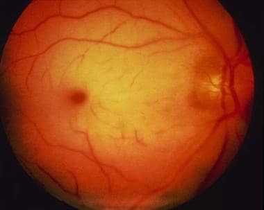
The cherry red spot of central retinal artery occlusion. |
 | View Full Size Image | |
Media type: Photo
|
- Biousse V, Calvetti O, Bruce BB, Newman NJ. Thrombolysis for central retinal artery occlusion. J Neuroophthalmol. Sep 2007;27(3):215-30. [Medline].
- Beiran I, Goldenberg I, Adir Y, Tamir A, Shupak A, Miller B. Early hyperbaric oxygen therapy for retinal artery occlusion. Eur J Ophthalmol. Oct-Dec 2001;11(4):345-50. [Medline].
- Atebara NH, Brown GC, Cater J. Efficacy of anterior chamber paracentesis and Carbogen in treating acute nonarteritic central retinal artery occlusion. Ophthalmology. Dec 1995;102(12):2029-34; discussion 2034-5. [Medline].
- Augsburger JJ, Magargal LE. Visual prognosis following treatment of acute central retinal artery obstruction. Br J Ophthalmol. Dec 1980;64(12):913-7. [Medline].
- Beiran I, Reissman P, Scharf J, et al. Hyperbaric oxygenation combined with nifedipine treatment for recent-onset retinal artery occlusion. Eur J Ophthalmol. Apr-Jun 1993;3(2):89-94. [Medline].
- Brown GC. Retinal artery obstructive disease. In: Ryan SJ, ed. Retina. Vol 2. St. Louis: Mosby; 1994:1361-77.
- Brown GC, Magargal LE, Shields JA, et al. Retinal arterial obstruction in children and young adults. Ophthalmology. Jan 1981;88(1):18-25. [Medline].
- Butz B, Strotzer M, Manke C, et al. Selective intraarterial fibrinolysis of acute central retinal artery occlusion. Acta Radiol. Nov 2003;44(6):680-4. [Medline].
- Cella W, Avila M. Optical coherence tomography as a means of evaluating acute ischaemic retinopathy in branch retinal artery occlusion. Acta Ophthalmol Scand. Nov 2007;85(7):799-801. [Medline].
- Ffytche TJ, Bulpitt CJ, Kohner EM, et al. Effect of changes in intraocular pressure on the retinal microcirculation. Br J Ophthalmol. May 1974;58(5):514-22. [Medline].
- Fraser S, Siriwardena D. Interventions for acute non-arteritic central retinal artery occlusion. Cochrane Database Syst Rev. 2002;CD001989. [Medline].
- Hayreh SS, Kolder HE, Weingeist TA. Central retinal artery occlusion and retinal tolerance time. Ophthalmology. Jan 1980;87(1):75-8. [Medline].
- Hertzog LM, Meyer GW, Carson S, et al. Central retinal artery occlusion treated with hyperbaric oxygen. J Hyperbaric Med. 1992;7:33-42.
- Klein R, Klein BE, Moss SE, Meuer SM. Retinal emboli and cardiovascular disease: the Beaver Dam Eye Study. Arch Ophthalmol. Oct 2003;121(10):1446-51. [Medline].
- Knoop K, Trott A. Ophthalmologic procedures in the emergency department--Part I: Immediate sight-saving procedures. Acad Emerg Med. Jul-Aug 1994;1(4):408-12. [Medline].
- Lacy C, Armstrong LL, Ingram N, et al. Drug Information Handbook. 4th ed. Hudson, Cleveland: Lexi-Comp Inc; 1996.
- Magargal LE, Goldberg RE. Anterior chamber paracentesis in the management of acute nonarteritic central retinal artery occlusion. Surg Forum. 1977;28:518-21. [Medline].
- Mangat HS. Retinal artery occlusion. Surv Ophthalmol. Sep-Oct 1995;40(2):145-56. [Medline].
- Mead GE, Lewis SC, Wardlaw JM, Dennis MS. Comparison of risk factors in patients with transient and prolonged eye and brain ischemic syndromes. Stroke. Oct 2002;33(10):2383-90. [Medline].
- Miyake Y, Horiguchi M, Matsuura M, et al. Hyperbaric oxygen therapy in 72 eyes with retinal arterial occlusion. 9th International Symposium on Underwater and Hyperbaric Physiology. 1987;949-53.
- Rumelt S, Brown GC. Update on treatment of retinal arterial occlusions. Curr Opin Ophthalmol. Jun 2003;14(3):139-41. [Medline].
- Schmidt D, Schumacher M, Wakhloo AK. Microcatheter urokinase infusion in central retinal artery occlusion. Am J Ophthalmol. Apr 15 1992;113(4):429-34. [Medline].
- Schmidt DP, Schulte-Monting J, Schumacher M. Prognosis of central retinal artery occlusion: local intraarterial fibrinolysis versus conservative treatment. AJNR Am J Neuroradiol. Sep 2002;23(8):1301-7. [Medline].
- Suri MF, Nasar A, Hussein HM, Divani AA, Qureshi AI. Intra-arterial thrombolysis for central retinal artery occlusion in United States: Nationwide In-patient Survey 2001-2003. J Neuroimaging. Oct 2007;17(4):339-43. [Medline].
- Wray SH. The management of acute visual failure. J Neurol Neurosurg Psychiatry. Mar 1993;56(3):234-40. [Medline].
Retinal Artery Occlusion excerpt Article Last Updated: Jan 14, 2008
|