Background
Endophthalmitis is an inflammatory condition of the intraocular cavities (ie, the aqueous and/or vitreous humor), usually caused by infection. Noninfectious (sterile) endophthalmitis may result from various causes, such as retained native lens material after an operation or from toxic agents. Panophthalmitis is inflammation of all coats of the eye, including intraocular structures.
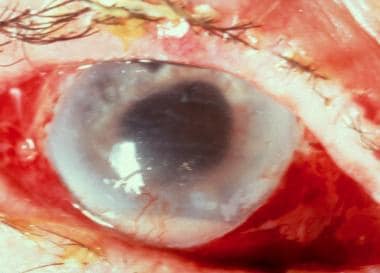 Severe endophthalmitis. Courtesy of Ron Afshari Adelman MD, MPH, MBA, FACS, Yale Medical Group.
Endophthalmitis can be broadly classified by the source of infection into endogenous and exogenous. Endogenous endophthalmitis results from the hematogenous spread of organisms from a distant source of infection. Exogenous endophthalmitis occurs with direct inoculation of an organism from the outside as a complication of ocular procedures, foreign bodies, and/or blunt or penetrating ocular trauma.
Endophthalmitis may be as subtle as white nodules on the lens capsule, iris, retina, or choroid. It also can be as obvious as inflammation of all the ocular tissues, leading to a globe full of purulent exudate. In severe cares, inflammation can spread to involve the orbital soft tissues.
Pathophysiology
Under normal circumstances, the blood-eye barrier provides a natural resistance against invading organisms.
In endogenous endophthalmitis, blood-borne organisms (seen in patients who are bacteremic or fungemic) arrive at the eye via the highly vascular choroid and permeate the blood-eye barrier. This leads to direct seeding of the eye and microbial penetration into deeper intraocular spaces. Destruction of intraocular tissues may be due to direct invasion by the organism and/or from inflammatory mediators of the immune response.
Exogenous endolphtalmitis results from microbial inoculation from an external source or the ocular surface. This happens due to breaks in globe integrity, such as those seen during ocular procedures or as a result of direct trauma to the eye. Common ocular procedures associated with endophthalmitis include cataract surgery, intravitreal infections, keratoplasty, and filtering blebs.
Epidemiology
Frequency
United States
Endogenous endophthalmitis is rare, occurring in only 2-8% of all cases of endophthalmitis. [1, 2] The overall incidence rates are reported to be between 0.04-0.4%. [3] In recent years, endogenous endophthalmitis cases have risen with intravenous drug use. [4]
In unilateral cases, the right eye is twice as likely to become infected as the left eye, probably because of its more proximal location to direct arterial blood flow from the right innominate artery to the right carotid artery. [5] The number of people at risk may be increasing because of the more frequent use of immunosuppressive agents (eg, for autoimmune diseases, cancer therapies, transplant antirejection therapies) or in cases of pathogenic immunocompromise (cancer, uncontrolled HIV). [2]
Most cases of exogenous endophthalmitis occur after intraocular procedures, particularly cataract surgery. When surgery is implicated in the cause, endophthalmitis usually begins within 1 week after surgery. In the United States, post-cataract surgery endophthalmitis is the most common form, with approximately 0.1% of operations having this complication. [6] Endophthalmitis occurs in approximately 0.2% of corneal transplantation (keratoplasty) cases in the acute postoperative period and up to 0.7% if later cases are included. [7] Endophthalmitis also may occur after intravitreal injections, although this risk usually is lower than with cataract surrgery. The incidence of endophthalmitis after intravitreal injections has been estimated at approximately 0.029% per injection. [8] Another postoperative cause of endophthalmitis is vitrectomy, with an incidence between 0.02-0.06%. [9]
Posttraumatic endophthalmitis comprises 25-30% of all endophthalmitis cases and occurs in up to 2-7% of cases of penetrating injury to the globe. [10, 11, 12, 13] Incidence of endophthalmitis with penetrating injuries in rural settings is higher when compared with nonrural settings. [14] Delay in the repair of a penetrating globe injury >24 hours, disruption of the lens, and retained intraocular foreign body are correlated with increased risk of developing endophthalmitis. [15, 16, 17] The incidence of endophthalmitis was found to be very low in injuries caused by explosives (eg, bombs or mines), believed to be the result of the high temperature of the foreign body, such that it passes through the eye in a relatively sterile manner. [18]
Mortality/Morbidity
Decreased vision and permanent loss of vision are common complications of endophthalmitis. Patients may require enucleation to alleviate a blind and painful eye.
Mortality is related to the patient's comorbidities and the underlying medical problem, especially when considering the etiology of hematogenous spread in endogenous infections.
Age
An association appears to exist between the development of endophthalmitis in cataract surgery and age greater than or equal to 85 years. [19] In addition, increasing age is associated with declining immune function and other comorbidities that may increase the risk for endogenous infections.
Prognosis
The prognosis is variable because of the variety of organisms and inoculation mechanisms involved. Visual acuity at the time of the diagnosis and causative agent are most predictive of outcome. [7] More virulent organisms and lower visual acuity at diagnosis are associated with worse visual outcomes.
In post-operative endophthalmitis, streptococcal infections lead to the worst outcomes. [20] The best outcomes are observed in cases of coagulase-negative staphylococci or cases that are culture negative. [20] In the Endophthalmitis Vitrectomy Study, about half of eyes afflicted by this condition eventually recovered 20/40 vision, but 10% of eyes remained with 20/800 vision or worse. [21]
Patients with traumatic endophthalmitis, especially that caused by Bacillus infection, typically have a poor visual outcome. Most eyes lose vision even with prompt treatment. [22]
The outcome of endogenous endophthalmitis generally is worse than exogenous due to the profile of the organisms and patients typically involved with this form (ie, more virulent organisms, immunocompromised host, delay in diagnosis). [23]
Regardless of inoculation route, fungal endophthalmitis caused by molds is associated with poor outcomes. Comparatively, patients with Candida endophthalmitis tend to fare better. In a case series of patients with endogenous fungal endophthalmitis, none of the eyes infected with mold species achieved a visual acuity of 20/200 or better, whereas 53% of the cases with Candida achieved 20/200 vision or better. [24]
-
Severe endophthalmitis. Courtesy of Ron Afshari Adelman MD, MPH, MBA, FACS, Yale Medical Group.
Tables

What would you like to print?
- Multifocal Choroidopathy Syndromes
- Inherited Retinal Diseases: Diagnosis and Treatment
- Fast Five Quiz: Inherited Retinal Diseases
- Fast Five Quiz: Management of Inherited Retinal Diseases
- Fast Five Quiz: Diagnosis of Inherited Retinal Diseases
- Inherited Retinal Diseases: Early Signs and Advances in Treatment
- Could Retinal Changes Be a Harbinger of Parkinson's?
- AI Identifies Retinal Biomarkers for Preeclampsia
- Combining Retinal Scanning and Liver Screening in T2D
-
 Association for Research in Vision and Ophthalmology (ARVO) 2025 Annual Meeting
-
Immunoglobulin A Nephropathy (IgAN): 5 Differential Diagnoses to Know
-
Conference MDAngle: 2024 AAOWet Age-Related Macular Degeneration


