Background
Patients with foreign bodies in the gastrointestinal (GI) tract commonly present to the emergency department (ED). Foreign bodies in the upper GI tract are usually swallowed, purposefully or accidentally. The presentation is usually straightforward but on occasion can be extremely subtle. A foreign body in the GI tract is shown in the radiograph below.
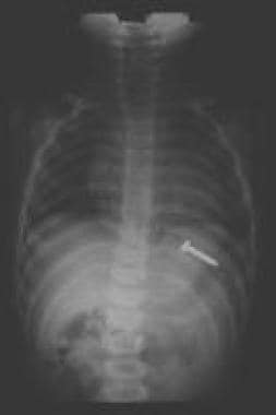 A screw in the stomach; peristaltic action will carry the screw through the GI tract with the blunt end (head) leading and the sharp end trailing.
Most of the literature covering GI foreign bodies is anecdotal, with the exception of some recent studies on esophageal foreign body removal techniques.
Pathophysiology
Foreign bodies may involve the entire upper GI tract. The oropharynx is well innervated, and patients can typically localize oropharyngeal foreign bodies. Scratches or abrasions to the mucosal surface of the oropharynx can create a foreign body sensation. Chronic foreign bodies or perforations can cause infections in surrounding soft tissues of the throat and neck.
The esophagus is a tubular structure approximately 20-25 cm in length. Patients can usually localize foreign bodies in the upper esophagus but localize them poorly in the lower two thirds of the structure. The esophagus has three areas of narrowing where foreign bodies are most likely to become entrapped: the upper esophageal sphincter (UES), which consists of the cricopharyngeus muscle; the crossover of the aorta; and the lower esophageal sphincter (LES). Structural abnormalities of the esophagus, including strictures, webs, diverticula, and malignancies, increase the risk of foreign body entrapment, as do motor disturbances such as scleroderma, diffuse esophageal spasm, or achalasia.
After reaching the stomach, a foreign body has greater than an 80% chance of passage. [1] Coins reaching the stomach are very likely to pass into the small bowel. Objects larger than 2 cm in diameter are less likely to pass the pylorus, and objects longer than 6 cm may become entrapped at either the pylorus or the duodenal sweep. Objects reaching the small bowel occasionally are impeded by the ileocecal valve. Rarely, a foreign body may become entrapped in a Meckel diverticulum.
Occasionally objects such as bones or toothpicks may pass all the way to the rectum, where they become entrapped, causing a foreign body sensation and, potentially, a perforation.
Swallowed magnets from toys and household items have become a serious health hazard in children. Buckey-ball magnets are small round magnets in the shape of ball-bearings that are especially strong and are used to make toys of various shapes. If these small magnets are ingested, especially at various times, they can adhere across layers of bowel and lead to pressure necrosis, fistula, volvulus, perforation, infection, or obstruction. [2]
Etiology
Children
The most common causes of GI foreign bodies are food boluses and accidental swallowing of other objects.
Young children often put any object they find into their mouths and may accidentally swallow them. Although less common, older children also put smooth objects, such as coins or marbles, in their mouths and swallow them. However, the larger diameter esophagus in this age group results in fewer entrapped foreign bodies compared to young children.
Children who are abused may present with GI foreign bodies after being forced to swallow objects; however, this is rare.
Adults
The most common cause of GI foreign bodies in adults involves food that does not pass through the esophagus because of underlying mechanical problems.
In adults, accidental swallowing often involves toothpicks and dentures.
Psychiatric patients may swallow a wide variety of objects, including multiple objects, large objects, and bizarre items.
Prisoners may swallow objects either to hide them from authorities or to seek medical care. In the case of razor blades, they often tape the sharp edge to avoid injury.
Drug smugglers may swallow multiple condoms (usually double wrapped) filled with cocaine or heroin. This is called "body packing," as opposed to "stuffing," which occurs when the patient attempts to elude arrest by swallowing packets of drugs in their possession.
Epidemiology
United States data
The incidence of foreign body ingestions in children and adults is unknown. Data are largely anecdotal.
One study suggested approximately 1671 ingested magnet injuries annually. This was expected to decrease as sales of these small toy magnets have been banned by the Consumer Protection Agency because of safety concerns. [3]
International data
In a retrospective study (2002-2015) that evaluated the effectiveness of a mandatory product recall on the frequency of multiple mini-magnet ingestion at a large tertiary Canadian pediatric hospital, investigators noted a significant reduction for all magnet ingestions and multiple magnet ingestions in the 2 years after the recall (2014-2015) compared to the 2 years before the recall (2011-2012). [4]
In another retrospective study (1995-2013) of pediatric esophageal foreign bodies (age ≤12 years) in King Khalid University Hospital, Riyadh, Saudi Arabia, investigators identified 70 children, of whom 53 presented within 24 hours of ingestion and 13 had underlying predisposing factors, with a coin being the most common foreign object. [5]
Race-, sex-, and age-related demographics
No differences in race or nationality have been noted.
In children with swallowed foreign bodies, the incidence in males and females is equal. [6, 7, 8] In adults, the incidence of accidentally swallowed foreign bodies is slightly higher in men than in women, and the incidence of intentionally swallowed foreign bodies is much higher in men than in women.
Patients with foreign bodies in the upper GI tract usually fall into 1 of 3 categories: (1) children, (2) psychiatric patients and prisoners, and (3) edentulous patients.
Children account for 75-85% of patients with foreign bodies in the upper GI tract, with a preponderance at age 18-48 months.
The objects involved also differ by age group. [8, 9, 10] Children typically ingest objects they pick up and place in their mouths, such as coins, buttons, marbles, crayons, and similar items. [8] In contrast, adults are more prone to ingest food boluses, chicken or fish bones, fruit pits, dentures, or toothpicks. [9] Prisoners and psychiatric patients may present with bizarre objects, as well as multiple objects. [11] Nonfood-type items are common in young and elderly patients. [10]
The site of entrapment of esophageal foreign bodies also differs with age groups, with about 75% of children having entrapment at the upper esophageal sphincter (UES) and about 70% of adults having entrapment at the lower esophageal sphincter (LES). [8, 9, 12, 13, 14] Except for geriatric patients (> 65 years), the tonsils appear to be the most common anatomic site of foreign body impaction, and the stomach and esophagus are also common locations in children younger than 10 years and those older than 65 years. [10]
Prognosis
Morbidity/mortality
An estimated 1500 deaths occur annually from foreign bodies in the upper GI tract. [12]
Complications
Complications of GI foreign bodies include the following:
-
Potential complications of oropharyngeal foreign bodies include esophageal or pharyngeal abrasions, lacerations, and punctures, with associated abscesses (eg, retropharyngeal abscess), perforations, and soft-tissue infections.
-
Esophageal foreign bodies can also cause mucosal scratches or abrasions, punctures, and perforations [15] , with resultant injuries or infections to surrounding structures, including abscesses, pneumomediastinum or mediastinitis [16] ; pericarditis/tamponade, pneumothorax, pneumomediastinum, tracheoesophageal fistula, or even vascular injuries to the aorta (aortoesophageal fistulas) [17] ; or pulmonary vasculature. [18] Additionally, button batteries can rapidly create esophageal necrosis. [19] Esophageal strictures may also occur.
-
Complications from foreign bodies in the stomach and small intestine typically involve perforation and associated infection, including peritonitis and sepsis. In general, perforation occurs in less than 1% of cases. [20] Small-bowel obstruction may also occur
-
Swallowed toy magnets that adhere across layers of bowel can cause pressure necrosis, fistula, volvulus, perforation, infection, or obstruction. [2]
-
Coin (quarter) lodged at the level of the cricopharyngeus muscle.
-
Coin lodged at the level of the aortic crossover.
-
Coin lodged at the lower esophageal sphincter.
-
A screw in the stomach; peristaltic action will carry the screw through the GI tract with the blunt end (head) leading and the sharp end trailing.
