Practice Essentials
Carcinoma of the temporal bone is rare, accounting for fewer than 0.2% of all tumors of the head and neck. Only 200 new cases of temporal bone cancer may be diagnosed each year across the United States. This number includes cancers arising from skin of the pinna that spread to the temporal bone; primary tumors of the external auditory canal (EAC), middle ear, mastoid, or petrous apex; and metastatic lesions to the temporal bone.
See the image below.
Malignancies of the temporal bone arise most commonly from the pinna and lateral concha because these sites are likely to have undergone many years of sun exposure. In these areas, basal cell carcinoma and squamous cell carcinoma are most common. [1, 2] If neglected, these tumors may spread medially to the EAC. The most common type of primary cancer in the EAC is squamous cell carcinoma, and squamous cell carcinoma of the temporal bone may originate from the EAC or middle ear where chronic otorrhea and inflammation, cholesteatoma, or both may be associated risk factors.
Adenocarcinoma, melanoma, rhabdomyosarcoma, osteosarcoma, lymphoma, adenoid cystic carcinoma, and acinic cell carcinoma are other types of malignancies that may arise in the temporal bone. In children, rhabdomyosarcoma is the most common malignancy of the temporal bone. About 10% of all rhabdomyosarcomas occur in the ear. Tumors, such as meningioma, chordoma, parotid malignancy, and nasopharyngeal carcinoma, may spread to the temporal bone from contiguous sites. The temporal bone may also be a site for metastasis from lymphoma or malignant tumors of the breast, lung, kidney, or prostate. Lesions of the temporal bone are summarized as follows:
-
Benign
Osteoma
Neurofibroma
Paraganglioma
Adenoma
Schwannoma
Chordoma
Hemangiopericytoma
Lipoma
-
Malignant
Squamous cell carcinoma
Basal cell carcinoma
Adenocarcinoma
Acinic cell carcinoma
Adenoid cystic carcinoma
Melanoma
Osteosarcoma
Chondrosarcoma
Rhabdomyosarcoma
Metastatic carcinoma
Lymphoma
Malignant neuroma
Malignant paraganglioma
CNS malignancy
In a retrospective study of 20 patients with temporal bone metastasis, Song et al reported that lung cancer was the most common primary malignancy, being found in 45% of the group. The investigators also determined that metastasis to the EAC and the middle ear/mastoid occurred more frequently with hematologic malignancies than with solid tumors. In addition, metastasis to the temporal bone tended to be a late event, subsequent to metastasis of the primary malignancy to other parts of the body. [3]
Signs and symptoms of malignant tumors of the temporal bone
These include the following:
-
Otalgia (80-85%)
-
Otorrhea (40-75%)
-
Facial paralysis (25%)
-
Hearing loss (45-80%)
-
Tinnitus (8-10%)
-
Vertigo
-
Auricular lesion
-
External canal mass (10%)
-
Parotid mass (19%)
-
Skin lesions
-
Cranial nerve (CN) V, IX, I, XI deficits (30%)
Workup in malignant tumors of the temporal bone
Routine preoperative testing includes complete blood counts (CBCs), electrolyte level tests, renal function tests, liver function tests, and coagulation studies (if warranted based on the patient's history of bleeding and current medications).
Imaging studies can include the following:
-
Computed tomography (CT) scanning of the temporal bone and neck
-
Magnetic resonance imaging (MRI)
-
Chest radiography - If the histology indicates squamous cell carcinoma, obtain plain radiographs or CT scans of the chest to rule out metastasis
-
CT scanning of the chest, abdomen, or pelvis - This is not necessary unless the biopsy specimen of the temporal bone tumor reveals a tumor with a known propensity for metastasis
-
Carotid angiography with balloon occlusion Xenon test - If the carotid artery is suspected to be involved
Other tests include the following:
-
Audiometry - An audiogram is obtained prior to performing any major procedure on the ear or temporal bone; audiograms provide baseline hearing thresholds for future comparison
-
Electrocardiography
In addition, obtain a biopsy to determine whether the lesion in the ear is benign or malignant.
Histologic examination is important because, although CT scanning provides important preoperative staging information, systematic pathologic evaluation of the specimen is crucial for staging and treatment.
Management of malignant tumors of the temporal bone
Primary radiation is ineffective for curative treatment. In the most extreme cases in which contraindications to surgery are serious deterrents to an operation, palliative radiation and chemotherapy may be offered. The literature supports a beneficial effect of adjunctive radiation on survival, but no well-controlled studies have been performed. Postoperative radiation treatment may be indicated in advanced disease. Most authors advocate full course postoperative radiation to stage T3 or T4 tumors as defined by the University of Pittsburgh staging system. Some authors also recommend radiation for T2 disease. [4]
In general, all patients who are medically able should undergo surgical treatment. The optimal surgery removes all of the cancer en bloc because positive margins are associated with poor survival rates. [5] The resection procedures that can be performed for the temporal bone include the following:
-
Modified lateral temporal bone resection
-
Lateral temporal bone resection
-
Subtotal temporal bone resection
-
Total temporal bone resection
Etiology
Since temporal bone cancer is so rare, measuring specific etiologic factors for cancers in this area is very difficult. However, fair-skinned whites are more prone to nonmelanomatous skin cancers in other areas, especially areas exposed to ultraviolet radiation. A genetic predisposition to skin cancer may also exist, manifested as the development of skin cancers in sites not exposed to sunlight as well as sun-exposed areas. Chronic otitis media and cholesteatoma are common in patients with temporal bone cancers and have been implicated as etiologic factors. [6, 7] Chronic suppurative otitis media and the resulting chronic inflammation may lead to squamous metaplasia. Human papillomavirus has been implicated in squamous cell carcinomas of the middle ear. [8] Lim et al (2000) reported a series of temporal bone cancers in 7 patients who had undergone radiotherapy for nasopharyngeal carcinoma. [9] These patients had a particularly poor outcome.
Pathophysiology
The complex anatomy of the temporal bone makes tumor spread difficult to predict. Tumors of the skin around the auricle may extend along the soft tissues of the neck and ear. The soft tissues are a poor barrier against tumor spread, and eventually the tumors may extend along the conchal bowl and into the EAC. The cartilage of the EAC provides minimal resistance to tumor spread. The fissures of Santorini, foramen of Huschke, and bony-cartilaginous junctions are a source of direct access to the periparotid tissues and temporomandibular joint.
Cancer in the external auditory meatus can invade posteriorly through the soft tissue into the retroauricular sulcus over the mastoid cortex. The bony canal is more resistant to cancer extension; however, erosion through the posterior bony canal provides access to the mastoid cavity. Tumor growth medially along the EAC can extend through the tympanic membrane and bony tympanic ring, allowing invasion into the middle ear. Once a tumor enters the middle ear, the hard bone of the otic capsule provides a more effective barrier against tumor spread.
In the middle ear or mastoid, tumors spread easily via the eustachian tube, round and oval windows, neurovascular structures, and extensive air spaces of the mastoid cavity. The eustachian tube and neurovascular structures of the middle ear are potential means of tumor spread beyond the temporal bone to the infratemporal fossa, nasopharynx, or neck.
Aggressive tumors can erode through the tegmen tympani or mastoid into the middle or posterior fossa. The sigmoid sinus may become involved. The dura, although somewhat resistant to invasion, portends a grave prognosis if involved. The facial nerve and the stylomastoid foramen are metastatic routes to the soft tissues of the neck and the parotid. Proximal extension along the facial nerve leads toward the inner ear and posterior fossa. Leonetti et al (1996) offer an excellent review of the invasion patterns of temporal bone cancer. [10]
Nodal metastasis is uncommon in early disease but may occur in 10-20% of cases of advanced disease. [11, 6] The lymphatic drainage of the auricle and EAC extends anteriorly to the periparotid lymph nodes and parotid gland. [12] Drainage may also occur to the jugular chain or nodes overlying the mastoid. Lymphatic drainage of the medial EAC and middle ear is to the retropharyngeal nodes or deep jugular nodes. The lymphatic drainage of the inner ear is unknown.
Distant metastasis is rare.
Presentation
Patients with cancer of the temporal bone most often present when aged 60 years or older, although any age group, including children, can be affected. Common presenting symptoms include chronic otalgia, otorrhea, bleeding, and hearing loss. Physical findings include otorrhea, a mass lesion, facial swelling, facial paresis, and other cranial nerve (CN) deficits. Patients often present after many years of symptoms. In a series from the authors' institution, the average time from the onset of symptoms to the time of primary treatment for cancer was 3.9 years. [11] Nodal disease is present in 10-20% of patients. [11, 6] Symptoms and signs of temporal bone lesions are summarized as follows:
-
Otalgia (80-85%)
-
Otorrhea (40-75%)
-
Facial paralysis (25%)
-
Hearing loss (45-80%)
-
Tinnitus (8-10%)
-
Vertigo
-
Auricular lesion
-
External canal mass (10%)
-
Parotid mass (19%)
-
Skin lesions
-
CN V, IX, I, XI deficits (30%)
The aforementioned study by Song et al found that in cases of temporal bone metastasis, facial palsy and hearing loss were the most frequent otologic symptoms, with each occurring in 30.5% of patients. [3]
Physical examination in cases of temporal bone cancer should include inspection of the pinna, EAC, and middle ear for ulcers, mass lesions, soft tissue swelling or induration, old scars (eg, previously excised skin cancers may have been forgotten by the patient), and otorrhea. Perform a thorough CN examination. Close inspection for facial weakness is crucial. Perform audiography if hearing loss is suspected. As always, perform a complete head and neck examination. The patient's general medical condition should also be evaluated because it may greatly impact treatment options and outcome.
Indications
In general, all patients who are medically able should undergo surgical treatment. Primary radiation is ineffective for curative treatment; however, in the most extreme cases in which contraindications to surgery are serious deterrents to surgery, palliative radiation and chemotherapy may be offered.
Relevant Anatomy
The temporal bone is a complex structure comprised of 3 parts: the squamous, tympanic, and petrous portions. The squamous portion of the temporal bone forms a small portion of the bony EAC, the zygomatic process (and mandibular fossa), and a portion of the mastoid process. It has a superior portion that protects the temporal lobe and articulates with the parietal and occipital bones. The tympanic portion forms most of the bony canal and the posterior wall of the mandibular fossa. The middle ear is a space between the squamous and temporal portions laterally and the petrous portion medially. The petrous portion of the temporal bone contains the otic capsule and the internal auditory canal.
The EAC extends from the concha to the tympanic membrane. The lateral cartilaginous portion meets the bony portion at a bony-cartilaginous junction located about one third of its total length from the lateral aspect. The anterior cartilaginous wall contains small cartilage defects filled with connective tissue called fissures of Santorini, which are direct routes of tumor spread into the periparotid tissues. Within the bony portion is another potential route for tumor extension at the foramen of Huschke, a defect of the tympanic ring located inferiorly. The anterior wall of the canal is closely associated with the temporomandibular joint, and the anterior-inferior wall is close to the parotid gland.
The temporal bone contains or abuts many vital structures, including the internal carotid artery, jugular bulb, cavernous sinus, and sigmoid sinus. A thin layer of bone separates the middle ear and mastoid cavities from the middle and posterior fossae dura. Other important structures that lie within the temporal bone include the ossicles, the cochlea, and the eustachian tube and the cochlear, vestibular, facial, trigeminal, caroticotympanic, chorda tympani, and petrosal nerves.
Contraindications
In general, no contraindications specific to tumors of the temporal bone exist; all patients who are medically able should undergo surgical treatment. However, advanced tumors with intracranial invasion have a grave prognosis, and treatment should probably be limited to palliation with less extensive (and less morbid) surgical procedures.
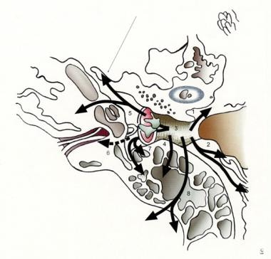
-
Coronal image that represents the following potential routes of spread of temporal bone cancer: 1) Anteriorly into the soft tissues of the temporomandibular joint and parotid through the fissures of Santorini. 2) Laterally via the soft tissue and cartilage of the external auditory canal to the concha and pinna. 3) Medially through the tympanic membrane to the middle ear, inner ear, and mastoid. 4) Posteriorly into the mastoid, epitympanum, and middle fossa. 5) Spread can continue through the Eustachian tube to the infratemporal fossa. 6) Spread through the round window, oval window, or facial nerve into the internal auditory canal and cerebellopontine angle. 7) Along the facial nerve through the mastoid and into the stylomastoid foramen and beyond. 8) Via the mastoid into posterior fossa 9) Along structures of the skull base, including the carotid, jugular bulb, and clivus.
-
Axial image that shows the spread of temporal bone cancer.
-
Axial T1 MRI with gadolinium of right T4 squamous cell carcinoma.
-
Axial T1 precontrast image of the same patient from Image 3.
-
Axial CT scan of the same patient from Image 3 with T4 squamous cell carcinoma.
-
Axial CT scan of a patient with recurrent basil cell carcinoma of the pinna, spread to the left inferior temporal bone.
