Background
Escherichia coli (E coli) are facultative anaerobic gram-negative bacteria that are part of the normal gastrointestinal system. [1] These organisms mainly are found within the large intestine and frequently are implicated as causes of bacterial infections. These infections can stem from disruption of the gut mucosal membrane leading to local tissue invasion and potential distant tissue seeding through bacteremia. Urinary tract infections are thought to occur via bacterial migration proximally up the ureter, causing colonization and potential infection of the bladder and more proximal structures. Common infections with E coli as a pathogen include cholecystitis, bacteremia, cholangitis, urinary tract infection (UTI), traveler's diarrhea, pneumonia, and neonatal meningitis.
The genus Escherichia is named after Theodor Escherich, the individual who isolated the type species of the genus. These organisms are gram-negative facultatively anaerobic bacilli, may exist singly or in pairs, utilize both fermentative and respiratory metabolism for energy, and either are nonmotile or motile by peritrichous flagella. E coli has a rapid reproduction time as low as 20 minutes in laboratory conditions, and virulence depends on what types of capsular antigens, flagellar antigens, and somatic polysaccharides each strain possesses. [2]
See Travelers' Diarrhea: Antimicrobial Therapy and Chemoprevention for more detail.
Pathophysiology
Acute bacterial meningitis
Most cases of neonatal meningitis are caused by group B streptococcal infections and E coli (50% and 20% respectively). Pregnant individuals are at a higher risk for colonization with the K1 capsular antigen strain of E coli. Approximately 80% of the E coli strains that cause neonatal meningitis feature the K1 capsular antigen. This K1 capsular antigen is similar to the group B Neisseria meningitidis capsule, which provides protection from phagocytosis. [3] This strain commonly is observed in neonatal sepsis, which carries a mortality rate of up to 50% if untreated and up to 10% if treated.
Some 25-50% of survivors have subsequent neurologic deficits and up to 20% demonstrated developmental abnormalities at 5 years post infection. Low birth weight and a positive cerebrospinal fluid (CSF) culture result portend a poor outcome. [4] In adults, E colimeningitis is rare but occasionally can be seen in those with neurosurgical trauma or complications of neurosurgical procedures with prosthetic device infections. E coli meningitis in an individual with no significant medical comorbidities or those receiving immunosuppression, particularly steroids, should raise suspicion of Strongyloides stercoralis. [5]
Pulmonary (lung) infections
E coli respiratory tract infections are extremely uncommon due to this bacteria’s normal habitat of the large intestine being located remotely from the lungs. E coli pneumonia is thought to be secondary to aspiration or microaspiration, which can be seen in those with neurological disorders that affect the swallowing mechanism and protection of the airway. [6] Due to the structure of the lungs, predominant lung fields affected are the lower lobes with right greater than left affected. If an individual aspirates while supine, the right upper field may develop pneumonia due to the change in dependent area of the airways.
A parapneumonic effusion and empyema may be secondary to an untreated E coli pneumonia. Lung abscesses from septic emboli may develop from an E coli bacteremia. This mechanism is different from a pneumonia, as the primary route of infection is seeding the lungs through the blood stream and not an infection through the alveoli as seen in pneumonia. The primary etiology of the bacteremia generally is pyelonephritis or intraabdominal infection.
Unless from a sputum culture isolated during pneumonia, blood culture during bacteremia, or wound culture aspirated from a lung abscess that identifies E coli, it is impossible to distinguish lung pathology caused by this organism from other enteric gram-negative organisms.
Intra-abdominal Infections
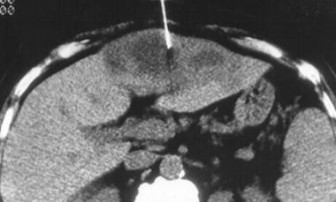
E coli intra-abdominal infections often result from damage to the gut mucosal barrier. [7] This leads to localized infections (eg, diverticulitis, appendicitis) or geographically distant infections (transient splanchnic vein bacteremia leading to pyogenic liver abscesses) (see image below). Complete disruption of the gastrointestinal tract can be spontaneous, traumatic, or anastomotic (prior surgical reconnection site of bowel with failure to heal) in origin with subsequent spillage of gastrointestinal contents, subsequent peritonitis, and complicated by abscess formation. Intra-abdominal abscesses often are polymicrobial as they derive mainly from the gastrointestinal tract that harbors millions of different gram-positive, gram-negative, and anaerobic species. Therefore, E coli plays a component role in these infections but is not the sole cause unless isolated via culture from a sterile space.
Obstruction of flow of different parts of the gastrointestinal system can lead to subsequent bacterial superinfection (eg, cholecystitis, ascending cholangitis) leading to severe illness and sepsis.
Enteric infections
E coli enteric infections are molecularly and clinically identified through their pathogenicity mechanisms. These strains generally only can be differentiated through molecular mechanisms or presumed through the patient’s clinical syndrome. In total, there are 6 different distinct mechanisms for which E coli can be differentiated – enterotoxigenic E coli (ETEC), Shiga toxin-producing E coli (STEC), enteropathogenic E coli (EPEC), enteroinvasive E coli (EIEC), enteroaggregative E coli (EAEC), and diffusely adherent E coli (DAEC). [8]
Despite naming conventions, there are no differences in antimicrobial susceptibilities of these different E coli bacteria. Thus, antibiotics that target E coli would treat all these organisms if they were identified in the same patient. Care must be given in identifying hematochezia or gross blood in the stool, as this may be secondary to dysentery caused by STEC with lysis of these bacteria through antibiotic treatment potentially leading to release of their toxins and clinical deterioration of the patient.
As a cause of enteric infections, 6 different mechanisms of action of 6 different varieties of E coli have been reported. ETEC is a cause of traveler's diarrhea [1] ; EPEC is a cause of childhood diarrhea; EIEC causes a Shigella-like dysentery; STEC causes hemorrhagic colitis that can lead to a diffuse systemic illness of hemolytic-uremic syndrome (HUS); EAEC primarily is associated with persistent diarrhea in children in developing countries, and DAEC is a cause of childhood diarrhea and traveler's diarrhea in Mexico and North Africa. ETEC, EPEC, EAEC, and DAEC colonize the small bowel, and EIEC and STEC preferentially colonize the large bowel prior to causing diarrhea.
STEC is among the most common causes of foodborne diseases. This organism is responsible for several GI illnesses, including nonbloody and bloody diarrhea. Patients with these diseases, especially children, may be affected by neurologic and renal complications, including HUS. Strains of STEC serotype O157:H7 have caused numerous outbreaks and sporadic cases of bloody diarrhea and HUS.
Kappeli et al looked at 97 non-O157 STEC strains in patients with diarrhea and found that HUS developed in 40% of patients; serotype O26:H11/H most often was associated with this syndrome. [2] Although strains associated with HUS were more likely to harbor STX 2 and EAE compared with those associated with bloody diarrhea, only 5 of the 8 patients with HUS had the STX2 gene; among the 3 patients with EAE -negative, STX2 -negative strains, only STX1 or STX1 and EHXA caused the HUS.
Urinary tract infections
The urinary tract is the most common site of E coli infection, and more than 90% of all uncomplicated urinary tract infections (UTIs) are caused by E coli infection. E coli UTIs are caused by uropathogenic strains of E coli. E coli causes a wide range of UTIs, including uncomplicated urethritis cystitis, symptomatic cystitis, pyelonephritis, acute prostatitis, prostatic abscess, and sepsis from an ascending urinary tract infection. Uncomplicated cystitis occurs primarily in females who are sexually active and are colonized by a uropathogenic strain of E coli. Subsequently, the periurethral region is colonized from contamination of the colon, and the organism reaches the bladder during sexual intercourse. The recurrence rate after a first E coli infection is 44% over 12 months. [9]
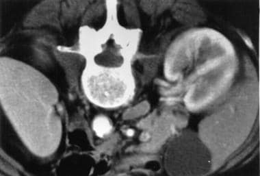
Uropathogenic strains of E coli have an adherence factor called P fimbriae, or pili, which binds to the P blood group antigen. These P fimbriae mediate the attachment of E coli to uroepithelial cells. Thus, patients with intestinal carriage of E coli that contains P fimbriae are at greater risk of developing UTI than the general population. Complicated UTI and pyelonephritis are observed in elderly patients with structural abnormalities or obstruction such as prostatic hypertrophy or neurogenic bladders or in patients with indwelling urinary catheters. E coli right sided pyelonephritis is visualized in the image below.
E coli bacteremia usually is associated with UTIs, especially in cases of urinary tract obstruction of any cause. The systemic reaction to endotoxin (cytokines) or lipopolysaccharides can lead to disseminated intravascular coagulation and death. E coli is a leading cause of nosocomial bacteremia from a GI or genitourinary source.
Other infections
Other miscellaneous E coli infections include septic arthritis, endophthalmitis, suppurative thyroiditis, sinusitis, osteomyelitis, endocarditis, and skin and soft-tissue infections (especially in patients with diabetes).
Epidemiology
Frequency
United States
E coli is the leading cause of both community-acquired and nosocomial UTI. [10] Up to 50% of females eventually experience at least 1 episode of UTI, and up to 10% of post-menopausal individuals report having a UTI within the past year. E coli causes 12-50% of nosocomial infections and 4% of cases of diarrheal disease.
International
E coli is the most common cause of traveler’s diarrhea. The attack rate is 10-40% among travelers. Worldwide, ETEC causes 30-60% of all cases of traveler's diarrhea and is the most isolated pathogen. EAEC also has been implicated, but less frequently. [11]
Mortality/Morbidity
E coli neonatal meningitis carries a mortality rate of up to 10%, and most survivors have neurological or developmental abnormalities.
The mortality and morbidity associated with E coli bacteremia is the same as that for other aerobic gram-negative bacilli.
Race
E coli infections have no recognized racial predilection.
Sex
E coli UTI is more common in females than in males because of differences in anatomic structure and changes during sexual maturation, pregnancy, and childbirth.
Men older than 45 years with prostatic hypertrophy are at an increased risk for UTI due to related bladder stasis.
Among neonates, E coli UTI is more common in boys than in girls, but circumcision reduces the risk.
Age
E coli is an important cause of meningitis in neonates.
Children are at high risk for traveler’s diarrhea due to oral sensory seeking and indiscretion with personal hygiene. [11]
-
Escherichia coli liver abscess.
-
Escherichia coli right pyelonephritis.
-
Escherichia coli on Gram stain. Gram-negative bacilli.
-
Escherichia coli culture on MacConkey agar.
Tables
Organism |
Ind* |
Urease |
Motility |
Glu Ferm† |
Lact Ferm‡ |
Sucr Ferm§ |
Malt Ferm|| |
Esc Hyd¶ |
Hyd Sulf TSI# |
Oxidase |
Orn Dec** |
Lys Dec†† |
|
E coli |
+ |
- |
+ |
+ |
+ |
+/- |
+ |
- |
- |
- |
+/- |
+ |
|
Klebsiella pneumoniae |
- |
+/- |
- |
+ |
+ |
+ |
+ |
+ |
- |
- |
- |
+ |
|
P mirabilis |
- |
+ |
+ |
+ |
- |
- |
- |
- |
+ |
- |
+ |
- |
|
Proteus vulgaris |
+ |
+ |
+ |
+ |
- |
+ |
+ |
+/- |
+ |
- |
- |
- |
|
Pseudomonas aeruginosa |
- |
+/- |
+ |
+ (ox)‡‡ |
- |
- |
- |
- |
- |
+ |
- |
- |
|
Enterobacter aerogenes |
- |
- |
+ |
+ |
+ |
+ |
+ |
+ |
- |
- |
+ |
+ |
|
Enterobacter cloacae |
- |
- |
+ |
+ |
+ |
+ |
+ |
- |
- |
- |
+ |
- |
|
Salmonella typhi |
- |
- |
+ |
+ |
- |
- |
+ |
- |
+ |
- |
- |
+ |
|
Citrobacter freundii |
+/- |
- |
+ |
+ |
+ |
+ |
+ |
- |
+/- |
- |
- |
- |
|
Serratia marcescens |
- |
+/- |
+ |
+ |
- |
+ |
+ |
+ |
- |
- |
+ |
+ |
|
*Indole †Glucose fermentation ‡Lactose fermentation §Sucrose fermentation ||Maltose fermentation ¶Esculin hydrolysis #Hydrogen sulfite on TSI **Ornithine decarboxylase ††Lysine decarboxylase ‡‡Oxidative |
|||||||||||||

What would you like to print?
- Overview
- Presentation
- DDx
- Workup
- Treatment
- Guidelines
- Guideline Summary
- International clinical practice guidelines for the treatment of acute uncomplicated cystitis and pyelonephritis in women updated in 2010 by IDSA and the ESMID
- A clinical practice guideline for the management of asymptomatic bacteriuria by the Infectious Diseases Society of America (IDSA) updated in 2019
- 2024 Guidance on the Treatment of Antimicrobial-Resistant Gram-Negative Infections by the Infectious Diseases Society of America (IDSA)
- Show All
- Medication
- Follow-up
- Media Gallery
- Tables
- References