Practice Essentials
Kawasaki disease (KD), also known as mucocutaneous lymph node syndrome and Kawasaki syndrome, is an acute febrile illness of early childhood characterized by vasculitis of the medium-sized arteries. Given its predilection for the coronary arteries, there is a potential for the development of coronary artery aneurysms (CAAs) and thus sudden death. [1] CAAs develop in approximately 25% of untreated cases; appropriate treatment decreases this risk to 3-5%. [2] Echocardiography is the study of choice to evaluate for CAAs. [3] KD is the leading cause of acquired heart disease in developed nations. [4]
The incidence of KD in the continental United States is approximately 25/100,000 children under 5 years of age; in Japan, the incidence has been estimated at approximately 250/100,000 children < 5 years of age. [2]
The etiology of this disorder remains unknown.
Diagnosis of Kawasaki disease
There are two forms of KD: complete and incomplete. Diagnosis of complete KD requires fever of at least 5 days' duration along with 4 or 5 of the principal clinical features. The principal clinical features are as follows:
-
Extremity changes
-
Polymorphous rash
-
Oropharyngeal changes
-
Bilateral, nonexudative, limbic sparing, painless bulbar conjunctival injection
-
Acute unilateral nonpurulent cervical lymphadenopathy with lymph node diameter greater than 1.5 cm
The acronym "FEBRILE" is used to remember the criteria as follows:
-
Fever
-
Enanthem (mucous membrane rash)
-
Bulbar conjunctivitis
-
Rash
-
Internal organ involvement (not part of the criteria)
-
Lymphadenopathy
-
Extremity changes
Incomplete KD is diagnosed when a patient presents with fever for 5 days or longer, 2 or 3 of the principal clinical features, and laboratory findings suggestive of the disease or echocardiographic abnormalities. Suggestive laboratory findings include elevated erythrocyte sedimentation rate (ESR), elevated C-reactive protein (CRP), hypoalbuminemia, anemia, elevated alanine aminotransferase (ALT), thrombocytosis, leukocytosis, and pyuria. The American Heart Association (AHA) suggests an algorithm for the diagnosis of incomplete KD in the most recent guideline. [4]
Echocardiography is the study of choice to evaluate for CAAs. Serial echocardiograms should be obtained as follows:
-
At the time of KD diagnosis
-
1-2 weeks after the onset of the illness
-
5-6 weeks after the onset of the illness
See Clinical Presentation and Workup for more details.
Management of Kawasaki disease
The principal goal of treatment is to prevent coronary artery disease. Intravenous immunoglobulin (IVIG), a purified preparation of gamma globulin, and aspirin are the mainstays of treatment. Patients should be treated with IVIG within 10 days after the onset of fever to prevent the development of cardiac sequelae. [5, 6, 7]
Other medications that are used variably as adjunctive treatments or for IVIG-resistant KD include corticosteroids, infliximab, cyclophosphamide, methotrexate, and ulinastatin. In addition to aspirin, other anticoagulants are sometimes utilized, including clopidogrel, dipyridamole, warfarin, and heparin. [8]
Guidelines
Clinical guidelines include the following:
See Treatment for more details.

Background
KD is an acute febrile vasculitic syndrome of early childhood. The disorder has also been called mucocutaneous lymph node syndrome and infantile periarteritis nodosa. It was first described in 1967 by Dr Tomisaku Kawasaki, who reported 50 cases of a distinctive illness in children seen at the Tokyo Red Cross Medical Center in Japan. [9] These children presented with fever, rash, conjunctival injection, cervical lymphadenopathy, inflammation of the lips and oral cavity, and erythema and edema of the hands and feet. In 1976, Melish et al first reported KD in the United States, in a group of 12 children from Honolulu. [10] KD is now recognized worldwide, although the greatest number of cases has been in Japan.
The illness was initially thought to be benign and self-limited. However, subsequent reports indicated that nearly 2% of patients with KD later died from the illness. These children died while they were improving or after they had seemingly recovered. Postmortem examinations revealed complete thrombotic occlusion of CAAs, with myocardial infarction (MI) as the immediate cause of death. It is now recognized as the leading cause of acquired heart disease in children in the developed world, surpassing rheumatic fever, and is a risk factor for adult ischemic heart disease.
The photographs below depict various manifestations of KD.
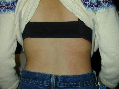 Kawasaki disease: Patchy generalized macular erythema, which is also typical of some viral exanthems.
Pathophysiology
Despite the prominent mucocutaneous clinical findings that define the illness, KD is best regarded as a generalized vasculitis that involves medium-sized arteries. Although the vascular inflammation is most pronounced in the coronary vessels, vasculitis can also occur in veins, capillaries, small arterioles, and larger arteries.
In the earliest stages of the disease, the endothelial cells and the vascular media become edematous, but the internal elastic lamina remains intact. Then, approximately 7-9 days after the onset of fever, an influx of neutrophils occurs, which is quickly followed by a proliferation of CD8+ (cytotoxic) lymphocytes and immunoglobulin A–producing plasma cells. The inflammatory cells secrete various cytokines (tumor necrosis factor, vascular endothelial growth factor, monocyte chemotactic and activating factor), interleukins (IL-1, IL-4, IL-6), and matrix metalloproteinases (primarily MMP3 and MMP9) that target the endothelial cells and result in a cascade of events that lead to fragmentation of the internal elastic lamina and vascular damage. [11] In severely affected vessels, the media develops inflammation with necrosis of smooth muscle cells. The internal and external elastic laminae can split, leading to aneurysms.
Over the next few weeks to months, the active inflammatory cells are replaced by fibroblasts and monocytes, and fibrous connective tissue begins to form within the vessel wall. The intima proliferates and thickens. The vessel wall eventually becomes narrowed or occluded owing to stenosis or a thrombus. [12, 13, 14, 15, 16] Cardiovascular death may occur from a myocardial infarction secondary to thrombosis of a coronary aneurysm or from rupture of a large coronary aneurysm. The period during of the greatest vascular damage is when a concomitant progressive increase in the serum platelet count occurs, and this is the point of the illness when the risk of death is most significant.
Etiology
The etiology of KD remains unknown. There has been a strong suspicion that the etiology of KD is infectious; however, no single infectious agent has been implicated. However, autoimmune reactions and genetic predisposition have been suggested as possible etiologic factors. By 2014, six genetic loci were linked to KD through genome-wide studies. However, the etiology of KD is complex, and these genetic factors still need to be fully applied to diagnosis and treatment. [17]
Infection
Features of KD that raise concern for an infectious etiology include the occurrence of epidemics primarily in late winter and spring, with 3-year intervals, and the wavelike geographic spread of those epidemics. KD is unusual in infants younger than 4 months, suggesting that maternal antibodies may provide passive immunity. Epidemiologic data suggest, however, that person-to-person transmission of the disease is unlikely.
Some authors have proposed a controversial association of KD with recent carpet shampooing, flooding, the use of a humidifier in the room of a child with an antecedent respiratory illness, [18] and locations near bodies of water. [19] These data have led to a waterborne vector hypothesis.
The overall clinical presentation of patients with KD is similar to that of patients with a viral or superantigenic disease. However, investigations have shown that the immune response in KD is oligoclonal, which is seen as a response to a conventional antigen, rather than polyclonal, as would be found in a superantigen-driven response. [20, 21]
Over the years, multiple infectious agents have been implicated; however, to date, no single microbial agent has surfaced as the prevailing cause. [22, 23] Suspected pathogens and infections have included the following:
-
Parvovirus B19
-
Neisseria meningitidis
-
Bacterial toxin–mediated superantigens
-
Mycoplasma pneumoniae
-
Klebsiella pneumoniae
-
Adenovirus
-
Cytomegalovirus [24]
-
Parainfluenza type 3 virus
-
Rotavirus
-
Measles
-
Epstein-Barr virus
-
Human lymphotropic virus
-
Mite-associated bacteria
-
Tick-borne diseases
-
Rickettsia
-
Propionibacterium acnes
Using light and electron microscopy, researchers have identified cytoplasmic inclusion bodies containing RNA in 85% of acute- and late-stage KD fatalities and 25% of adult controls. Based on this finding, it is hypothesized that the KD infective agent could be a ubiquitous RNA virus that results in asymptomatic infection in most individuals, but leads to KD in a subset of genetically predisposed individuals. It is also possible that many infectious agents trigger one final common pathway in susceptible hosts, which leads to KD. [2]
Genetic factors associated with Kawasaki disease
A genetic predilection to KD has long been suspected. [25, 26] Siblings of affected children have a 10-20 times higher probability of developing KD than the general population, and children in Japan whose parents had KD seem to have a more severe form of the disease and to be more susceptible to recurrence. [27] This risk of 2 family members having KD is greatest in twins, for whom the rate is approximately 13%. [28]
In 1978, Kato et al discovered that patients with KD are more likely to express HLA-Bw22J2, which is a major histocompatibility complex antigen seen predominantly in Japanese populations. This further implicated a genetic influence to the increased susceptibility to KD in Japanese patients. [29, 30] A genome-wide linkage analysis of affected sibling pairs was performed in Japan, and a multipoint linkage analysis identified evidence of linkage on chromosome 12q24. [31]
Dergun et al, Newburger et al, and Burns et al described families with multiple members affected with Kawasaki disease. [32, 33, 34] In these families, KD occurred in 2 generations or in multiple siblings. No clear pattern of inheritance could be deduced from these pedigrees. Therefore, multiple polymorphic alleles likely influence KD susceptibility.
A functional polymorphism of the inositol 1,4,5-triphosphate 3-kinase C (ITPKC) gene on band 19q13.2 has been found to be significantly associated with an increased susceptibility to developing KD. In addition, this polymorphism was associated with an increased risk of coronary artery lesions in both Japanese and American children. [35]
In a Dutch cohort, Breunis et al observed an association of KD with common genetic variants in the chemokine receptor gene-cluster CCR3-CCR2-CCR5. [36] The association of CCR2-CCR5 haplotypes and CCL3L1 copy number with KD, coronary artery lesions, and responses to IVIG have been reported in Japanese and other children. [37]
Genetic factors may influence the development of coronary artery lesions in Kawasaki disease. [38] In one study, genomic DNA was extracted from whole blood collected from 56 patients with Kawasaki disease who received IVIG treatment, and the genotypes for Fcg RIIIb-NA(1,2), Fcg RIIa-H/R131, and Fcg RIIIa-F/V158 were determined. About 23% of patients with the HH allele for the Fcg RIIa polymorphism developed coronary artery lesions, compared with 60% with the HR and RR alleles. HR and RR alleles may be a predictor of the progression of coronary lesions in KD before the start of IVIG.
Epidemiology
United States statistics
The incidence of KD in the continental United States is approximately 25 per 100,000 children under 5 years of age. A 2010 article retrospectively examined all hospitalizations of children younger than 18 years who had KD and found that the rate of hospitalization in the United Sates from 1997-2007 remained relatively stable, except for a slight increase in 2005. The hospitalization rate for children younger than 5 years was 20.8 cases per 100,000 children in 2006 and demonstrated a slight male predilection. [39]
International statistics
KD is most frequently observed in Japan, Taiwan, and Korea. The highest incidence of KD has been reported in Japan, where the frequency of the disease is 10 to 20 times higher than in western countries. [35] Although the birth rate has declined, the numbers of patients diagnosed as having KD and the incidence rate in Japan have risen rapidly since the 1990s. The incidence in 2000 was 134.2 cases per 100,000 children younger than 5 years; in 2012, the incidence was 264.8 per 100,000 children younger than 5 years. [40] Epidemics occurred in Japan during the years 1979, 1982, and 1986. No epidemics have occurred since that time.
Marked spatial and temporal patterns have been noted in both the seasonality and deviations from the average number of KD cases in Japan. Seasonality is bimodal, with peaks in January and June and/or July and a nadir in October. This pattern was consistent throughout Japan during an entire 14-year period which was studied. Very high or low numbers of cases were reported in certain years, but the overall variability was consistent throughout the entire country. Temporal clustering of KD cases was detected with nationwide outbreaks. [41]
A study by Ae et al showed that the number of patients in Japan who received a diagnosis of KD decreased from 17,347 in 2019 to 11,173 in 2020 (a 35.6% reduction); however, there was no evidence that this decrease resulted from a delay in seeking treatment during the global COVID-19 pandemic. [42] Investigators in other countries reported a similar decline in the incidence of KD during the COVID-19 pandemic. [43, 44]
Park et al noted the average annual rate of incidence of KD in South Korea was 105 cases per 100,000 in children younger than 5 years, which was the second highest reported rate in the world. [45] On average, the approximate annual incidence of KD in various Asian populations per 100,000 children younger than 5 years is 54.9 cases for Taiwan, 25.4 cases for Hong Kong, 16.8-36.8 cases for Shanghai, and 18.2-30.6 cases for Beijing.
The annual incidence reported in white populations outside the United States is similar to that reported in the US population, with 11.3-14.7 cases per 100,000 children younger than 5 years in Canada and 3.6 cases per 100,000 children younger than 5 years in Australia. [46, 47] From 1999-2000, the incidence in the United Kingdom was 8.1 cases per 100,000 children. [48]
Ontario has the highest rate of KD outside of Asia, with a yearly incidence of 26.2 cases per 100,000 population younger than 5 years. The incidence significantly increased from 1995 to 2006, with more patients diagnosed with incomplete KD. A reduction in coronary abnormalities was also seen during this period and was attributed to better recognition and treatment of the disease. [49]
Racial and sexual differences in incidence
Although KD has been reported in children of all ethnic origins, it occurs most commonly in Asian children, especially those of Japanese descent. Rates are intermediate among African Americans, Polynesians, and Filipinos, and are lowest among Caucasians. [50]
KD is slightly more common in males than in females. The male-to-female ratio ranges from 1.3-1.83:1 depending on the country from which the statistics are reported. Arthritis appears to be more common in girls than in boys. Death and serious complications are more common in boys than in girls. [39]
Age-related differences in incidence
Approximately 85-90% of KD cases occur in children younger than 5 years [24] ; 90-95% of cases occur in children younger than 10 years. In the United States, the incidence peaks in children aged 18-24 months. In Japan, the incidence peaks in children aged 6-12 months.
KD has rarely been reported in adolescents and adults, most of whom are between ages 18 and 30 years. [49] Fewer than 60 adult patients have been described in the literature for various geographic locations, including 25 in Europe, 23 in North America, 5 in Asia, 2 in South America, and 2 in Africa. [51] Kawasaki-like syndromes have been reported in adults infected with human immunodeficiency virus (HIV). However, it is unclear whether this is a variant of KD or an unrelated entity in the immunocompromised population. [52]
Children younger than 6 months and those older than 9 years are more likely to have an incomplete presentation, and are also more likely to have a suboptimal outcome. [53]
Prognosis
The prognosis relies on the extent and severity of cardiac disease. KD has surpassed rheumatic heart disease as the leading cause of acquired heart disease in the United States in children under the age of 5 years. Cardiovascular complications include the following:
-
Diffuse coronary artery ectasia and aneurysm formation, including giant aneurysms
-
Clinically significant heart failure or myocardial dysfunction (unlikely to occur once fever is resolved)
-
Myocardial infarction
-
Myocarditis (common, but rarely causes chronic heart failure)
-
Valvulitis, usually mitral (only occurs in 1% of patients and rarely requires valve replacement)
-
Pericarditis with small pericardial effusions (occurs in 25% of patients with acute illness)
-
Systemic arterial aneurysms
-
Rupture of coronary artery aneurysms with hemopericardium
Approximately 20-25% of untreated patients develop cardiac sequelae, including CAAs. Aneurysms develop in 3-5% of patients treated with IVIG before the 10th day of illness. [54]
Risk factors for CAAs include the following:
-
Fever >8 days
-
Recurrence of fever after an afebrile period of at least 48 hours
-
Male sex
-
Cardiomegaly
-
Age younger than 1 year
-
Asian and Pacific Islander race
-
Hispanic ethnicity
The most important predictor is total duration of fever longer than 8 days. [55] Incomplete Kawasaki disease may also be an independent predictor of CAA development. [56] Wilder et al reported that delayed diagnosis contributes to the development of these aneurysms. [57] Studies have shown that even in children who do not form aneurysms, up to 50% show a decrease in ventricular function and/or mild valvular regurgitation on echocardiograms.
Patients who do not develop CAAs recover fully. In those who develop CAAs, the severity of aneurysms determines the prognosis. More than half of all aneurysms resolve within 2 years. Endovascular ultrasonography has shown that, even when aneurysms resolve, marked intimal thickening is present in some. Vessel flow may be abnormal. These patients may have an increased risk of premature coronary atherosclerotic disease. [58]
Coronary artery bypass grafting (CABG) has been required in some children with severe perfusion deficits. Follow-up of children and adolescents 20-25 years after CABG has shown a 95% survival rate, though some patients have required repeat CABG or percutaneous coronary intervention. [59] Transplant has been performed in some children who had large aneurysms in vessels not amenable to bypass.
The greatest risk of cardiac damage occurs in children younger than 1 year and in older children, which may be related to the incomplete presentation often seen in the older age group that leads to delay in diagnosis and, consequently, treatment. Providers must be aware of both the complete and incomplete presentations of this disease to ensure timely diagnosis and treatment. Therapy with IVIG should be started within 10 days, and ideally within 7 days, of fever onset to prevent cardiac complications. [60]
Recurrence of KD is unusual: the recurrence rate in Japan is 3%, and it is approximately 1% in North America. Most relapses occur within 2 years from the initial episode. The highest incidence is in younger children and those who had cardiac sequelae from the initial episode.
The mortality from KD is low at less than 0.5%, with the highest risk in the first year after disease onset. Death is typically due to acute MI in the setting of giant aneurysms. Aneurysm rupture is rare and typically occurs within the first few months after the illness began. In the first week, severe myocarditis leading to hemodynamic instability or arrhythmias can occur. [2]
Patient Education
Patient and family education on KD should include the following key points:
-
KD causes swelling and damage to the blood vessels in the body.
-
The cause is not known, but it is not believed to spread from person to person.
-
KD is treated with an IV medication called IVIG, which is infused over 8-12 hours. It is also treated with aspirin. If the patient still has fevers 36 hours after IVIG, the patient will likely receive a second dose, and may receive other medications as well.
-
The patient will need pictures of the heart taken to look for swelling of the blood vessels of the heart and other heart abnormalities, which are dangerous complications of KD. With treatment with IVIG, this occurs in only 3-5 out of 100 patients with KD. Patients with KD who do not receive treatment are at much higher risk for heart problems. If heart problems are not present 6-8 weeks after symptoms began, it is very likely that the patient will have a full recovery with no complications.
-
The patient will need to follow up with a primary care doctor and a cardiologist after discharge from the hospital. This is very important so that the patient is appropriately treated and monitored for heart complications.
-
Kawasaki disease: Patchy generalized macular erythema, which is also typical of some viral exanthems.
-
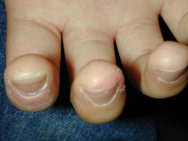
Kawasaki disease: Peeling and erythema of the fingertips.
-
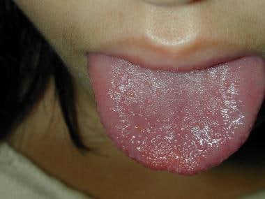
Kawasaki disease: Strawberry tongue.
-
Pediatrics, Kawasaki disease. Note the appearance of the hand and lips. Photo courtesy of Sam Richardson, MD.
-
Clinical manifestations and time course of Kawasaki disease.
-
Oral manifestations of Kawasaki disease: red lips and strawberry tongue.
-
Video overview of Kawasaki disease pathophysiology, symptoms, diagnosis, and treatment.
