Practice Essentials
The term hepatic cyst usually refers to solitary nonparasitic cysts of the liver, also known as simple cysts. However, several other cystic lesions must be distinguished from true simple cysts. Cystic lesions of the liver include the following:
-
Simple cysts
-
Multiple cysts arising in the setting of polycystic liver disease (PCLD)
-
Parasitic or hydatid (echinococcal) cysts
-
Cystic tumors
These conditions can usually be distinguished on the basis of the patient's symptoms, the clinical history, and the radiographic appearance of the lesion. Ductal cysts, choledochal cysts, and Caroli disease are differentiated from hepatic cysts by involvement of the bile ducts and are not reviewed in this article.
In patients with simple liver cysts, it is generally agreed that laparoscopic unroofing offers the best balance between efficacy and safety. How patients with PCLD should be treated remains less clear because the failure rates for laparoscopic unroofing and fenestration are high. Liver resection, though more effective, carries higher risks. Treatment of hydatid cysts continues to be controversial. As more experience is reported in the literature, indications for PAIR (puncture, aspiration, injection, reaspiration) versus surgery are delineated. [1]
Pathophysiology and Etiology
Simple cysts
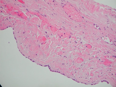
The cause of simple liver cysts is not known, but they are believed to be congenital in origin. The cysts are lined by biliary-type epithelium (see the image below) and perhaps result from progressive dilatation of biliary microhamartomas. Because these cysts seldom contain bile, the current hypothesis is that the microhamartomas fail to develop normal connections with the biliary tree. Typically, the fluid within the cyst has an electrolyte composition that mimics plasma. Bile, amylase, and white blood cells are absent. The cyst fluid is continually secreted by the epithelial lining of the cyst. For this reason, needle aspiration of simple cysts is not curative, and recurrence is the norm.
Polycystic liver disease
Adult PCLD (AD-PCLD) is congenital and is usually associated with autosomal dominant polycystic kidney disease (AD-PKD). Mutations in the genes PKD1 and PKD2 have been identified in these patients. Occasionally, PCLD has been reported in the absence of polycystic kidney disease (PKD). In these patients, a third gene, protein kinase C substrate 80K-H (PRKCSH), has been identified. Despite these differences in genotype, patients with PCLD are similar phenotypically. [2]
In patients with PKD, the kidney cysts usually precede the liver cysts. PKD often results in renal failure, whereas liver cysts only rarely are associated with hepatic fibrosis and liver failure.
Neoplastic cysts
Liver tumors with central necrosis visualized on imaging studies are often misdiagnosed as liver cysts. True intrahepatic neoplastic cysts are rare. The cause of cystadenomas and cystadenocarcinomas is unknown, but they may represent proliferation of abnormal embryonic analogues of the gallbladder or biliary epithelium. These cystic tumors are lined with biliary-type cuboidal or columnar cells and are surrounded by ovarianlike stroma. Cystadenoma is a premalignant lesion with neoplastic transformation to cystadenocarcinoma confirmed by tubulopapillary architecture and invasion of the basement membrane.
In a retrospective study, Kim et al investigated the value of quantitative color mapping of the liver’s arterial enhancement fraction (AEF) in the detection of hepatocellular carcinoma (HCC). [3] The investigators determined that when the color maps were analyzed in combination with multiphasic computed tomography (CT) scans, the mean sensitivity for HCC detection reached 88.8%, in comparison with 71.7% sensitivity for HCC detection using the multiphasic CT scans alone.
Hydatid cysts
Hydatid cysts are caused by infestation with the parasite Echinococcus granulosus. This parasite is found worldwide, but it is particularly common in areas of sheep and cattle farming.
The adult tapeworm lives in the digestive tract of carnivores, such as dogs or wolves. Eggs are released into the stool and are inadvertently ingested by the intermediate hosts, such as sheep, cattle, or humans. The egg larvae invade the bowel wall and mesenteric vessels of the intermediate host, allowing circulation to the liver.
In the liver, the larvae grow and become encysted. The hydatid cyst develops an outer layer of inflammatory tissue and an inner germinal membrane that produces daughter cysts. When carnivores ingest the liver of the intermediate host, the scolices of the daughter cysts are released in the small intestines and grow into adult worms, thus completing the life cycle of the worm. [4, 5]
Hepatic abscesses
Hepatic abscesses can be amebic or bacterial in origin. Entamoeba histolytica is the causative agent in amebic abscesses. It is contracted by ingestion of food or water contaminated by the cyst stage of the parasite. Amebiasis generally only involves the intestine but can invade the mesenteric venules resulting in liver abscesses. Its only host is the human. Pyogenic abscesses can be a result of instrumentation but are most often caused by ascending cholangitis in the setting of biliary obstruction. Microorganisms isolated are most often bowel flora. Other routes of contamination include the portal vein and hepatic artery.
Patients with intra-abdominal infections may present with liver abscesses with extension of bacteria through the portal venous system. Hematogenous spread via the hepatic artery in patients with bloodstream infection is rare.
Epidemiology
The precise prevalence and incidence of liver cysts are not known, because most do not cause symptoms; however, liver cysts have been estimated to occur in 5% of the population. No more than 10-15% of these patients have symptoms that bring the cyst to clinical attention. Hepatic cysts are usually found as an incidental finding on imaging or at the time of laparotomy. Most series in the literature are relatively small, typically reporting fewer than 50 patients each.
Prognosis
Several small series of patients undergoing laparoscopic unroofing of simple hepatic cysts have reported cure rates of 90% or higher. Kneuertz et al reported improvements in quality of life. [6]
Patients with PCLD have lower cure rates (see Table 1 below).
Table 1. Series of Patients Undergoing Laparoscopic Unroofing of Liver Cysts (Open Table in a new window)
First Author (Year) |
Institution |
Number of Patients |
Success Rate, Simple Cysts |
Success Rate, Polycystic Liver Disease |
Morino (1994) [7] |
University of Torino, Italy |
17 |
100% |
40% |
Krahenbuhl (1996) |
University of Bern, Switzerland |
8 |
100% |
N/A |
Hansen (1997) [8] |
University of California, San Francisco, United States |
19 |
94% |
0% |
Emmerman (1997) |
Eppendorf University, Hamburg, Germany |
18 |
89% |
N/A |
Fabiani (1997) [9] |
University of Nice, France |
10 |
100% |
N/A |
Martin (1998) [10] |
Royal Infirmary, Edinburgh, Scotland |
38 |
92% |
39% |
Katkhouda (1999) [11] |
University of Southern California, United States |
25 |
100% |
89% |
Zacherl (2000) [12] |
University Clinic of Surgery, Vienna, Austria |
11 |
86% |
N/A |
Fiamingo (2003) [13] |
University Hospital, Via Giustiniani, Italy |
15 |
89% |
50% |
Robinson (2004) [14] |
University of Colorado, United States |
11 |
N/A |
45% |
Konstadoulakis (2005) [15] |
Athens University, Greece |
9 |
N/A |
78% |
Fabiani (2005) [16] |
University Nice, France |
38 |
96%* |
N/A |
*Clinicoradiologic recurrence |
||||
Investigating optimal treatments for nonparasitic hepatic cysts, Mazza et al analyzed the outcomes associated with various surgical procedures used to treat these lesions. [17] The study involved the evaluation of data from 131 patients (78 with simple cysts, 53 with PCLD) treated at an institution where the authors practiced.
Mazza et al reported that laparoscopic unroofing or marsupialization (66 patients) completely relieved symptoms from either simple lesions or PCLD, with the procedure's morbidity, mortality, and recurrence rates being, respectively, 2%, 0%, and 2% for patients with simple cysts, and 25%, 0%, and 5% for patients with PCLD. [17] For infected cysts, the investigators' procedure of choice was percutaneous drainage (19 patients), the morbidity, mortality, and recurrence rates for this procedure being, for simple cysts, 0%, 0%, and 75%, respectively, and for PCLD, 0%, 0%, and 20%, respectively.
-
Histology demonstrating biliary epithelium lining simple cyst.
-
Ultrasonographic appearance of large simple hepatic cyst.
-
Computed tomography (CT) scan appearance of large hepatic cyst.
-
Computed tomography (CT) scan of polycystic liver disease curiously limited to right liver.
-
Hepatic cysts. Sagittal magnetic resonance imaging (MRI) reconstruction in patient with large echinococcal cyst; note daughter cysts in interior.
-
Computed tomography (CT) appearance of biliary cystadenoma.
-
Resection of involved liver in polycystic liver disease.
-
Laparoscopic view of initial hepatic cyst puncture, before unroofing. Lesion is located high in right liver near the diaphragm.
-
Laparoscopic view of beginning of unroofing of large simple hepatic cyst near dome of right liver.
-
Drawing of final result of laparoscopic unroofing of a large simple hepatic cyst in right liver.
-
Initial penetration of hepatic cyst with drainage of cyst fluid.
-
Unroofing of hepatic cyst.
-
Omentum sutured to excised margin.


