Practice Essentials
Imperforate hymen is at the extreme of a spectrum of variations in hymenal configuration. Variations in the embryologic development of the hymen are common and result in fenestrations, septa, bands, microperforations, anterior displacement, and differences in rigidity and/or elasticity of the hymenal tissue. Inspection of the external genitalia and anus are important components of the routine physical examination of the female neonate and child. [1]
However, the anatomic variants of this structure preclude any correlation to prior sexual activity. The International Federation of Pediatric and Adolescent Gynecology (FIGIJ) advocates that the terms "virginity" and "virgin" be removed from legal assessments, as they are not meaningful medical terms, and that so-called "virginity testing" is considered gender-based violence that should be eliminated. [2]
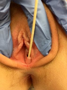
While a routine examination can and should be accomplished by the pediatrician or primary clinician at well-child examinations, the observant delivering obstetrician can learn much about the normal variations in genital configuration by examining the female neonate in the delivery room, keeping in mind the influence and structural changes induced by maternal estrogens. Under this influence, the labia majora are plump, the hymen is elastic and often fimbriated, and the mucosal surfaces (ie, introitus, fossa navicularis, vaginal vestibule) are pale pink.
Problem
Imperforate hymen has been diagnosed with prenatal ultrasound documentation of bladder outlet obstruction due to hydrocolpos or mucocolpos. [3] However, in spite of the recommendations for inspection of the external genitalia during the neonatal and early childhood period, variations in hymenal anatomy commonly escape diagnosis until puberty or the expected time of menarche, when a hematometrocolpos develops and obstruction produces pain and pressure symptoms. See the image below.
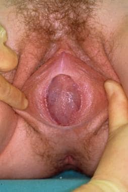 Imperforate hymen, classic appearance of bulging, blue-domed, translucent membrane.
Different normal variants in hymenal configuration are described, varying from the common annular, to crescentic, to navicular ("boatlike" with an anteriorly displaced hymenal orifice). Hymenal variations are rarely clinically significant before menarche. In the case of a navicular configuration, urinary complaints (eg, dribbling, retention, urinary tract infections) may occasionally result. Sometimes, a cribriform (fenestrated), septate, or navicular configuration to the hymen can be associated with retention of vaginal secretions and prolongation of the common condition of a mixed bacterial vulvovaginitis.
Occasionally, a hymenal tag will protrude from the vaginal vestibule, leading to concerns about a tumor or other significant pathology. These hymenal tags are of no clinical significance, and they do not require therapy if hymenal origin can be confirmed based on findings from a careful examination.
Imperforate hymen in infancy or childhood
On occasion, an infant or young child may be thought to have an imperforate hymen. However, after the neonatal period, when maternal estrogen levels have declined, examination of the area may be challenging owing to the small area involved. Careful examination with lateral labial retraction and pressure applied to the fourchette may reveal microperforations, sometimes with an anteriorly displaced opening just beneath the urethra. Capraro described a surgical technique similar to a perineotomy to correct such a defect; however, in asymptomatic patients, waiting until puberty is generally recommended before deciding whether such a technique is necessary.
The hymenal changes that result from estrogenization (increased elasticity and redundancy) may reveal the hymen to be open and obviate the need for surgery. With estrogen stimulation, the hymen could be described as having the appearance of an annular "scrunchie" (ie, a fabric-covered elastic hair tie). In addition, surgical procedures to the vagina and hymen during childhood, when endogenous estrogen levels are low, may result in scarring and the need for subsequent surgical revision. Thus, surgery during this time should generally be avoided if possible. If the hymen is suspected to be imperforate during childhood, re-examination should be performed after the onset of estrogen production, as signaled by breast development. If required, surgery can be performed at this time when healing is optimal and prior to the accumulation of a hematocolpos.
In a review of 23 cases of imperforate hymen, Posner et al emphasizes the ease of making a diagnosis of imperforate hymen by routine genital examinations in childhood. [4] The authors compared the significant delays and difficulties in making the diagnosis after the onset of puberty, primarily because the diagnosis was not considered, with the simplicity of making the diagnosis in asymptomatic prepubertal children by a simple genital examination.
Sexual abuse
Accurate description of the morphology and integrity of the hymen is critical in the diagnosis of female sexual abuse. Imperforate hymen has been described as occurring as a result of scarring from penetration and abuse, thus emphasizing the importance of an early examination to document the congenital, rather than acquired, etiology. [5]
Updated guidelines for the medical assessment and care of children who may have been sexually abused have been published, based on consensus from a group of specialists in child abuse pediatrics who reviewed the published research on the topic. [6] This document includes a table of medical findings including normal variations in the appearance of the hymen, findings associated with medical conditions other than trauma or sexual contact, conditions commonly mistaken for abuse, as well as findings caused by trauma and/or sexual contact. Concerns about hymenal disruption and lacerations associated with sexual abuse with digital or penile penetration have led to discussions of the normal hymenal diameter. However, this concept has now largely been abandoned. [7]
Experts in sexual abuse assessment have used unaided visual examination and colposcopy to examine the integrity of the hymenal ring. A normal examination or nonspecific findings are commonly found in cases of alleged sexual abuse unless the abuse and injuries are quite recent. [8]
Lacerations through the hymen into the fossa navicularis and introitus suggest a penetrating injury. Frequently, sexual abuse evaluations are conducted at some time remote from the immediate injury; thus, normal findings or healed or healing lacerations may be noted.
Muram concluded that the use of the colposcope by an experienced examiner adds little to an evaluation by an experienced examiner with expertise in abuse. [9] In addition, Muram proposed a scale that the examiner can use to evaluate physical findings as normal, abnormal and nonspecific, abnormal and suggestive of abuse, and definitive for abuse. [9] That last category includes only the situation in which sperm are found during the examination. Additional aids to the examination of the hymen have been described, including the procedure of inserting a Foley catheter into the vagina and inflating the balloon behind the hymen to stretch the hymenal margin and allow for a better examination or “floating” the hymen with water or saline. [10]
Anatomic anomalies
The classic image of an imperforate hymen is noted at the time of typical diagnosis: after the onset of menses, when a hematometrocolpos is present (see the image below).
Imperforate hymen, classic appearance of bulging, blue-domed, translucent membrane.
Consider anatomic anomalies that can be confused with imperforate hymen in the differential diagnosis. [11] These anomalies include the following:
-
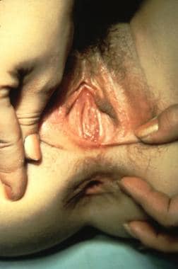
Acquired labial adhesions (see image below)
-
Obstructing or partially obstructing vaginal septa (longitudinal or transverse)
-
Vaginal cyst or hymenal tag/cyst
-
Hymenal variants, including a hymenal band/septum or navicular (“boatlike”) configuration to the hymen
-
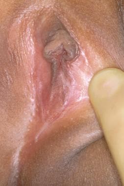
Vaginal agenesis (Mayer-Rokitansky-Kuster-Hauser syndrome) with or without the presence of a uterus or functional endometrium (see image below)
-
Complete androgen insensitivity syndrome (previously termed testicular feminization)
Epidemiology
Imperforate hymen is likely the most frequent obstructive anomaly of the female genital tract, but estimates of its frequency vary from 1 case per 1000 population to 1 case per 10,000 population. A population-based study estimated the frequency at 0.5 case per 1000 women (95% confidence interval, 0.3-0.7). [12]
Heger et al examined 147 premenarchal girls with a mean age of 63 months to collect normative data on genital anatomy; an imperforate hymen was found in only one patient (< 1%) and hymenal septa were found in 3 (2%). [13]
Imperforate hymen usually occurs sporadically, but a handful of cases have been reported to be familial, including a report of the occurrence in a mother and her twin premature daughters. [14, 15] Examination of first-degree relatives/female siblings of affected individuals has been recommended. [15]
Pathophysiology
Imperforate hymen and related genital tract anomalies result from abnormal or incomplete embryologic development.
The genital tract develops during embryogenesis, from 3 weeks' gestation to the second trimester. The initial development of both the male and female genital tracts is identical and is referred to as the indifferent stage of development. Note the following:
-
Paired wolffian (mesonephric) ducts connect the mesonephric kidney to the cloaca. The metanephric or true kidney derives from the ureteric bud (arising from the mesonephric duct) at about the fifth embryonic week.
-
The paramesonephric or müllerian ducts can be identified during the sixth week of embryologic development and lie lateral to the wolffian ducts until they reach the caudal end of the mesonephros, where they come toward the midline.
-
During the seventh week, the urorectal septum forms to separate the rectum from the urogenital sinus.
-
By the ninth week, the müllerian ducts move caudally to reach the urogenital sinus, forming the uterovaginal canal and inserting into the urogenital sinus.
By the 12th week, the paired müllerian ducts have fused into a single tube (ie, primitive uterovaginal canal). Two solid evaginations from the distal aspects of the müllerian tubercle form the sinovaginal bulbs (of urogenital sinus origin) or vaginal plate. The initial or cephalad portion of the müllerian ducts forms the fimbria and fallopian tubes; the more distal segment forms the uterus and upper vagina. The canalization of the paramesonephric ducts and/or upper vagina joins with the vaginal plate, which canalizes beginning caudally and creates the lower vagina. By the fifth month of gestation, the canalization of the vagina is complete. The hymen itself is formed from the proliferation of the sinovaginal bulbs, becoming perforate before or shortly after birth. An imperforate hymen results when this "sheet" of tissue fails to completely canalize. Varying degrees of perforation result in findings such as a cribriform or septate hymen.
Gonadal development
The development of the gonads occurs from the migration of primordial germ cells to the genital ridge, while the genital tract itself develops from the müllerian ducts (paramesonephric ducts), urogenital sinus, and vaginal plate. Thus, anomalies of the vagina, hymen, and uterus are not accompanied by abnormalities of ovarian development.
In girls with hymenal anomalies, hormonal and endocrinologic function is normal, leading to expected pubertal breast and pubic hair development. In cases of uterovaginal agenesis, imaging may fail to detect ovaries in the normal location (they may be located high and/or lateral in the pelvis), leading to unnecessary concern that the ovaries may be absent. Patients and families can be easily reassured that given both embryologic development and normal hormonal function (evidenced by the presence of normal breast development), the ovaries are present and functioning appropriately.
Because the mesodermal layer contributes to the development of the kidneys, gonads, and ductal structures, defects or insults in embryologic development may result in congenital defects of the kidneys or ureters that accompany abnormalities of the vagina and uterus. These anomalies should be considered with vaginal and uterine anomalies. However, given the embryologic origins of hymenal anomalies, urologic abnormalities are not associated.
The lining of the urethra and urinary bladder derives from endoderm, and the urogenital sinus forms the urethra and vestibule in females. The ectoderm fuses with the endoderm to contribute to the patency and canalization of the genital tract. Defects in this process lead to fusion failures and imperforate and obstruction defects.
Familial occurrence
Familial occurrence, although rare, is reported and screening by history or examination of family members is warranted. [14] Dominant transmission (either sex-linked or autosomal) and sibships suggesting a recessive mode of inheritance are described. [16] The inheritance of müllerian defects likely is polygenic or multifactorial, although some syndromes of heritable disorders are described with associated genital and nongenital anomalies.
Anomalies of the female reproductive tract
Anomalies of the female reproductive tract can result from agenesis or hypoplasia, vertical fusion and/or canalization defects, lateral fusion and/or duplication abnormalities, or failure of resorption, resulting in septa. Reports have noted the concurrent presence of lateral fusion defects with imperforate hymen. [17]
Presentation
Prenatal diagnosis
Rarely, diagnosis of imperforate hymen in the fetus has been made with obstetric ultrasonography. In such cases, the anomaly is visible on the imaging study because of hydrocolpos, hydrometrocolpos, or mucocolpos. [18, 19]
Diagnosis in infancy or childhood
The diagnosis is infrequently made during infancy in the neonatal nursery. The infant may have a bulging, yellow-gray mass at or beyond the introitus. Several case reports describe the presence of an abdominal mass in association with urinary obstruction.
Ultrasonography is an essential first step in diagnosis, precluding unwise and unplanned surgical intervention with resultant injury to the urethra or other pelvic structures, and excluding other more complicated anomalies.
Routine examination of the female genitalia by primary care clinicians during childhood is strongly recommended so that genital abnormalities can be diagnosed early. [1] Observation throughout childhood, with a planned hymenotomy after the onset of puberty is a reasonable course of action in most cases diagnosed in infancy or childhood, assuming no urinary symptoms or obstruction is present. Surgery in the presence of adequate estrogenization avoids scarring and the potential need for a repeat surgery that can occur to correct scarring when surgery is performed on the unestrogenized hymen and vagina.
If the diagnosis is equivocal (ie, imperforate hymen vs labial adhesions vs late-onset congenital adrenal hyperplasia), referral to a pediatric gynecologist may be warranted. Typically, a mucocele is not present even if the condition is noted at birth. If a patient is diagnosed with an asymptomatic imperforate hymen in infancy or childhood beyond the neonatal period, the optimal time for surgical repair is after the onset of puberty and prior to menarche.
Diagnosis and surgical repair in adolescence
Diagnosis of imperforate hymen depends on an awareness of the condition as a possible anomaly and surveillance with well-child care. The typical presenting complaint is primary amenorrhea, but this is a late presentation of a condition that should have been diagnosed at an earlier time. Textbooks frequently state that amenorrhea is not pathologic until age 16 years. Statistically, this statement is not evidence-based, as age 15 years represents the 98th percentile for menarche in girls in the United States and other developed countries. [20]
Additionally, failure to menstruate beyond 2-3 years from the onset of breast development, thelarche, is also statistically uncommon, and should be investigated to determine a cause. Imperforate hymen is one uncommon, but important, anatomic cause of primary amenorrhea.
When the condition presents as abdominal pain or an abdominal mass (see image below), diagnostic testing is often extensive because the condition is not considered. [1] An abdominal mass may prompt the consideration of an ovarian tumor and tumor markers may be obtained. While a false-positive elevation of CA-125 in premenopausal women has numerous causes, and testing has thus been discouraged, elevated CA-125 and 19.9 have been described with imperforate hymen, and may delay the diagnosis. [21]

Surgical repair after the onset of puberty but before menarche is optimal. The most common scenario is that in which a young woman presents with increasingly severe intermittent abdominal and pelvic pain due to a large hematocolpos and hematometra. This situation is preventable, as routine examinations of the genitalia can detect this obstruction and allow correction before menarche.
Walsh and Shih present a case of a 14-year-old elite athlete who presented to the emergency department and her pediatrician on multiple occasions over several months with cyclic abdominal pain, urinary retention, and constipation due to hematocolpos and hematometra. [22] This is an all too common presentation. In this case, even after placement of a Foley catheter for urinary retention on 2 separate occasions, the diagnosis of imperforate hymen was missed. The initial misdiagnosis of hematometrocolpos due to an imperforate hymen as constipation and subsequently as an ovarian mass has also been reported. [23]
Laasri et al describe a case of an 11-year-old girl who presented with suprapubic discomfort and acute urinary retention due to a hematocolpos that resulted from an imperforate hymen. [24] Russell et al report a case of a premenarchal 13-year-old girl with chronic constipation, who presented with symptoms that mimicked those of acute appendicitis. The diagnosis of imperforate hymen and retrograde menstruation was made during laparoscopy. [25]
While these young adolescents typically present to an emergency department with relatively acute pain, this condition should generally not be managed emergently until the definitive diagnosis is made. Defining the anatomy with appropriate imaging techniques and arranging for the most skilled and experienced gynecologist to perform surgery on a scheduled rather than emergent basis is essential. If necessary, menstrual suppression with gonadotropin-releasing hormone (GnRH) analogs can minimize pain pending appropriate imaging and clarification of anatomy. This is more likely to be necessary with complex genital anomalies than with a straightforward imperforate hymen.
Urinary pressure and even retention, with hydroureter and/or hydronephrosis, may occur due to the mass effect and resultant obstruction. Vaginal and rectal pressure is typically present. Severe constipation and low-back pain are described as presenting symptoms. The laborlike menstrual cramps may be severe and cyclic, although the cyclic nature of the symptoms may not be easily or immediately appreciated by the young woman or her family.
Unfortunately, the typical findings at diagnosis may include a large collection of blood within the uterus (hematometra) and an even larger collection of blood within the more distensible vagina (hematocolpos). Additional findings may include blood-filled fallopian tubes (hematosalpinges) and signs of retrograde menses, occasionally to the point of the development of intra-abdominal endometriosis and severe pelvic adhesions. The classic teaching is that endometriosis associated with obstructive anomalies resolves spontaneously and does not cause problems with subsequent pain and infertility compared with endometriosis arising spontaneously; however, this assertion is anecdotal rather than evidence-based.
Clinically, families are often concerned about whether the ovaries are normal when vaginal or hymenal anomalies are present; the course of separate embryologic development allows assurance of normal hormonal function without any need for hormonal testing or ovarian imaging. The exception to this is the diagnosis of androgen insensitivity syndrome with XY chromosomal complement in which the gonads require removal to prevent malignant transformation.
Differential diagnosis
The differential diagnosis of an imperforate hymen includes many conditions, some rare and others relatively common. Absolute confirmation of the diagnosis of an imperforate hymen is imperative prior to any attempted surgical repair in order to prevent vaginal scarring that can occur if a thick vaginal septum is inadvertently confused with a thin imperforate hymen.
Labial adhesions
See the image below.
The presence of acquired labial adhesions in a prepubertal girl is a common situation that is often confused with absence of the vagina. Labial adhesions, sometimes incorrectly termed vaginal adhesions, are not congenital and result from acquired labial agglutination most commonly due to inflammation. Small areas of labial adhesions can be managed expectantly. Extensive labial adhesions or those associated with such symptoms as recurrent urinary tract infections, urinary dribbling, or recurrent vulvovaginitis can be managed easily using the topical application of estrogen cream for 2-6 weeks. Alternative therapy, using topical steroid ointment has also been shown to be effective. [26] Such treatment results in marked thinning of the adhesions, often with spontaneous resolution.
Separation of thick adhesions is possible in an office setting with a child who can be restrained; however, this procedure ultimately is counterproductive because the examination frequently is difficult and traumatic, resulting in the subsequent inability to adequately examine the genital area due to the child's refusal because of memories of pain. Such traumatic lysis should be avoided. General anesthesia in an operative setting may thus be required.
Management of labial adhesions can be problematic as recurrence is common. Parents or caretakers must be instructed on how to ensure the child maintains excellent perineal hygiene and avoids vulvovaginitis. Families are often incorrectly encouraged to avoid baths in favor of showers. While bubble baths may occasionally contribute to vulvar inflammation, a plain water bath with soaking and cleansing of the interlabial folds using a washcloth without soap is preferable to a shower, which makes interlabial cleansing more difficult. The daily application of a topical emollient (such as A&D ointment) helps reduce the risk of recurrence until endogenous pubertal estrogen stimulation alleviates the risk. Thus, the application of a topical emollient should be continued until the child shows signs of estrogen-stimulated breast development.
Rarely, an adopted child will be found to have what appears to be labial adhesions, and these may be suggestive of female genital mutilation that occurred at a young age. The thick adhesions that result from this trauma may require surgical separation and management by a gynecologist with experience in managing female genital mutilation.
Labial adhesions may be confused with posterior labial fusion encountered in persons with congenital adrenal hyperplasia and may be differentiated by careful physical examination with attention to the presence or absence of clitoromegaly. This abnormality is noted at birth, rather than acquired.
The differential diagnosis for a cystic mass at the hymen includes ectopic ureter, hymenal cyst, hymenal skin tag, periurethral cyst, and vaginal cyst. [27]
Incomplete hymenal obstruction
In the case of incomplete hymenal obstruction due to a cribriform hymen or hymenal band, the typical presenting symptom is difficulty inserting a tampon or even the inability to achieve vaginal intercourse in an adolescent or young adult. Anatomic variations must be distinguished from involuntary vaginismus or contraction of the perineal and pelvic musculature or levator ani muscles, which can be associated with the learning process of tampon insertion, becoming a vicious cycle when persistent insertion is attempted without success and causes pain.
Hymenotomy occasionally may be indicated in the case of a rigid inelastic hymen, particularly for young female athletes (eg, swimmers, divers, gymnasts, cheerleaders) who may be hypoestrogenic, leading to the rigid hymenal configuration. As athletes, these girls are often eager to use tampons. A reasonable alternative to surgical correction involves the use of progressive dilation in a motivated young woman, along with topical estrogen. In these athletes with a rigid hymen, an evaluation for hypoestrogenism associated with overly vigorous physical activity should be considered; if present, estrogen replacement improves the hymenal characteristics and increases hymenal elasticity.
Hymenal bands
This condition is typically amenable to division using a local anesthetic in the office; however, the young woman's age and tolerance of such an office procedure must be predicted and judged. Her degree of motivation for tampon use or intercourse impacts the timing at which she requests such a procedure. A typical presenting history of an individual with a hymenal band is the ability to insert a tampon but extreme difficulty removing it. The author has encountered a patient in whom the tampon string became wrapped around the hymenal band, leading to marked edema and pain when removal was attempted.
Obstructing longitudinal or transverse septa
These conditions require careful preoperative evaluation to define the anatomy prior to any attempted surgical reconstruction. The repair of such complicated anomalies should usually be referred to a gynecologist at a tertiary care center where these cases are not a rarity. MRI is the optimal imaging modality for defining complicated female reproductive anatomy. [28]
Vaginal agenesis or androgen insensitivity
The evaluation and management of vaginal agenesis or androgen insensitivity syndrome is beyond the scope of this article, but these conditions should be considered in the differential diagnosis. Like imperforate hymen, primary amenorrhea is typically the presenting complaint
Androgen insensitivity is diagnosed based on findings of a blind vaginal pouch, with an XY chromosomal complement. Mayer-Rokitansky-Kuster-Hauser syndrome (uterovaginal agenesis) may include uterine remnants, some containing endometrium as well as myometrium. These patients should be referred to a gynecologist who specializes in adolescents and who has experience in managing these conditions.
Others
The presentation of an abdominal mass must be differentiated from urinary obstruction or tumors such as sacrococcygeal teratoma with abdominal extension, ovarian tumor, or other masses like mesenteric cysts or anterior meningoceles. [27]
Neovagina options
For girls with vaginal agenesis, the options for creation of a neovagina are nonoperative using progressive dilators (preferred approach) or operative, such as a McIndoe, Davydov, Vecchietti, or Williams procedures. [29]
Nonoperative management, using progressively larger Lucite dilators, is generally thought to be the first-line approach to management, with excellent success rates when appropriate support and encouragement is also provided. [30] Nonsurgical management at a time when the young woman is motivated to use vaginal dilators minimizes the potential for scarring and has high rates of success. [29]
Coital dilation has also been observed to be successful and may be offered as a management strategy.
Indications and Contraindications
Indications
An imperforate hymen at the time of puberty must be corrected surgically. The surgical decision-making process should focus on appropriate diagnosis and timing of surgical repair. While the patient may present with acute pain, the repair should not be performed emergently before carefully defining the anatomy, as obstructing vaginal septa require a different surgical approach. The surgery should be performed by a gynecologist who is skilled and experienced in the care of adolescents with genital anomalies.
Contraindications
The contraindications for a surgical repair of an imperforate hymen relate to the surgeon's inexperience with this condition, failure to adequately consider the alternative diagnoses, or failure to carefully define the anatomy.
Relevant Anatomy
An imperforate hymen presenting after the onset of menstrual shedding is visible upon examination as a translucent thin membrane just inferior to the urethral meatus that bulges with the Valsalva maneuver. This bluish discoloration is due to the presence of a hematocolpos visible behind the translucent hymenal membrane. Vaginal septa do not typically appear translucent (see the image below).
Imperforate hymen, classic appearance of bulging, blue-domed, translucent membrane.
Depending on the size and volume of the hematometra, hematocolpos, or hematosalpinges, a pelvic or abdominal mass may be palpable during abdominal or rectal examination. See the image below.
Radiographic documentation must demonstrate that the true diagnosis is not an obstructing transverse vaginal septum or other anomaly. Pelvic ultrasonography via the transabdominal, transperineal, or transrectal route is indicated as the initial diagnostic test, followed by MRI if any questions remain about the anatomy. Transperineal ultrasonography can be helpful in measuring the thickness of a transverse vaginal septum. Because renal and urologic abnormalities are associated with müllerian abnormalities, imaging of the upper urinary tract can help diagnose ipsilateral renal agenesis, duplex collecting systems, and other complex renal anomalies if there are uterovaginal anomalies other than imperforate hymen.
The prevalence of renal agenesis is estimated at 1 case per 600-1200 persons in patients with müllerian anomalies on the basis of autopsy studies. As many as 25-90% of women with renal anomalies are suggested to have concurrent genital anomalies; thus, abdominal and pelvic imaging is also warranted for such patients.
-
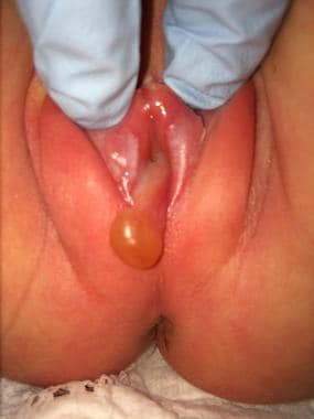
Abdominal mass with imperforate hymen.
-
Imperforate hymen, classic appearance of bulging, blue-domed, translucent membrane.
-
Extensive labial adhesion. Not to be confused with imperforate hymen.
-
Vaginal agenesis. Not to be confused with imperforate hymen.
-
Diagram of hematometra and hematocolpos with imperforate distal transverse vaginal septum.
-
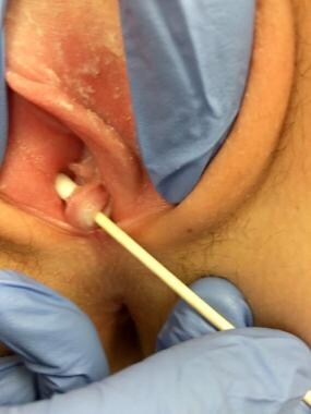
Hymenal cyst/tag in a 2-day-old newborn.
-
Hymenal band.
-
“Navicular” or boat-shaped hymen.


