Overview
The delivery of a full-term newborn refers to delivery at a gestational age of 37-42 weeks, as determined by the last menstrual period or via ultrasonographic dating and evaluation. The Naegel rule is a commonly used formula to predict the due date based on the date of the last menstrual period. This rule assumes a menstrual cycle of 28 days and mid-cycle ovulation. Ultrasonographic dating can be more accurate, especially when it is performed early in pregnancy and is used to corroborate or modify a due date based on the last menstrual period.
Approximately 11% of singleton pregnancies are delivered preterm, and 10% of all deliveries are postterm. Thus, nearly 80% of newborns are delivered at full term, although only 3-5% of deliveries occur on the estimated due date. [1, 2] Over the past few decades, the number of patients who go into spontaneous labor has decreased, and the percentage of inductions (iatrogenic labor) has increased to 22% of all pregnancies. [3]
Labor and delivery is divided into 3 stages:
-
In the first stage, the cervix dilates as a result of progressive rhythmic uterine contractions. This is typically the longest stage of labor. Cervical effacement, or thinning, occurs throughout the first stage of labor, and is graded 0-100%.
Epidemiology
Birth and natality statistics for the end of 2017 are as follows [5] :
-
Number of births: 3,855,500
-
Birth rate: 11.8 births per 1000 population
-
Fertility rate: 60.3 births per 1000 women aged 15-44 years
-
Low birthweight rate: 8.3%
-
Preterm birth rate: 9.9%
-
Unmarried birth rate: 39.8%
-
Mean age at first birth: 26.8 years
Statistics for type of delivery (2017) are as follows [5] :
-
Vaginal deliveries: 2,621,010
-
Cesarean deliveries: 1,232,339 (32%)
Statistics for multiple births (2017) are as follows [5] :
-
Twin births: 128,310
-
Triplet births: 3,675
-
Quadruplet births: 193
-
Quintuplets and other higher order births: 49
Indications
Normal vaginal delivery of the newborn includes the following circumstances:
-
Spontaneous labor mediated by pituitary and placental hormone cascades
-
Rupture of amniotic and chorionic membranes (suggested by the presence of a watery vaginal discharge or new oligohydramnios on ultrasonograph)
-
Induction of labor (indicated if fetal or maternal medical conditions necessitate delivery)
While sporadic contractions may occur, and the cervix may begin to soften in anticipation of delivery, the presence of contractions that lead to active cervical change defines labor.
Not all vaginal fluid is amniotic fluid, and membrane rupture requires confirmation.
If the cervix is favorable, oxytocin is given to induce uterine contractions. A favorable cervix is defined by the Bishop score, which includes parameters like cervical dilation, softening, effacement, and station. If the cervix is not favorable and no contraindications are present, cervical ripening can be facilitated with intravaginal prostaglandins before oxytocin is initiated. [6]
There are several medications available for cervical ripening. Misoprostol or prostaglandin E1(Cytotec) is most often used for cervical ripening. Since 2002, it has been FDA approved for cervical ripening and induction of labor. Dosing is 25-50 mcg given vaginally, buccally, or sublingually. prostaglandin E2 (dinoprostone) can also be used for cervical ripening, although it is more expensive than misoprostol and has an increased rate of tachysystole (too many contractions). [3]
A balloon catheter can also be used for ripening. Pennell et al compared 3 methods of ripening the cervix in nulliparous women at term and found that the single-balloon catheter offers the best combination of safety and patient comfort. In a randomized controlled trial, 330 nulliparous women with unfavorable cervices induced at term were treated with 1 of 3 methods: double-balloon catheters, single-balloon catheters, or prostaglandin gel.
Cesarean delivery rates were high with all 3 methods. Single-balloon catheter use was associated with earlier delivery and with significantly less pain: 36% of patients had a pain score of ≥4, vs 55% of patients treated with double-balloon catheterization and 63% of those treated with prostaglandin gel (P< .001). Induction was complicated by uterine stimulation in 14% of patients in the prostaglandin arm, but none of those in the catheter arms, and mean cord arterial pH was lower in the prostaglandin arm (7.25 vs 7.26 in the catheter arms [P =.050]). [7]
For more information, see Cervical Ripening article.
Contraindications
While most full-term newborns in the United States are delivered vaginally, vaginal birth is contraindicated in some circumstances, including those described in this section. Among the contraindications are the following:
-
Cord prolapse
When cord prolapse is detected on pelvic examination, the clinician should leave the hand in place, applying pressure against the presenting fetal part to keep it as far out of the pelvis as possible to prevent cord compression.
The incidence of cord prolapse is directly proportional to cord length.
The treatment is immediate conversion to cesarean delivery. If not treated emergently, cord prolapse is associated with high perinatal mortality.
-
Brow presentation
This may convert to face or vertex presentation and may be managed expectantly.
If the patient is unstable or no conversion occurs, cesarean delivery is recommended.
-
Face presentation
Clinicians and patients may tolerate a trial of expectant management, if cephalopelvic disproportion is not suspected and if the face is in a mentum anterior or mentum transverse position.
If the face is mentum posterior (chin facing the maternal sacrum), a cesarean delivery is required.
-
Up to 5% of all fetuses and 1-3% of full-term pregnancies present in the breech position. Plan for abdominal delivery for a footling presentation. For frank breech (ie, hips flexed, knees extended) and complete breech (ie, hips and knees flexed) presentations detected before the onset of labor, manual pressure maneuvers called external cephalic version (ECV) may be performed to attempt conversion to a vertex presentation.
The success rates of ECV are greater than 50% in properly selected patients, but these maneuvers should be performed at term, as they may stimulate labor or result in complications that necessitate prompt delivery.
The American Congress of Obstetricians and Gynecologists (ACOG) recommends abdominal delivery if ECV fails or if a patient in labor presents with breech presentation, as the rates of fetal morbidity and mortality in these cases are increased with vaginal delivery. [8]
-
Malposition
Fetal positions compatible with vaginal delivery are occiput anterior (OA), right occiput anterior (ROA), and left occiput anterior (LOA).
The occiput posterior (OP) position can be unfavorable for passage through the birth canal. Labor progress should be monitored for progression. If the fetal station is high and without descent during labor, change to abdominal delivery should be considered.
Deep transverse arrest occurs when the fetal head remains in transverse position without descent. Unfavorable maternal pelvic anatomy is the most common cause; abdominal delivery should be considered promptly.
Shoulder presentation is a sign of a transverse fetal lie. If this presentation is detected prior to active labor, external rotation through ECV may be attempted. When this presentation is detected during labor, maternal risk for infection, uterine rupture, or both is high. Emergent abdominal delivery is indicated.
-
Twin pregnancy
If a nonvertex second twin presentation occurs, it is managed according to gestational age, maternal preference, and practitioner comfort. The exceptions to vaginal delivery include the following:
Presenting twin in breech position
Conjoined twin anatomy
Most cases of mono-amniotic twins
Signs of fetal distress or an abnormality that warrants abdominal delivery
-
Higher order births
In the United States, cesarean delivery is planned for higher order births.
-
Vaginal delivery after cesarean delivery [9]
While safe in most circumstances, vaginal delivery after previous cesarean delivery remains controversial because of the rare but serious complication of uterine rupture. The risk of uterine rupture is approximately 0.5% in patients who have had one prior low transverse cesarean delivery.
The success rate of this procedure is greater than 50%.
During the delivery, careful fetal and maternal monitoring are needed to detect early signs of dystocia or uterine rupture.
An in-house anesthesiologist and obstetrician should be available in case complications arise. This type of delivery is not offered in many small hospitals because of the inconsistent availability of anesthesia or operating room staff. This has led to an increase in the cesarean delivery rate to approximately 30% in 2006.
Vaginal birth after cesarean is contraindicated in cases of multiple prior cesarean deliveries (>2), a history of a classical or T-shaped uterine scar, the presence of placenta previa, the presence of other uterine scars, or concern for true cephalopelvic disproportion.
-
Nonreassuring fetal heart rate patterns
Hospital protocols in the United States recommend some form of fetal heart rate monitoring. The need for continuous fetal heart rate monitoring remains unproven in low-risk, full-term pregnancies; however, changes in fetal heart rate monitoring can signal fetal hypoxemia and may indicate the need for emergent abdominal delivery.
Causes of fetal hypoxemia include placental abruption, placental insufficiency, or a tight nuchal cord. Most cesarean deliveries undertaken for suspected fetal distress result in healthy birth outcomes.
-
Fetal weight greater than 4000-4500 g is associated with a higher risk of shoulder dystocia and birth trauma during vaginal delivery. [10]
Patients with diabetes have a higher incidence of macrosomia and risk of shoulder dystocia.
If the estimated fetal weight is greater than 4500 g in a patient with diabetes, ACOG recommends abdominal delivery.
If the patient does not have diabetes, abdominal delivery is not recommended until an estimated fetal weight of 5000 g.
-
Abnormal placentation
Placenta previa (the placenta implanted over the cervical os) is a contraindication to vaginal delivery because of the risk of hemorrhage as the cervix dilates.
Placenta previa complicates up to 2% of all pregnancies. Risk factors include artificial reproductive technology and prior cesarean delivery.
Known placenta accreta (the placenta invades at least the myometrium of the uterus) is also a contraindication to a vaginal delivery. Risk factors include prior cesarean delivery.
Anesthesia
The pain of labor and delivery is a result of muscular contractions and pelvic pressure from organ distention. In the first stage of labor, autonomic innervation of the visceral uterus senses pain from contractions and cervical dilation. In the second stage of labor, somatic innervation of the vagina, vulva, and perineum sense pressure pain from the newborn passing through the birth canal.
Regional epidural anesthesia
Regional epidural anesthesia is used in more than 50% of laboring persons in the United States. It is relatively easy to perform, generally low in risk for complications, and provides good pain control. ACOG guidelines recommend placement of epidural at maternal request regardless of cervical dilatation. [11]
Risks include short-term backache, puncture headache, hypotension, maternal fever, and delayed labor. [12] Another possible risk is increased rate of instrumental delivery. [13] In a Cochrane review, a subgroup analysis of studies published since 2005 showed epidural analgesia was not associated with an increase in assisted vaginal delivery. [14, 15]
Epidural anesthesia may be combined with a dose of spinal anesthesia; this is called combined spinal-epidural anesthesia. This permits delivery of a potent, fast-acting spinal anesthetic with the placement of a stable epidural catheter for subsequent anesthesia needs.
Pudendal block
The pudendal block is rarely used because it is not very effective for pain control. [16] It is a local anesthetic given during the second stage of labor for somatic sensory blockade. It may provide some degree of motor blockade of the levator ani, mediating relaxation of pelvic floor muscles.
Systemic analgesia
Narcotics are sometimes used for short-term pain control; they can all cross the placenta, but only some cross the fetal blood-brain barrier. Narcotic agonists and antagonists are most commonly used. Morphine crosses the fetal blood-brain barrier and is infrequently used.
Risks include hypotension, nausea, vomiting, respiratory depression, depressed mental status, and decreased GI motility.
If narcotics are used, resuscitation medication and equipment for the newborn should be readily available.
Nonpharmacologic pain management
Nonpharmacologic pain management can be used alone or in conjunction with pharmacologic options.
Nonpharmacologic options include the following:
-
Breathing and meditation methods
-
Hypnosis
-
Acupuncture
-
Labor exercise techniques (eg, walking, squatting)
-
Therapeutic massages
-
Social support, including a birth doula
-
Warm baths or showers
Equipment
Monitors
Monitors include the following:
-
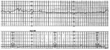
External fetal heart rate monitor (see normal tracing in image below)
Note the following:
Most labor and delivery units use continuous monitoring. The monitoring assesses the baseline, variability, presence, or absence of accelerations or decelerations. In 2008, the following consensus guidelines were developed to unify the interpretation of fetal heart tracings.
Category One: Normal fetal heart tracings. Continue expectant management.
Category Two: Indeterminate fetal heart tracings. These tracings require close observation or interventions to determine whether the fetus has acidemia.
Category Three: Abnormal fetal heart tracings. These tracings require immediate intervention. They are not reassuring and are indicative of fetal acidemia. If the strip does not improve with conservative measures, movement should be made toward delivery. [17]
Standard noninvasive labor monitoring includes the use of 2 sensors attached to the outside of the patient's abdomen. One sensor detects the fetal heart rate via ultrasonography, and the other monitors the timing and relative strength of contractions via a tocodynamometer.
The fetal heart rate is variable and ranges from 120-160 beats per minute (bpm). The heart rate may drop briefly to < 120 bpm, especially during contractions. Persistence of a fetal heart rate lower than 120 bpm defines fetal bradycardia; in labor, a heart rate >100 bpm with reassuring variation is not considered an emergency. Persistence of a rate >160 bpm is called fetal tachycardia.
-
Internal fetal heart rate monitor (fetal scalp electrode)
An internal fetal heart rate monitor may be placed to more accurately assess fetal heart rate patterns when the external monitor tracing may be inaccurate or difficult to trace.
A small electrode is passed through the cervix, after the membranes have ruptured, and placed on the fetal scalp.
-
Intrauterine pressure catheter (IUPC)
External monitoring of contractions only measures the timing of contractions. The strength of contractions can only be measured with an IUPC.
This catheter is placed in the uterus transcervically, next to the fetal head. It allows for more accurate measurement of strength and timing of contractions.
Delivery assistance (operative vaginal delivery)
Instruments used in delivery assistance include the following:
-
This is a handheld metal instrument with blade extensions that are applied to each side of the fetal head. The traction force of the blades aids in neonate delivery.
The use of forceps has decreased over the past several decades. [18]
The indications for forceps use include prolonged second stage of labor or ineffective maternal push power. The presenting part needs to be at +2 station before forceps should be applied. If the presenting part is at a higher station, abdominal delivery should be chosen.
Forceps use is associated with less fetal hematoma formation and quicker delivery times compared with vacuum assist [19] but is associated with increased maternal trauma and lacerations.
When compared with conversion to abdominal delivery, forceps use is associated with lower risk of maternal hemorrhage and a better chance that the patient will be able to deliver vaginally in subsequent pregnancies.
-
This instrument consists of a suction cup that attaches to the fetal head to assist with extraction. Traction pressure is created by a negative pressure handle system. Types include metal cup vacuums, plastic cup vacuums, and a mushroom-shaped vacuum cup that combines the advantages of the metal and plastic designs. [20]
Indications for use include the need for urgent delivery because of fetal distress, poor maternal push power, or maternal medical conditions that contraindicate strong pushing. Like forceps assistance, vacuum assistance should only be used when indicated, as it carries the risk of harm to the fetus and birthing parent.
Fetal complications from vacuum delivery include hematomas of the scalp, retina, and intracranium. Maternal complications are less than those with forceps but also include vaginal and perineal lacerations.
-
The decision to use forceps or a vacuum assistance is guided by the particular indication for an instrumented delivery and the clinician’s experience with each technique. In cases of a nonreassuring fetal tracing, the decision to perform an assisted vaginal delivery over rapid conversion to abdominal delivery is based on fetal position and presentation and the availability of personnel for emergency surgical delivery.
-
When comparing forceps to vacuum, the vacuum has less maternal morbidity, including need for anesthesia and trauma to birth canal; however, there are increased risks to the fetus, including increased risk of cephalohematoma, retinal hemorrhage, and neonatal jaundice. [21]
-
The combination of vacuum followed by forceps delivery carries increased risk of neonatal intracranial hemorrhage and should be avoided. This increased risk is also present if a failed operative vaginal delivery proceeds to a cesarean delivery. [21]
Positioning
First stage of labor
The patient may alternate positions frequently and is permitted to be out of bed if not under anesthesia motor blockade. Taking walks during this time can ease pain. Some clinicians report that labor may be shorter when the patient is intermittently upright. Swaying motions, such as rocking or slow dancing, may be soothing.
Second stage of labor
The patient may choose a delivery position that is most comfortable and still conducive for clinical monitoring. Most commonly, patients assume a partially sitting position, with the knees flexed and the back supported. The gravity advantage of being at least partially upright can help during delivery.
Other acceptable delivery positions include the following:
-
Squatting
-
Dangling and supported by the arms of a partner
-
Kneeling on the knees or on both the hands and knees
-
Lying on one side with the upper leg supported
In some circumstances, repositioning of the patient may be indicated during delivery. Such circumstances include the following:
-
Maternal back pain
-
Shoulder dystocia
-
Posterior presentation of the occiput
Clinicians are also becoming more familiar with water immersion and water birthing. In a Cochrane review of 11 trials on this topic, 6 reported that water immersion during the first stage of labor significantly reduced regional analgesia without increasing duration of labor, operative delivery rates, or neonatal outcome. One study showed that immersion in water during the second stage of labor increased women's reported satisfaction with pushing. [22]
Technique
First stage of labor
The first stage of labor includes the following:
-
Take a complete history and perform a complete physical examination. The physical examination should include a vaginal examination to assess the cervix. If the patient is not ruptured, a sterile digital examination should be performed.
-
If the membranes may be ruptured, minimize digital examinations. Membrane rupture should be confirmed by at least two of the following:
Positive nitrazine pH test results
Evidence of microscopic ferning pattern of the dried fluid (positive fern test)
Observation of amniotic fluid in the vaginal vault (pooling)
-
Assess fetal and maternal vital signs.
Obtain an external fetal heart monitor strip.
A duration of 20-30 minutes is standard to assess fetal well-being and to record contraction patterns.
Provide continuous fetal heart rate monitoring for indicated maternal or fetal reasons. Intermittent monitoring may be used if the fetal strip is reassuring.
Monitor maternal vital signs regularly.
-
All patients should be screened for group B Streptococcus (GBS) colonization during pregnancy. A patient who is GBS-positive needs to receive antibiotics during labor. This applies to 10-30% of patients.
The first choice of antibiotic is penicillin. An acceptable alternative is ampicillin.
If the patient is allergic to penicillin, cefazolin is the next choice (if the patient did not have an anaphylactic response). Penicillin allergy testing is an alternative for patients who report a penicillin allergy, particularly an allergy that is likely to be IgE mediated or is of unknown severity.
If anaphylaxis occurs, evaluate sensitivities to clindamycin. If sensitivities are not performed or if resistance is exhibited to clindamycin, then vancomycin should be administered. [23, 24]
-
Monitor and chart cervical effacement and dilatation. Patients should be re-evaluated every few hours.
-
Review anesthesia options with the patient early so that appropriate plans can be made.
-
Record medications given. Consider the use of oxytocin in cases of prolonged labor.
-
Encourage frequent spontaneous bladder voiding or provide catheter drainage. This prevents bladder distension, especially in patients with an epidural, and allows for better abdominal palpation and external maneuvers in cases of dystocia.
-
Discuss positioning options for the upcoming second stage of labor.
-
Patients may ambulate and reposition themselves to maximize comfort.
-
They may also eat small amounts of food throughout this stage, unless concern exists for impending difficulty during vaginal delivery and the possible need to convert to abdominal delivery.
Second stage of labor
The second stage of labor includes the following:
-
Follow and chart fetal station as the neonate descends in the pelvis.
-
Assess fetal position by palpation or by inspection (as the head becomes visible).
-
Monitor fetal and maternal vital signs closely.
-
Reassess pain status frequently and provide anesthesia as indicated. Pudendal blocks may take 15 minutes to reach full effect.
-
Delivery is imminent at crowning (+5 station).
Crowning occurs when the fetal head bulges the perineum as the head moves through the birth canal.
Distention pressure on the perineum creates a tremendous urge to push for most patients.
If the patient does not instinctively feel when to push, as can occur with heavy anesthesia, instruct her to push with contractions to aid in expulsion.
-
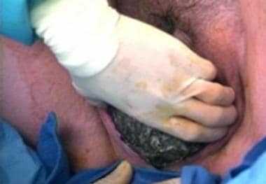
Delivery of the head
Drape and prepare for delivery when the fetal station is low.
Drapes and gowns protect the clinician from the fluid of delivery; sterile preparation is not required.
Use one hand to support and maintain the head in the flexed position as it delivers.
Use the other hand to support the perineum (see image below).
Control the pace of the delivery of the head. Maternal pushing is often helpful, but forceful pushing can cause the head to deliver too precipitously.
Have the patient momentarily withhold pushing once the head is delivered to check for nuchal cords.
Reduce nuchal cords (if present) if the patient and newborn are sufficiently stable to permit a pause in delivery.
Routine suctioning of the nares is no longer recommended by the AAP. [25]
-
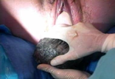
Delivery of the shoulders
With both hands on the head, support delivery of the shoulders one at a time as the patient pushes with a contraction.
Without pulling, apply gentle posterior traction of the head at an angle of 45° to deliver the anterior shoulder followed by gentle anterior traction of the head to deliver the posterior shoulder (see images below).
-
Delivery of the body
With one hand still holding the head, use the other hand to catch the newborn (see image below).
Guide the newborn’s body as it is delivered.
Clamp the umbilical cord in 2 locations, several centimeters apart. The clinician or the patient’s partner can cut the cord between the clamps. There has been increasing data over the past few years advocating for delayed cord clamping. It has been shown to decrease intraventricular hemorrhage and necrotizing enterocolitis in preterm infants, and to decrease anemia in term infants. Delayed cord clamping is defined as >30 seconds after delivery. [26, 27]
-
After delivery
Clean the newborn or place directly with the birthing parent, assuming a normal appearance and Apgar evaluation.
If the newborn is given directly to the birthing parent, wrap the newborn and place on the patient’s bare chest; the newborn's wet skin or the patient’s wet clothes, combined with exposure to ambient air, lead to significant heat loss. Encourage skin to skin contact between the birthing parent and newborn as much as possible. [28]
Continue to monitor the patient during progression to the third stage of labor.


-
Placental separation is evidenced by the following:
An increase in umbilical cord slack
A bolus of blood from the uterus
Superior migration of the uterus within the abdomen with an increase in uterine firmness
-
The clinician can facilitate placental delivery.
Apply gentle traction on the umbilical cord with one hand.
Apply vertical pressure just superior to the pubic symphysis with the other hand to prevent inversion of the uterus.
Administer intravenous oxytocin to expedite the third stage of labor. Oxytocin should be started at delivery of the anterior shoulder.
-
Inspect the placenta after delivery.
Manually explore the uterus if the placenta is not intact.
Retained placenta fragments increase the risk of postpartum hemorrhage.
Pearls
The medical view regarding the best position for delivery has evolved over time. Patient preference should influence positioning as much as possible.
Epidural anesthesia is the most common form of obstetric anesthesia and is used in over half of deliveries in the United States.
Contraindications to vaginal delivery include cord prolapse, persistent fetal distress on monitoring, placental abruption when delivery is not imminent, placenta previa, suspected or confirmed cephalopelvic disproportion, fetal malpresentation, maternal instability, a history of multiple prior abdominal deliveries or of a vertical uterine scar, or active genital herpes.
Controlled maternal pushing helps prevent deep perineal tearing. Prophylactic episiotomy is not recommended for routine births.
The incidence of shoulder dystocia is increasing. A higher incidence is associated with macrosomia, although most cases occur in infants of normal birth weight. The McRobert and suprapubic pressure maneuvers are successful in nearly 50% of cases.
Indications for a forceps or vacuum assist include development of fetal distress when delivery is imminent or an inability of the patient to push secondary to fatigue, anesthesia effect, or a medical condition that contraindicates strong pushing. For more information, see Special Procedures section below.
An essential part of the third stage of labor is assessing the integrity of the placenta to rule out a retained placental fragment.
Blood loss in excess of 500 mL from vaginal delivery is abnormal. The most common causes for postpartum hemorrhage are uterine atony and deep tears within the birth canal.
Complications
Failure to progress
Dystocia is, literally, difficult labor. It is traditionally qualified as a problem of power (contractibility of the uterus), passage (maternal pelvic properties), or passenger (presentation or size of the fetus).
Power
-
On average, cervical dilation progresses at a rate of 1 cm per hour in nulliparous persons and 1.2 cm per hour in multiparous persons.
-
Multiple sites within the uterus can stimulate weak, uncoordinated contractions early in labor, but the pacing of contractions becomes centralized and more effective as labor progresses. If this does not happen, the contractile power needed to complete cervical dilation may be inadequate.
-
Nulliparous patients and those with anatomical uterine abnormalities have a higher risk for this type of dystocia.
Oxytocin
-
When needed, oxytocin improves the frequency and strength of contractions.
-
It may also cause uterine tachysystole (>5 contractions in 10 minutes); stopping the infusion works quickly to remove the medication effect if this occurs
-
Because oxytocin increases the strength of contractions, patients tend to report more pain while on oxytocin.
Passage and passenger
-
During the second stage of labor, the fetal head typically descends within the pelvis at a rate of 1 cm per hour. Abnormal fetal presentation or position or cephalopelvic disproportion (CPD) can slow this progress.
-
True CPD, due to a small pelvic outlet or fetal macrosomia, is rare. While macrosomia occurs in up to 10% of pregnancies in the United States, notably in patients who are delivering post term or who have diabetes, it does not always obstruct labor and cause CPD.
Nonreassuring fetal heart rate
Fetal heart rate monitoring is used to assess baseline heart rate, variability, and the presence of accelerations, and to compare deceleration patterns against the timing of maternal contractions. Indications for operative delivery for fetal well-being include abnormal fetal heart rate patterns suspicious for fetal hypoxia and persistent fetal heart rate decelerations in the context of a fetus remote from delivery.
Bradycardia is mediated by vagal tone. Preserved variability in the setting of mild bradycardia is reassuring. Significant bradycardia may result from cord compression, fetal cardiac anomalies, or fetal hypoxia. Infrequently, it may represent a deceased fetus with monitor capture of the underlying slower maternal heart rate. Ultrasonography can discriminate between fetal bradycardia and maternal heart rate.
Tachycardia is less specific than bradycardia for fetal distress. A high sympathetic tone drives tachycardia and may abolish vagally mediated heart rate variability. Causes of sympathetic surges include maternal fever, hypotension, the use of sympathomimetic drugs, and fetal anemia.
Decelerations are classified as early, late, or variable. Early decelerations are associated with uterine contractions (when compression of the fetal head causes an increase in vagal tone). Late decelerations are more concerning. They may represent uteroplacental insufficiency and signal fetal hypoxia. Variable decelerations vary in the timing of onset and length of duration; they represent cord compression. [29]
Premature rupture of membranes
Premature rupture of membranes (PROM) means rupture of membranes at term before onset of labor.
The most recent ACOG guidelines suggest that augmentation of labor should occur on presentation to the hospital. [30]
Antibiotic treatment is no longer routinely recommended unless the patient develops a fever >100.5 º F.
PROM is most concerning in preterm newborns (PPROM). In those cases, the risk of infection and of the loss of supportive amniotic fluid must be weighed against the risk of premature delivery.
Intrapartum hemorrhage
During labor and delivery, a small amount of blood may be mixed with amniotic fluid, creating a serosanguineous appearance. A bloody show may herald the onset of labor. Significant blood loss, however, is abnormal.
Causes of intrapartum hemorrhage include the following:
-
Placental abruption is the premature separation of the placenta from the uterus.
-
Placenta previa is when the placenta covers the cervical os. In the United States, where most birthing parents have prenatal ultrasonographic evaluations, placenta previa is usually diagnosed by ultrasonographic evaluation prior to labor onset.
-
Placenta accreta is the extension of the placenta into the uterine wall.
-
Ruptured vasa previa (abnormal fetal vessels covering the cervix)
-
Uterine rupture can also cause intrapartum hemorrhage.
Loss of >500 mL of blood during vaginal delivery is abnormal.
Uterine atony, or failure of the uterus to contract following delivery of the placenta, is the most common cause. The uterine blood vessels that are torn and exposed during placental separation are not adequately compressed and may bleed excessively.
Retained placental tissue, the use of uterine muscle relaxants during labor, prolonged labor, or an abnormally distended uterus are causes of uterine atony.
Deep vaginal or cervical lacerations are also a cause of postpartum hemorrhage.
Rarely, coagulopathies can cause postpartum hemorrhage. Von Willebrand disease is sometimes first noted in patients after a vaginal delivery.
To treat postpartum hemorrhage, perform bimanual uterine massage and start an oxytocin drip if uterine atony is suspected; misoprostol or other prostaglandins may also be indicated. If these interventions do not control bleeding, reexplore the vagina, cervix, and uterus for tears or for retained products of conception. If this cannot be accomplished safely in the delivery room, the patient should be moved to the operating room for further evaluation.
Special Procedures
Episiotomy
The decision to perform an episiotomy is often made as the newborn crowns. Until recently, episiotomies were routinely performed during most deliveries with the assumption that this minimized deep traumatic tearing. Evidence, however, does not support the routine practice of episiotomy. [8]
In 2000, episiotomies were performed in approximately 27.5% of deliveries; it is one of the most common obstetrical surgical procedures. [31] By 2006, the national episiotomy rate was 9%. [32]
When indicated, episiotomies are made in a midline (or mediolateral) position. The depth of the incision is directly proportional to how precipitous the delivery is and to the stiffness of the perineum. The procedure for episiotomy is as follows:
-
Make a 0.75-1.5-in incision from the midpoint of the posterior fourchette, directing back toward the rectum (see image below).
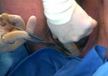
The Cochrane database compared restrictive use to liberal use of episiotomy. There is an increased risk of anterior perineal trauma with restrictive use. The advantages, however, include decreased posterior perineal trauma, fewer sutures, and fewer healing complications. There has been no quality research on midline vs mediolateral episiotomy. [32]
Amniotomy
Amniotomy is a procedure by which the provider artificially ruptures the fetal membranes to induce or expedite labor.
-
Advance the amniohook until it touches the membranes.
-
Once the hook is engaged, pull back slightly; fluid should slowly leak out.
-
Inspect for the presence of meconium, a discolored (yellow to green) fluid due to the presence of fetal stool.
ACOG recommends amniotomy for patients undergoing augmentation or induction of labor to shorten the duration of labor. [33]
External cephalic version
External cephalic version (ECV) is a prelabor maneuver used to convert a breech fetus to a vertex presentation. ECV may reduce the rate of abdominal delivery; success rates in carefully selected full-term patients approach 60%. [34]
Risks include premature rupture of membranes, inadvertent induction of labor, fetal distress or demise, and maternal pain.
Contraindications include multiple gestations or placental, fetal, or maternal abnormalities.
The procedure for ECV is as follows:
-
Position the patient supine.
-
Liberally lubricate the abdomen.
-
Attempt a forward roll first. To do this, apply upward pressure on the breech while guiding the head gently downward to rotate the fetus clockwise.
-
Attempt a reverse, backward roll if the forward roll is unsuccessful.
Induction of labor
Induction of labor can be performed for maternal indications, fetal indications, or electively. [3, 33]
-
Maternal indications include hypertensive complications of pregnancy and exacerbation of maternal illness in pregnancy.
-
Fetal indications include postterm pregnancy, intrauterine growth restriction, fetal demise, oligohydramnios, abnormal fetal heart rate testing, history of stillbirth, and chorioamnionitis.
Elective induction of labor should not occur prior to 39 weeks' gestation because of an increased risk of respiratory problems for the newborn. There has been a concerted effort by the March of Dimes, ACOG, and other governing bodies to eliminate elective deliveries less than 39 weeks because of increased morbidity to the infant. [35]

-
Normal fetal heart rate tracing.
-
Perineal support during delivery of the head.
-
Delivery of the anterior shoulder.
-
Delivery of the posterior shoulder.
-
Episiotomy.
-
Delivery of the body.
-
Amniohook.
Tables

What would you like to print?
- Is immunotherapy for cancer safe in pregnancy?
- Does Endometriosis Complicate Pregnancy?
- Pregnancy: No Reason Not to Treat Migraine
-
 Continuous Glucose Monitoring in Clinical Type 2 Diabetes Practice: Benefits, Accessibility, and Patient Resistance
-
Recognizing and Treating Hyperprolactinemia
-
Lupus Rashes: What to Spot, What to Shield


