Continually Updated Clinical Reference
 
 
  All Sources     eMedicine     Medscape     Drug Reference     MEDLINE
 
eMedicine - IgA Nephropathy : Article by

Quick Find
Authors & Editors
Introduction
Clinical
Differentials
Workup
Treatment
Medication
Follow-up
Miscellaneous
Multimedia
References

Related Articles
Alport Syndrome

Glomerulonephritis, Acute

Glomerulonephritis, Chronic

Glomerulonephritis, Crescentic

Glomerulonephritis, Diffuse Proliferative

Glomerulonephritis, Membranoproliferative

Glomerulonephritis, Poststreptococcal

Glomerulonephritis, Rapidly Progressive

Nephritis, Lupus


Patient Education
Kidneys and Urinary System Center

Blood in Urine Overview

Causes of Blood in Urine

Blood in Urine Symptoms

Blood in Urine Treatment


Author: Mona Brake, MD, Assistant Professor, Department of Internal Medicine, Kansas University School of Medicine

Mona Brake is a member of the following medical societies: American College of Physicians and American Society of Nephrology

Coauthor(s): Douglas Somers, MD, Assistant Professor, Department of Internal Medicine, Division of Nephrology, University of Iowa Medical Center

Editors: James H Sondheimer, MD, FACP, Director of Hemodialysis Unit, Harper Hospital, Associate Professor, Department of Internal Medicine, Division of Nephrology, Wayne State University School of Medicine; Francisco Talavera, PharmD, PhD, Senior Pharmacy Editor, eMedicine; Christie Thomas, MD, FACP, FAHA, FASN, Department of Internal Medicine, Division of Nephrology, Professor, University of Iowa Hospitals and Clinics; Rebecca J Schmidt, DO, FACP, FASN, Clinical Associate Professor of Medicine, West Virginia School of Osteopathic Medicine; Professor of Medicine, Section Chief, Department of Medicine, Section of Nephrology, West Virginia University School of Medicine; Vecihi Batuman, MD, FACP, FASN, Professor of Medicine, Chief, Section of Nephrology, Tulane University School of Medicine; Chief, Renal-Hypertension Section, Department of Medicine, Tulane University Medical Center and Veterans Affairs Medical Center

Author and Editor Disclosure

Synonyms and related keywords: immunoglobulin A nephropathy, Berger disease, mesangial glomerulonephritis, mesangial proliferative glomerulonephritis, Berger's disease, glomerular mesangium, mucosal immune system, end-stage renal disease, ESRD, acute renal failure, chronic renal insufficiency, glomerular lesions, Henoch-Schönlein purpura, HSP, systemic lupus erythematosus, hepatitis, dermatitis herpetiformis, ankylosing spondylitis, episodic gross hematuria, persistent microscopic hematuria, synpharyngitic nephritis, Haemophilus parainfluenzae, H parainfluenzae, rapidly progressive glomerulonephritis, RPGN, renal transplantation, renal transplants, kidney transplantation, kidney transplants

Background

Immunoglobulin A (IgA) nephropathy (also known as Berger disease) was first described by Berger and Hinglais in 1968. IgA nephropathy is characterized by predominant IgA deposition in the glomerular mesangium. Long-term follow-up data illustrated that some patients with IgA nephropathy progress to end-stage renal disease (ESRD). IgA nephropathy is now the most common cause of glomerulonephritis in the world.

IgA nephropathy is highly variable, both clinically and pathologically. Clinical features range from asymptomatic hematuria to rapidly progressive glomerulonephritis (RPGN). IgA nephropathy is most often associated with microscopic hematuria or recurrent macroscopic hematuria, and spontaneously resolving acute renal failure can occur. The condition can sometimes lead to chronic kidney disease as well. Pathologically, a spectrum of glomerular lesions can be seen, but mesangial proliferation with prominent IgA deposition is observed in almost all biopsies.

Although IgA nephropathy is a limited nonsystemic renal disease, many systemic diseases are sporadically associated with mesangial IgA deposition. Henoch-Schönlein purpura (HSP), a systemic illness, has been closely linked to IgA nephropathy. Other systemic diseases in which mesangial deposits of IgA are regularly observed include systemic lupus erythematosus, hepatitis, dermatitis herpetiformis, and ankylosing spondylitis.

Pathophysiology

Almost 40 years after IgA nephropathy was first described, the pathogenesis of this disease remains incompletely understood. The characteristic pathologic findings by immunofluorescence microscopy of granular deposits of IgA and complement 3 (C3) in the glomerular mesangium suggest that this disease is the result of the deposition of circulating immune complexes leading to the activation of the complement cascade.

Deposited IgA is predominantly polymeric IgA1, which is mainly derived from the mucosal immune system. The association of some cases of IgA nephropathy with syndromes that affect the respiratory tract or gastrointestinal tract, such as celiac disease, led to the suggestion that IgA nephropathy is a disease of the mucosal immune system. This concept is also supported by the clinical observation that hematuria worsens during or after upper respiratory tract or gastrointestinal tract infections.

The role of the complement system in the pathogenesis of IgA nephropathy is controversial. While IgA antibodies cannot activate complement through the classic pathway, studies have shown that complement can be activated by the alternate pathway.

IgA in the mesangium is likely to be deposited from the circulation as IgA-containing immune complexes. This hypothesis is supported by the high recurrence rate of IgA nephropathy in renal transplant recipients who have IgA nephropathy and, conversely, by the disappearance of the deposits from donor kidneys with IgA nephropathy when transplanted into donors without the disease. Furthermore, the mesangial pattern of IgA deposits suggests that circulating IgA complexes are responsible for the disease.

Serum IgA levels are elevated in approximately half of patients with IgA nephropathy, but that increase is unlikely to play a role in the pathogenesis of the disease, as markedly elevated IgA levels are observed in patients with AIDS who do not have IgA nephropathy. However, IgA is probably accumulated and deposited because of a systemic abnormality rather than a defect intrinsic to the kidney.

Frequency

United States

Distribution of IgA nephropathy varies in different geographic regions throughout the world. The condition accounts for about 10% of biopsies performed for glomerular disease in the United States. Prevalence rates are lower in the United States compared to Asian countries. These lower rates may be influenced by a conservative approach by nephrologists in the United States who are reluctant to perform renal biopsies in asymptomatic patients with only mild abnormalities on urinalyses.

International

IgA nephropathy is observed in up to 40% of all biopsies performed for glomerular disease in Asia, compared to 20% in Europe and 10% in North America. High prevalence rates are observed in Singapore, Japan, Australia, Hong Kong, Finland, and southern Europe, whereas low prevalence rates are the rule in the United Kingdom, Canada, and the United States.

In Asia, routine urinalyses are performed for schoolchildren, and renal biopsies are performed for patients with asymptomatic hematuria, thus raising the reported prevalence of the disease.

Mortality/Morbidity

This disorder is thought to follow a benign course in most cases. However, many patients are at risk for slow progression to ESRD, which develops in approximately 15% of patients by 10 years and 20% by 20 years, though these percentages depend on how the disease is defined.

Race

IgA nephropathy is more common in whites and Asians and is rare in blacks, both in the United States and in Africa. The condition is frequently observed in American Indians of the Zuni and Navajo tribes.

Sex

IgA nephropathy is more common in males than in females. Virtually all studies show a male predominance of at least 2:1, with reported ratios of up to 6:1. The higher male predilection is observed in white patients in northern Europe and the United States.

Age

IgA nephropathy can affect all ages but is most common in the second and third decades of life. Eighty percent of patients are aged 16-35 years at the time of diagnosis. The condition is uncommon in children younger than 10 years.


History

Two common presentations of patients with IgA nephropathy are episodic gross hematuria and persistent microscopic hematuria. Recurrent macroscopic hematuria, usually associated with an upper respiratory tract infection, or, less often, gastroenteritis is the most frequent clinical presentation and is observed in 40-50% of presenting patients. In 30-40% of patients, the disease is asymptomatic, with erythrocytes (RBCs), RBC casts, and proteinuria discovered on urinalysis. Patients with IgA nephropathy can also present with acute or chronic renal failure.

  • Hematuria: Many patients present with episodes of recurrent macroscopic hematuria.


    • Eighty percent of these episodes are associated with upper respiratory tract infections, mainly acute pharyngotonsillitis. This synchronous association of pharyngitis and macroscopic hematuria has been dubbed synpharyngitic nephritis.


    • Gross hematuria usually appears simultaneously or within the first 48-72 hours after the infection begins; persists less than 3 days; and, in about a third of patients, is accompanied by loin pain, presumably due to renal capsular swelling.


    • Urine is usually brown rather than red, and clots are unusual.


    • The presenting illness of episodic, grossly visible hematuria is more common in younger people, whereas that of abnormal urine sediment is more frequent in older individuals.


    • Episodes of gross hematuria in IgA nephropathy have been associated with a variety of other infections.

      • Urinary tract infections


      • Pneumonia


      • Staphylococcal sepsis


      • Staphylococcal osteomyelitis


      • Acute gastroenteritis


      • Influenza


      • Infectious mononucleosis
         
    • Gross hematuria has also followed tonsillectomy, vaccinations, strenuous physical exercise, and trauma.


    • Between episodes of gross hematuria, many patients have persistent microhematuria, proteinuria, or both.

  • It is rare for proteinuria to occur without microscopic hematuria in IgA nephropathy.


    • Mild proteinuria is common.

    • Nephrotic range proteinuria is uncommon, occurring in only 5% of patients with IgA nephropathy, and is more commonly seen in children and adolescents. Nephrotic range proteinuria can be seen at different stages of the disease, both in patients early in the disease course and in patients with advanced disease.


    • Patients with heavy proteinuria and nephrotic syndrome are likely to have IgA deposition with diffuse proliferative glomerular lesions or minimal-change light microscopic findings.
       
  • Acute renal failure: Acute renal failure, with edema, hypertension, and oliguria, occurs in fewer than 5% of patients. It can develop from either of 2 distinct mechanisms.

    • Acute severe immune injury can manifest as necrotizing glomerulonephritis and crescent formation.


    • Alternatively, only mild glomerular injury is observed with gross hematuria, and renal failure is presumably due to tubular occlusion by RBCs. This is reversible, and renal function recovers with supportive measures.
       
  • Hypertension: Hypertension seldom occurs at the time of initial presentation but more commonly manifests as the course of the disease lengthens or when patients develop chronic renal insufficiency and ESRD.


  • Chronic renal failure: Chronic renal insufficiency seldom develops but is usually slowly progressive. Approximately 1-2% of all patients with IgA nephropathy develop ESRD each year.

Physical

A minority of patients have hypertension; otherwise, physical examination findings in patients with IgA nephropathy are usually unremarkable.

Causes

Most cases of IgA nephropathy are idiopathic, but the onset or exacerbation of the disease is often preceded by a respiratory tract infection. Association with some bacteria, such as Haemophilus parainfluenzae, has been reported. A variety of other disorders have also been linked with IgA nephropathy, as discussed below.

  • Cirrhosis and other liver diseases
    • Glomerular IgA deposition is a common finding in cirrhosis, occurring in up to a third of patients. Liver disease is accompanied by impaired removal of IgA-containing complexes by the Kupffer cells, predisposing patients to IgA deposition in the kidney.
    • Glomerular IgA deposits are common in advanced liver disease, but most adults have no clinical signs of glomerular disease, whereas up to 30% of children may have asymptomatic hematuria or proteinuria. Those abnormalities usually resolve after successful liver transplantation.
  • Gluten enteropathy
    • Glomerular IgA deposition occurs in up to a third of patients with gluten enteropathy.
    • Most of these patients have no clinical manifestations of the disease. However, IgA nephropathy and gluten hypersensitivity are associated, and withdrawal of gluten from the diet of these patients has resulted in clinical and immunological improvement of the renal disease.
  • HIV disease
    • IgA nephropathy has been reported in patients with HIV infection, both whites and blacks, despite the rarity of typical IgA nephropathy in the black population.
    • Clinically, patients have hematuria, proteinuria, and, possibly, renal insufficiency.
    • Histologically, findings range from mesangial proliferative glomerulonephritis to collapsing glomerulosclerosis with mesangial IgA deposits.
    • Several patients have circulating immune complexes containing IgA antibodies against viral proteins.
  • Familial IgA nephropathy
    • While IgA nephropathy is usually a sporadic disease, data suggest that genetic factors are important in susceptibility to development of mesangial glomerulonephritis. Several cases of familial disease have been reported in Italy and the United States, and an autosomal dominant form has been linked to band 6q22-23.
    • Additionally, increased frequency of specific HLA groups has been reported in some patients.


Alport Syndrome
Glomerulonephritis, Acute
Glomerulonephritis, Chronic
Glomerulonephritis, Crescentic
Glomerulonephritis, Diffuse Proliferative
Glomerulonephritis, Membranoproliferative
Glomerulonephritis, Poststreptococcal
Glomerulonephritis, Rapidly Progressive
Nephritis, Lupus

Other Problems to be Considered

Thin basement membrane disease


Lab Studies

  • The first step in confirming the diagnosis is a careful urinalysis of a first-void urine sample performed by an experienced urine analyst. Direct examination of the urine sediment is required to identify RBCs, leukocytes, and RBC casts, all of which indicate glomerular injury.


  • Quantitating proteinuria testing can be accomplished by a 24-hour measurement of urinary protein or semiquantitatively by measuring a urine protein/creatinine ratio. A normal ratio should be less than approximately 0.1. Also, adults older than 50 years with proteinuria should have a urine protein electrophoresis performed to exclude monoclonal light chains as a cause of proteinuria.


  • Assess renal function in patients with proteinuria or hematuria by a 24-hour creatinine clearance test, or the glomerular filtration rate (GFR) can be estimated using the Modification of Diet in Renal Disease (MDRD) formula.


  • Although the serum IgA level is elevated in up to half of patients, this finding is insensitive, nonspecific, and of no clinical utility.

Procedures

  • Diagnosis of IgA nephropathy is confirmed by renal biopsy.

Histologic Findings

Light microscopy

The most common light microscopy findings are focal or, more often, diffuse mesangial proliferation and extracellular matrix expansion (see Image 1). Morphology can range from normal to moderate or severe intracapillary or extracapillary proliferative lesions. While some patients have IgA deposits on immunofluorescence and little or no change by light microscopy, a few patients have segmental necrotizing lesions with crescent formation due to extensive disruption of the capillaries. Occasionally, patients have focal glomerular sclerosis indistinguishable from focal segmental glomerulosclerosis on light microscopy. A number of other findings can be observed in advanced disease, including interstitial fibrosis, tubular atrophy, and vascular sclerosis. These findings can be helpful prognostic tools in patients with IgA nephropathy.

Electron microscopy

Electron microscopy shows mesangial hypercellularity and increased mesangial matrix. The important finding is electron-dense deposits in the mesangium, but deposits in the subendothelial and subepithelial region of the glomerular capillary wall are found in a minority of patients, especially those with more severe disease (see Image 2).

Immunofluorescence

Immunofluorescence findings are the pathologic hallmark of this disease. IgA is deposited in a diffuse granular pattern in the mesangium and occasionally in the capillary wall (see Image 3). Immunoglobulin G (IgG) may accompany IgA, and C3 is often present.


Medical Care

IgA nephropathy is a common cause of glomerulonephritis. Although it is a benign disease in most patients, chronic renal failure and ESRD occur in about 20-40% of patients within 20 years of presentation. Currently, no cure exists for IgA nephropathy, but therapies that can delay the onset of need for dialysis and transplantation are available. Current recommendations include the following:

  • Monitor patients with isolated hematuria without proteinuria or hypertension with urinalysis, renal function testing, and blood pressure measurement.


  • Treat hypertension early and aggressively.


  • ACE inhibitors are the preferred agents for lowering blood pressure. They are beneficial in decreasing proteinuria and should be strongly considered even in normotensive patients with proteinuria.

    • The decrease in proteinuria with ACE inhibitors may be an effect of decreasing the intraglomerular pressure and of changing the glomerular size selectivity.


    • Recent reports have demonstrated that ACE inhibitors are more effective than other antihypertensive drugs in slowing the progression of proteinuric renal disease.
       
  • A recent randomized controlled trial followed patients for a mean of approximately 6 years. The group that received ACE-I had an improved renal survival rate compared to the group receiving other antihypertensive agents.


  • Angiotensin II receptor blockers should be used for patients who cannot tolerate ACE inhibitors. ACE inhibitors and angiotensin II receptor blockers may have an additive effect in decreasing proteinuria. Whether high doses of ACE inhibitors better preserve renal function than combined therapy with ACE inhibitors and angiotensin II receptor blockers is unknown.


  • The combination of an ACE-I and the angiotensin receptor blocker losartan has shown an additive urinary protein–lowering effect compared to doubling the dose of monotherapy. However, patients on combination therapy should be monitored closely for the development of hyperkalemia, and combination therapy should be avoided in patients with advanced kidney failure.


  • Administer prednisone for 4-6 months to patients who have IgA nephropathy with preserved renal function, nephrotic syndrome, and minimal-change findings on light microscopy.


  • Early treatment with prednisone in patients with proliferative IgA nephropathy has been shown to be effective in reducing proteinuria and improving histologic findings, such as proliferation and cellular crescents. Additionally, corticosteroids given for 6 months (1-3.5 g/d) were beneficial against deterioration in renal function in patients with moderate proteinuria.


  • A randomized, controlled, long-term study on the effectiveness of steroids in IgA nephropathy showed improved 10-year renal survival in the steroid treated group compared to the control group. Patients had a proteinuria of 1.9 g/d on average in the treatment group and 1.7 g/d in the control group. Steroids were given for 6 months.


  • Mycophenolate mofetil has been used in patients with IgA nephropathy associated with proteinuria, even though some reports have shown some benefit and others have not. The studies are of small size, and longer term studies are required for more information.


  • Patients with crescentic RPGN can be treated similarly to patients with idiopathic RPGN by using intravenous pulse prednisone followed by oral prednisone and cyclophosphamide.


  • Fish oil (omega-3 fatty acids) at a dose of 12 g/d has been used with controversial and conflicting results, but it is frequently used in patients with declining renal function. Deficiencies of essential fatty acids have been detected in IgA nephropathy, and fish oil is rich in long-chain omega-3-polyunsaturated fatty acids. These produce altered and less biologically effective prostaglandins and leukotrienes, as well as reduced platelet aggregation.


Surgical Care

  • Renal transplantation
    • Renal transplantation is effective in patients with IgA nephropathy. Survival of cadaveric kidney transplants in patients with IgA nephropathy is among the highest observed among common causes of ESRD.


    • IgA nephropathy frequently recurs after transplantation (20-60%). The higher recurrence rates in transplantation from living related donors suggest genetic susceptibility to the disease.


    • Some patients present with microscopic hematuria and proteinuria; others have only positive histologic findings.


    • The disease usually progresses slowly, similarly to the disease in the native kidneys, and graft loss due to recurrent disease occurs in fewer than 10% of patients.


Consultations

Patients should be seen by a nephrologist.

Diet

  • A low-antigen diet, which consists of restricting dietary gluten and avoiding meats and dairy products, has been recommended to decrease mucosal antigen exposure but has not been shown to preserve renal function.


  • Low-protein diets have been recommended to slow the rate of progression of many nephropathies. No large trial explicitly addresses the role of low-protein diets in slowing the decline in renal function in IgA nephropathy. The MDRD Study Group trial is the largest trial of low-protein diets to date, but it included patients with a variety of renal diseases. This trial was unable to determine whether a low-protein diet was beneficial. Although the meta-analysis of studies of low-protein diets suggests some benefits, the effects are subtle and difficult to apply to a given patient (Klahr, 1994; Levy, 1996).


Currently, no cure exists for IgA nephropathy. However, therapies that can delay the onset of need for dialysis and transplantation are available. Hypertension should be treated early and aggressively. ACE inhibitors are the antihypertensives of choice.

Drug Category: Angiotensin-converting enzyme inhibitors

Comparative studies show ACE inhibitors are more effective than other antihypertensives (ie, beta-blockers, calcium channel blockers) in reducing blood pressure and proteinuria, protecting renal function, and delaying onset of ESRD.

Drug NameBenazepril (Lotensin)
DescriptionPrevents conversion of angiotensin I to angiotensin II, which is a potent vasoconstrictor. Also causes lower aldosterone secretion, thus reducing systemic and glomerular capillary pressure.
Adult Dose20-40 mg/d PO qd or divided bid
Pediatric DoseNot established
ContraindicationsDocumented hypersensitivity; angioedema
InteractionsMay increase digoxin, lithium, and allopurinol levels; probenecid may increase levels; coadministration with diuretics increases hypotensive effects
PregnancyD - Unsafe in pregnancy
PrecautionsCaution in dehydration, hyperkalemia, renal impairment (adjust dose), valvular stenosis, or severe congestive heart failure (adjust dose)

Drug Category: Angiotensin II receptor antagonists

Reduce blood pressure and proteinuria, protect renal function, and delay onset of ESRD.

Drug NameLosartan (Cozaar)
DescriptionNonpeptide angiotensin II receptor antagonist that blocks the vasoconstrictor and aldosterone-secreting effects of angiotensin II. May induce a more complete inhibition of the renin-angiotensin system than ACE inhibitors, do not affect the response to bradykinin, and are less likely to be associated with cough and angioedema. For patients unable to tolerate ACE inhibitors.
Adult Dose25-100 mg PO qd/bid
Pediatric DoseNot established
ContraindicationsDocumented hypersensitivity
InteractionsCYP450-3A4 inducers (eg, carbamazepine, rifampin, phenobarbital) may increase metabolism and decrease effects; CYP450-3A4 and -2C9 inhibitors (eg, cimetidine, sulfaphenazole, fluoxetine, ketoconazole) may increase effects by decreasing clearance
PregnancyD - Unsafe in pregnancy
PrecautionsCaution in patients with unilateral or bilateral renal artery stenosis

Drug Category: Corticosteroids

Prednisone should be used in patients with nephrotic syndrome and minimal histologic findings. When treated with corticosteroids, patients with proteinuria and preserved renal function (ie, CrCl >70 mL/min) have shown significant delay of disease progression compared to patients not receiving corticosteroids.

Drug NamePrednisone (Deltasone, Meticorten, Orasone, Sterapred)
DescriptionImmunosuppressant for treating autoimmune disorders. Decreases inflammation by reducing capillary permeability and suppressing PMN activity. Stabilizes lysosomal membranes and suppresses lymphocyte and antibody production.
Adult Dose1 mg/kg/d PO for 6-8 wk initially, then qod and gradual tapering in responsive patients; total treatment period of 6 mo
Pediatric Dose1-2 mg/kg/d PO for 6-8 wk initially; gradually taper in responsive patients to lowest effective dose; typical treatment period of 4-6 mo
ContraindicationsDocumented hypersensitivity; active infection
InteractionsCoadministration with estrogens may decrease clearance; concurrent use with digoxin may cause digitalis toxicity secondary to hypokalemia; phenobarbital, phenytoin, and rifampin may increase metabolism of glucocorticoids (consider increasing maintenance dose); monitor for hypokalemia with coadministration of diuretics; may decrease effectiveness of vaccine and toxoids
PregnancyB - Usually safe but benefits must outweigh the risks.
PrecautionsAbrupt discontinuation of glucocorticoids may cause adrenal crisis; hyperglycemia, edema, osteonecrosis, myopathy, peptic ulcer disease, hypokalemia, osteoporosis, euphoria, psychosis, myasthenia gravis, growth suppression, and infections may occur with glucocorticoid use

Drug Category: Omega-3 polyunsaturated fatty acids

Orphan drug indicated for treatment of IgA nephropathy. Used in patients with proteinuria and decreased renal function.

Drug NameOmega-3 polyunsaturated fatty acid (Fish oil)
DescriptionMay be of benefit by decreasing mediators of glomerular injury and decreasing platelet aggregation.
Adult Dose4-12 g/d PO divided tid/qid
Pediatric DoseNot established
ContraindicationsDocumented hypersensitivity
InteractionsMonitor for enhanced effect with antiplatelet agents; coadministration with other oils (eg, olive oil) may interfere with effect
PregnancyC - Safety for use during pregnancy has not been established.
PrecautionsMay cause fishy odor, diarrhea, hyperglycemia, and bleeding due to decreased platelet aggregation; caution in bleeding disorders or diabetes

Drug Category: Immunosuppressive agents

Cyclophosphamide is used in nonmalignant renal diseases for its immunosuppressive effects.

Drug NameCyclophosphamide (Neosar, Cytoxan)
DescriptionChemically related to nitrogen mustards. As an alkylating agent, the mechanism of action of the active metabolites may involve cross-linking of DNA, which may interfere with growth of normal and neoplastic cells. May be administered PO or IV. Short-term advantages of intravenous pulse of cyclophosphamide may include fewer toxic effects on the bladder and a lower risk of infection.
Adult Dose1-2 mg/kg/d PO; alternatively, 600 mg/m2 IV qmo for ~6 mo
Pediatric DoseNot established
ContraindicationsDocumented hypersensitivity; severely depressed bone marrow function
InteractionsAllopurinol may increase risk of bleeding or infection and enhance myelosuppressive effects; may potentiate doxorubicin-induced cardiotoxicity; may reduce digoxin serum levels and antimicrobial effects of quinolones; chloramphenicol may increase half-life while decreasing metabolite concentrations; may increase effect of anticoagulants; coadministration with high doses of phenobarbital may increase rate of metabolism and leukopenic activity; thiazide diuretics may prolong cyclophosphamide-induced leukopenia and neuromuscular blockade by inhibiting cholinesterase activity
PregnancyD - Unsafe in pregnancy
PrecautionsRegularly examine hematologic profile (particularly neutrophils and platelets) to monitor for hematopoietic suppression; regularly examine urine for RBCs, which may precede hemorrhagic cystitis


Prognosis

  • Natural history of IgA nephropathy: Although this disease usually follows a benign course, ESRD occurs in 15-20% of patients within 10 years of onset and in about 25-30% of patients by 20 years. However, these observations are obtained from biopsy-proven disease and do not include patients with isolated hematuria, who typically do not have a biopsy performed and have a good prognosis.


  • Predictors of disease progression: IgA nephropathy is characterized by a highly variable clinical course. Many efforts have been made to determine clinical and histological features associated with progression to ESRD.
    • Clinically, patients with microscopic hematuria tend to have a higher risk than those with macroscopic hematuria. This phenomenon could be due to the ability to identify patients with gross hematuria at an earlier stage of disease. Sustained hypertension, impaired renal function, persistent hematuria, and proteinuria above 1 g/d are also poor prognostic markers.


    • Histologically, interstitial fibrosis, tubular atrophy, and glomerular scarring predict a worse outcome. As with other glomerular diseases, the risk of progression is more closely correlated with tubulointerstitial findings than with glomerular changes.


    • A study published in 2002 by Italian investigators showed a worse prognosis for patients with familial IgA nephropathy, as compared to sporadic IgA nephropathy.


Patient Education


Medical/Legal Pitfalls

  • Failure to monitor patients with isolated hematuria or hypertension with urinalysis, renal function testing, and blood pressure measurement.
  • Failure to treat hypertension early and aggressively.

Special Concerns

  • Pregnancy: Women with IgA nephropathy tolerate pregnancy well. Only women with glomerular filtration rates lower than 70 mL/min, uncontrolled hypertension, or severe tubulointerstitial disease on renal biopsy are at risk for worsening renal function.
  •  


Media file 1:  
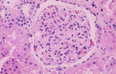
Light microscopy of a glomerulus from a patient with immunoglobulin A nephropathy showing increased mesangial matrix and cellularity.
Click to see larger pictureClick to see detailView Full Size Image
Media type:  Photo

Media file 2:  Electron microscopy showing large dark mesangial deposits.
Click to see larger pictureClick to see detailView Full Size Image
Media type:  Photo

Media file 3:  
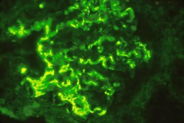
Immunofluorescence microscopy demonstrating large mesangial immunoglobulin A (IgA) deposits diagnostic of IgA nephropathy.
Click to see larger pictureClick to see detailView Full Size Image
Media type:  Photo


  • Barratt J, Feehally J. IgA nephropathy. J Am Soc Nephrol. Jul 2005;16(7):2088-97. [Medline].
  • Berger J, Hinglais N. [Intercapillary deposits of IgA-IgG]Les Depots Intercapillaires d'IgA-IgG. J Urol Nephrol (Paris). Sep 1968;74(9):694-5. [Medline].
  • D'Agati V, Appel GB. HIV infection and the kidney. J Am Soc Nephrol. Jan 1997;8(1):138-52. [Medline].
  • Dillon JJ. Fish oil therapy for IgA nephropathy: efficacy and interstudy variability. J Am Soc Nephrol. Nov 1997;8(11):1739-44. [Medline].
  • Dillon JJ. Treating iga nephropathy. J Am Soc Nephrol. Apr 2001;12(4):846-7. [Medline].
  • Donadio JV Jr, Grande JP. Immunoglobulin A nephropathy: a clinical perspective. J Am Soc Nephrol. Aug 1997;8(8):1324-32. [Medline].
  • Donadio JV Jr, Larson TS, Bergstralh EJ, Grande JP. A randomized trial of high-dose compared with low-dose omega-3 fatty acids in severe IgA nephropathy. J Am Soc Nephrol. 12(4):791-9. [Medline].
  • Falk RJ, Jennette JC, Nachman PH. IgA nephropathy. In: Brenner & Rector's The Kidney. 6th ed. WB Saunders; 2000:1302-1310.
  • Galla JH. IgA nephropathy. Kidney Int. Feb 1995;47(2):377-87. [Medline].
  • Gharavi AG, Yan Y, Scolari F, et al. IgA nephropathy, the most common cause of glomerulonephritis, is linked to 6q22-23. Nat Genet. 26(3):354-7. [Medline].
  • Ibels LS, Gyory AZ, Caterson RJ, et al. Primary IgA nephropathy: natural history and factors of importance in the progression of renal impairment. Kidney Int Suppl. 61:S67-70. [Medline].
  • Klahr S, Levey AS, Beck GJ, et al. The effects of dietary protein restriction and blood-pressure control on the progression of chronic renal disease. Modification of Diet in Renal Disease Study Group. N Engl J Med. 330(13):877-84. [Medline].
  • Kloke HJ, Branten AJ, Huysmans FT, Wetzels JF. Antihypertensive treatment of patients with proteinuric renal diseases: risks or benefits of calcium channel blockers?. Kidney International. 53(6):1559-1573. [Medline].
  • Levey AS, Adler S, Caggiula AW, et al. Effects of dietary protein restriction on the progression of advanced renal disease in the Modification of Diet in Renal Disease Study. Am J Kidney Dis. 27(5):652-63. [Medline].
  • Nolin L, Courteau M. Management of IgA nephropathy: evidence-based recommendations. Kidney Int Suppl. Jun 1999;70:S56-62. [Medline].
  • Pozzi C, Andrulli S, Del Vecchio L, et al. Corticosteroid effectiveness in IgA nephropathy: long-term results of a randomized, controlled trial. J Am Soc Nephrol. Jan 2004;15(1):157-63. [Medline].
  • Pozzi C, Bolasco PG, Fogazzi GB, et al. Corticosteroids in IgA nephropathy: a randomised controlled trial. Lancet. 353(9156):883-7. [Medline].
  • Radford MG Jr, Donadio JV Jr, Bergstralh EJ, Grande JP. Predicting renal outcome in IgA nephropathy. J Am Soc Nephrol. 8(2):199-207. [Medline].
  • Rodicio JL. Idiopathic IgA nephropathy. Kidney Int. Apr 1984;25(4):717-29. [Medline].
  • Russo D, Minutolo R, Pisani A, et al. Coadministration of losartan and enalapril exerts additive antiproteinuric effect in IgA nephropathy. Am J Kidney Dis. Jul 2001;38(1):18-25. [Medline].
  • Russo D, Pisani A, Balletta MM, et al. Additive antiproteinuric effect of converting enzyme inhibitor and losartan in normotensive patients with IgA nephropathy. Am J Kidney Dis. May 1999;33(5):851-6. [Medline].
  • Sayegh MH. IgA nephropathy: recurrence after transplantation. Up-to-Date. 2000;8.
  • Schena FP, Cerullo G, Rossini M, et al. Increased risk of end-stage renal disease in familial IgA nephropathy. J Am Soc Nephrol. Feb 2002;13(2):453-60. [Medline].
  • Scolari F, Amoroso A, Savoldi S, et al. Familial clustering of IgA nephropathy: further evidence in an Italian population. Am J Kidney Dis. May 1999;33(5):857-65. [Medline].
  • Shoji T, Nakanishi I, Suzuki A, et al. Early treatment with corticosteroids ameliorates proteinuria, proliferative lesions, and mesangial phenotypic modulation in adult diffuse proliferative IgA nephropathy. Am J Kidney Dis. 35(2):194-201. [Medline].
  • van Es LA. IgA nephropathy. In: Massry and Glossock's Textbook of Nephrology. Lippincott Williams & Wilkins; 2001:733-741.
  • van Es LA. Pathogenesis of IgA nephropathy. Kidney Int. Jun 1992;41(6):1720-9. [Medline].
  • Wyatt RJ, Julian BA, Baehler RW, et al. Epidemiology of IgA nephropathy in central and eastern Kentucky for the period 1975 through 1994. Central Kentucky Region of the Southeastern United States IgA Nephropathy DATABANK Project. J Am Soc Nephrol. May 1998;9(5):853-8. [Medline].

IgA Nephropathy excerpt

Article Last Updated: Apr 9, 2007