Practice Essentials
The vein of Galen is located under the cerebral hemispheres and drains the anterior and central regions of the brain into the sinuses of the posterior cerebral fossa. The vein of Galen aneurysmal malformation (VGAM) is a choroidal type of arteriovenous malformation involving the vein of Galen forerunner and is distinct from an arteriovenous malformation with venous drainage into a dilated, but already formed, vein of Galen.
VGAMs typically result in high-output congestive heart failure or may present with developmental delay, hydrocephalus, and seizures. [1]
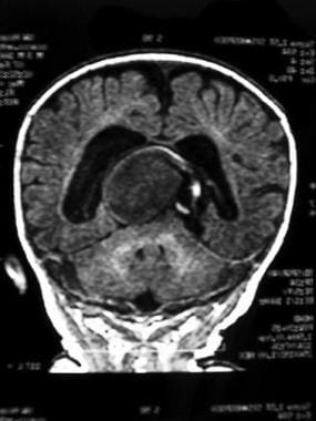 Coronal MRI of head showing large central vein of Galen malformation with moderate degree of hydrocephalus.
VGAM commonly presents in the neonatal period, although it may present later, in early childhood. Typically, in the neonatal period, VGAM presents with congestive heart failure, a cranial bruit, and marked carotid pulses.
Diagnosis
Imaging studies localize or identify the lesion and help confirm the diagnosis and define the degree of involvement.
In patients being considered for surgery or for occlusive therapy, cranial angiography is required to define the extent of aneurysmal dilatation and details for arterial feeders.
Management
Endovascular embolization is the first option for treatment of VGAM.
Cardiac management of high-output heart failure is essential.
Seizures should be managed with antiepileptic medications.
Neurosurgical procedures to relieve hydrocephalus are important. [2] A ventriculoperitoneal shunt may be required in some infants. [3, 4, 5, 6]
Background
The vein of Galen is located under the cerebral hemispheres and drains the anterior and central regions of the brain into the sinuses of the posterior cerebral fossa. The vein of Galen aneurysmal malformation (VGAM) is a choroidal type of arteriovenous malformation involving the vein of Galen forerunner and is distinct from an arteriovenous malformation with venous drainage into a dilated, but already formed, vein of Galen.
VGAMs typically result in high-output congestive heart failure or may present with developmental delay, hydrocephalus, and seizures. [1]
Pathophysiology
Vein of Galen aneurysmal malformation (VGAM) results from an aneurysmal malformation with an arteriovenous shunting of blood. The congenital malformation develops during weeks 6-11 of fetal development as a persistent embryonic prosencephalic vein of Markowski; thus, VGAM is actually a misnomer. The vein of Markowski actually drains into the vein of Galen.
VGAM usually causes high-output heart failure in the newborn resulting from the decreased resistance and high blood flow in the lesion. Associated findings include cerebral ischemic changes such as strokes or steal phenomena that result in progressive hemiparesis. Hemorrhage from the malformation can occur, although this is not a common finding. Finally, the malformation may result in mass effects, causing progressive neurological impairment. Alternatively, the malformation may cause obstruction of the cerebrospinal fluid (CSF) outflow and result in hydrocephalus. [7]
Vein of Galen malformation has been associated with capillary malformation-arteriovenous malformation (CM-AVM), [8] which is a newly recognized autosomal dominant disorder, caused by mutations in the RASA1 gene in 6 families. The authors report severe intracranial AVMs, including vein of Galen aneurysmal malformation, which was symptomatic at birth or during infancy, extracranial AVM of the face and extremities, and Parkes Weber syndrome, previously considered sporadic and nongenetic. [9]
Epidemiology
Frequency
The incidence of the vein of Galen aneurysmal malformation (VGAM) is unknown.
A prospective epidemiologic study in Germany estimated the incidence of VGAM ito be 1:58,100 live births. [10]
Mortality/morbidity
Infants often die if the high-output congestive heart failure is the presenting feature.
Macrocephaly usually improves following shunting for hydrocephalus.
Demographics
VGAM is a congenital malformation; therefore, it may present at birth or in early childhood. It occurs in all races, and boys and girls are affected equally.
Prognosis
Fetuses with prenatally diagnosed vein of Galen aneurysmal malformation (VGAM) have unexpectedly poor outcomes in the presence of cardiac or cerebral anomalies, while those with strictly isolated VGAM tend to have more favorable outcomes. Of 21 cases of prenatally diagnosed VGAM, 4 (19.0%) cases were isolated and 17 (81.0%) were associated with other anomalies. There were nine terminations (42.9%) and six neonatal deaths (28.6%). [11]
A retrospective study of 52 patients with VGAM (17 females, 32 males [data missing for n = 3]) with at least one Francophone parent (5 fetuses and 47 children) found that 33 patients were alive at the long-term evaluation time-point, whereas 19 patients had died. Risk of postnatal death was associated with severe neonatal cardiac failure (p = 0.007) or isosystemic or suprasystemic pulmonary hypertension (p = 0.014). Among survivors, 19 had a good outcome with normal schooling, however a large number had neurodevelopmental alterations; 14 patients with normal schooling had a poor outcome. [12]
-
Cerebral MRI showing large flow void in the central region with enlarged straight sinus.
-
Coronal MRI of head showing large central vein of Galen malformation with moderate degree of hydrocephalus.
-
Cranial MRI showing flow void in the sagittal plain and drainage to the straight and transverse sinuses.
-
Sagittal cerebral MRI with gadolinium showing the relationship of a vein of Galen malformation to the corpus callosum.
-
MRI venogram showing vein of Galen malformation with draining veins.
-
Venogram showing the draining vasculature for the vein of Galen malformation.
-
Skull radiograph showing coils that have been placed during an intravascular embolization of a vein of Galen malformation. Note the ventriculoperitoneal shunt catheter in the anterior head region to relieve hydrocephalus.
