Overview
Thyroid-associated orbitopathy (TAO) is part of an autoimmune process that can affect the orbital and periorbital tissue, the thyroid gland, and, rarely, the pretibial skin or digits (thyroid acropachy). [1, 2, 3] This condition is the most common autoimmune disease of the orbit. [4, 5] Although the terms Graves ophthalmopathy, thyroid ophthalmopathy, and thyroid eye disease are pervasive, these are misnomers. [6] The disease process is a primary orbitopathy in which the orbital and periocular soft tissues are primarily affected, with secondary effects on the eye. See the images below.
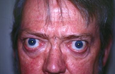 Thyroid-Associated Orbitopathy. This image demonstrates long-standing thyroid-associated orbitopathy with typical features of lid retraction (upper and lower) and scleral show with proptosis. This patient's chief complaint was binocular vertical diplopia. A small right hypotropia was observed on alternate cover testing.
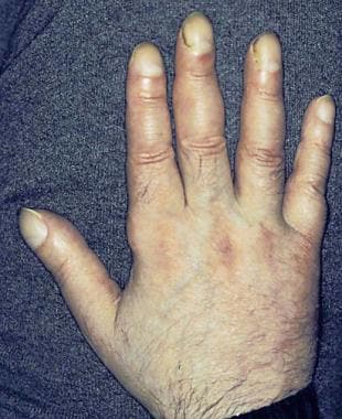 Thyroid-Associated Orbitopathy. Thyroid acropachy imitates the appearance of clubbing and is an uncommon finding in patients with thyroid-associated orbitopathy. This patient required bilateral orbital decompression and strabismus surgery.
Thyroid-associated orbitopathy is usually a self-limited disease characterized by an early inflammatory (active) phase that becomes quiescent over 9-12 months. Thyroid-associated orbitopathy is frequently associated with Graves disease, and the vast majority of patients are hyperthyroid. A systematic review of the world literature found that in patients with thyroid-associated orbitopathy 10% were hypothyroid and 8% were euthyroid. [7]
Thyroid-associated orbitopathy may precede, coincide, or follow the systemic complications of dysthyroidism. Risk factors for thyroid-associated orbitopathy include increased age of onset, duration of Graves hyperthyroidism, and smoking. [8] The ocular manifestations of thyroid-associated orbitopathy include eyelid retraction, proptosis, chemosis, periorbital edema, and altered ocular motility with significant functional, social, and cosmetic consequences. Of affected patients, 20% indicate the ocular morbidity of this condition is more troublesome than the systemic complications of dysthyroidism.
The annual incidence rate of thyroid-associated orbitopathy was estimated at 16 cases per 100,000 women and 2.9 cases per 100,000 men in one rural Minnesota community. [9] There appears to be a female preponderance, with women are affected 2-6 times more frequently than men; however, severe cases occur more often in men than in women. [5] In addition, most patients are aged 30-50 years, with severe cases appearing to be more frequent in those older than 50 years.
Early diagnosis and appropriate monitoring of thyroid-associated orbitopathy may decrease corneal exposure and compressive optic neuropathy. Although most cases of thyroid-associated orbitopathy do not result in visual loss, this condition can cause vision-threatening exposure keratopathy, troublesome diplopia, and compressive optic neuropathy. Therefore, although the prognosis is generally favorable for patients with this condition, and most patients do not require surgical intervention, [10, 11] all clinicians should be able to recognize thyroid-associated orbitopathy.
Patients with thyroid-associated orbitopathy should be counseled that smoking exacerbates the condition. The traditional treatment options for moderate to severe thyroid-associated orbitopathy include pulse glucocorticoids, orbital radiation, and surgery. Teprotumumab is a relatively new targeted medication for moderate to severe thyroid-associated orbitopathy, but it is expensive and unavailable in some countries. If teprotumumab is unavailable, and the patient does not respond to conventional treatment, medications such as tocilizumab or rituximab may be considered—however, these are off-label treatments. Unless the patient has compressive optic neuropathy or severe corneal exposure, surgery for thyroid-associated orbitopathy is usually delayed until the quiescent phase of the disease.
See also the following Medscape Drug & Diseases topics:
Pathophysiology
In the simplest terms, the underlying pathophysiology of thyroid-associated orbitopathy (TAO) is thought to be an antibody-mediated reaction against the thyroid-stimulating hormone (TSH) receptor [12] with orbital fibroblast modulation of T-cell lymphocytes. T-cell lymphocytes are believed to react against thyroid follicular cells with shared antigenic epitopes in the retroorbital space. An active phase of inflammation usually occurs initially followed by a quiescent phase.
In thyroid-associated orbitopathy, the orbital fibroblast is both the target and effector cell. Interaction of the TSH receptor with growth factors such as insulin-like growth factor (IGF) and platelet-derived growth factor, as well as leukocyte interactions with costimulatory proteins such as CD40, activate or sustain reactions of the orbital fibroblasts to produce cytokines and glycosaminoglycans, or induce adipocyte transformation.
Lymphocytic infiltration, fibroblast reaction, and increased orbital volume
Lymphocytic infiltration of the orbital tissue causes a release of cytokines (eg, tumor necrosis factor [TNF], interleukin 1 [IL-1]) from CD4+ T cells stimulating the orbital fibroblasts to produce mucopolysaccharides, which, by hyperosmotic shift, cause tissue edema in the extraocular muscles.
The orbital fibroblasts are the target and effector cells in thyroid-associated orbitopathy. Fibroblasts are extremely sensitive to stimulation by cytokines and other soluble proteins and immunoglobulins that are released in the course of an immune reaction. The cytokines activate previously quiescent fibroblasts to secrete hyaluronic acid, a glycosaminoglycan. Doubling the hyaluronic acid content in the orbital tissue causes a 5-fold increase in the tissue osmotic load. In addition, preadipocyte fibroblasts are influenced to transform into adipocytes, especially in young patients.
The orbit can be described as a pear-shaped box with an anterior opening; the stalk of the pear represents the optic nerve. In thyroid-associated orbitopathy, the increase in orbital volume from the extraocular muscles and fat causes forward protrusion (proptosis or exophthalmos) and, occasionally, optic nerve compression at the narrow posterior apex of the orbit. The edema results in tissue damage and fibrosis, with restriction in extraocular motility and lagophthalmos.
Usually within 1-2 years of the onset of orbital involvement, the inflammation settles to a more quiescent, fibrotic phase predominated by scarring of the orbital tissues.
Potential pathoimmunology
Thyroid-associated orbitopathy may be part of a more generalized disorder of connective tissue and striated muscle. [13] A more extensive discussion on the pathoimmunology of thyroid-associated orbitopathy is beyond the scope of this article. However, some of the research in this field is outlined below.
Elevated levels of IGF 1 receptor (IGF-1R) are found in orbital fibroblasts, T cells, and B cells of patients with thyroid-associated orbitopathy; they may play a role in the promotion of T-cell recruitment and the presence of circulating activating autoantibodies. T-helper 2 cytokines (IL-4 and IL-13) may induce the expression of 15-lipoxygenase-1, with upregulation in the production of 15-hydroxyeicosatetraenoic acid (15-HETE), causing tissue activation and remodeling. The IGF1-R site is targeted by the medication teprotumumab.
Cyclooxygenase 2 (COX-2) is expressed at higher levels in the orbital fibroadipose tissues of thyroid-associated orbitopathy. There is a positive correlation with increasing severity of orbital disease, suggesting a possible relationship with COX-2 expression and orbital inflammation in thyroid-associated orbitopathy.
Variants in the IL-23R gene are strongly associated with thyroid-associated orbitopathy. These variants may predispose to this condition by changing the expression and/or the function of IL-23R, thereby promoting a proinflammatory signaling cascade.
The role of clathrin-mediated signalling pathways, [14] palmitate, [15] and Thy-1 surface markers [16] on orbital fibroblasts as they relate to the pathogenesis of thyroid-associated orbitopath remains to be seen. More recently, a proteomics study has identified three new potential biomarkers (cystatin c, alpha-1 antichymotrypsin, retinal dehydrogenase) in the tears of patients with thyroid-associated orbitopathy that may aid in the diagnosis in future. [4, 5]
Peroxisome proliferator-activated receptor (PPAR)-γ expression has been shown in thyroid tissue and extraocular muscles. [17] PPAR-γ agonists, such as the oral hypoglycemic pioglitazone, expand the orbital fat in diabetic patients with or without thyroid-associated orbitopathy. Although pioglitazone (a PPAR-γ agonist) has been suggested as a treatment for autoimmune thyroid disease, [17] orbital fat expansion (via adipocyte proliferation) in patients with thyroid-associated orbitopathy may be problematic.
Etiology
The thyroid gland itself does not cause thyroid-associated orbitopathy (TAO), and regulation of thyroid function does not abort this condition. Rather, the thyroid gland, eye muscles, and pretibial skin are especially subject to the autoimmune attack. However, restoration of the euthyroid state (with antithyroid drugs and thyroxine) and avoidance of hypothyroidism may improve the orbital status to some extent.
Thyroid state and irradiation
Most patients with thyroid-associated orbitopathy are hyperthyroid, but euthyroidism/hypothyroidism, Hashimoto thyroiditis, thyroid carcinoma, and neck irradiation are also associated with this condition. Even if the patient is euthyroid, thyroid-associated orbitopathy may progress. In patients who are hyperthyroid, the eye signs of thyroid-associated orbitopathy usually develop within 18 months of dysthyroidism; very often, they develop concurrently.
Radioactive iodine
Although somewhat controversial, some publications have suggested that thyroid ablation with orally ingested radioactive iodine-131 (RAI) (131I) may exacerbate thyroid-associated orbitopathy compared with antithyroid drugs or surgical ablation; at least one meta-analysis suggests RAI increases the risk of thyroid-associated orbitopathy progression. [18] However, other studies have not shown that radioiodine is a significant risk for initiation or progression of mild thyroid-associated orbitopathy. [19, 20]
131I is believed to cause a release of thyroid antigens. In a study by Bartalena et al, approximately 15% of patients treated with only radioactive iodine developed or had transient worsening of thyroid-associated orbitopathy. [21] However, some authors feel the threshold for diagnosis of thyroid-associated orbitopathy was low (eg, ocular irritation). In contrast, none of the patients treated with both radioactive iodine and prednisone had progression of thyroid-associated orbitopathy, and two thirds showed improvement. [21] Only 3% of patients treated with methimazole showed worsening of thyroid-associated orbitopathy.
Diseases associated with thyroid-associated orbitopathy
Autoimmune diseases such as myasthenia gravis, Addison disease, vitiligo, and pernicious anemia have been described with thyroid-associated orbitopathy. In one study, 8% of patients with this condition had positive acetylcholine receptor antibodies [22] ; however, at 4.5-year follow-up visits, none of the patients with positive serology was identified clinically to have myasthenia gravis. There has also been a case report of a co-occurrence of ocular myasthenia gravis and thyroid-associated orbitopathy in a 35-year-old male hypothyroid patient. [23]
Yersinia enterocolitica infection has been also associated with thyroid-associated orbitopathy. [24]
Rarely, immune checkpoint inhibitors used for cancer treatment have been associated with thyroid-associated orbitopathy. [25]
Smoking
Thyroid-associated orbitopathy is associated strongly with smoking [8, 26, 27] ; the more severe the eye disease, the stronger the association. Thus, smoking is the strongest modifiable risk factor for thyroid-associated orbitopathy. In one study, smokers of European ethnicity had a 2.4 times increased risk for this condition compared with their Asian counterparts. Active smokers require more strabismus surgery than nonsmokers, independent of orbital decompression surgery. [28] A higher incidence of thyroid-associated orbitopathy in children older than 11 years may be caused by an increasing prevalence of active smoking in adolescents. [29]
The possible mechanisms by which smoking exacerbates thyroid-associated orbitopathy include direct effects of cigarette toxins, nonspecific suppression of T-cell activation, reduction of natural killer T cells, impairment of humoral and cell-mediated immunity, or trauma from heated transmitted from the ethmoid sinuses. [30]
Clinical Evaluation
Thyroid-associated orbitopathy (TAO) usually has a self-limited course over 12-18 months. Stable disease can occasionally reactivate.
Signs and symptoms may vary and depend on the stage that the patient is experiencing. Initially, an acute or subacute stage of active inflammation occurs. Later, the patient progresses to a more quiescent stage, which is characterized by fibrosis. [31]
Symptoms
Patients may complain of the following ocular symptoms:
-
Dry eyes
-
Puffy eyelids
-
Angry-looking eyes
-
Bulging eyes
-
Diplopia
-
Visual loss
-
Field loss
-
Dyschromatopsia
-
Photopsia on upgaze
-
Ocular pressure or pain
Hyperthyroidism symptoms include the following:
-
Tachycardia/palpitations
-
Nervousness
-
Diaphoresis
-
Heat intolerance
-
Skeletal muscle weakness
-
Tremor
-
Weight loss
-
Hair loss
-
Irritability
-
Goiter
Hypothyroidism symptoms include the following:
-
Bradycardia
-
Drowsiness
-
Poor mentation
-
Muscle cramps
-
Weight gain
-
Dry skin
-
Husky voice
-
Depression
-
Cold intolerance
Numerous eponymous signs are associated with thyroid-associated orbitopathy, including the following:
-
Vigouroux sign (eyelid fullness)
-
Stellwag sign (incomplete and infrequent blinking)
-
Grove sign (resistance to pulling down the retracted upper lid)
-
Joffroy sign (absent creases in the forehead on superior gaze)
-
Möbius sign (poor convergence)
-
Ballet sign (restriction of one or more extraocular muscles)
Proptosis and pseudoptosis
Thyroid-associated orbitopathy is the most common cause of unilateral and bilateral proptosis in adults. Proptosis or exophthalmos occurs, because the orbital contents are confined within the bony orbit, and decompression can only occur anteriorly. Unilateral proptosis of thyroid-associated orbitopathy usually reflects asymmetric muscle involvement.
Retropulsion (digital palpation of the globes through closed eyelids) is a useful test; it is decreased in patients with severe thyroid-associated orbitopathy. As noted earlier, thyroid-associated orbitopathy can be asymmetric. One study found asymmetry in 20% of euthyroid/hypothyroid patients with thyroid-associated orbitopathy and in 6.1% of hyperthyoid patients with thyroid-associated orbitopathy. [32] In addition, optic nerve compression in thyroid-associated orbitopathy can occur in the absence of obvious proptosis; for this reason, always check for retropulsion. Various exophthalmometers can be used to measure orbital protrusion.
Pseudoptosis and true ptosis may be seen in patients with thyroid-associated orbitopathy. Pseudoptosis may be observed if contralateral lid retraction is present. Ptosis may occur with thyroid-associated orbitopathy if levator dehiscence is present. Patients with thyroid-associated orbitopathy may have concurrent myasthenia gravis, which may lead to ptosis.
Lacrimal gland enlargement is not uncommon.
Lid retraction, lid lag, and glabellar furrows
Normally, the upper lid is located 1-1.5 mm below the superior limbus, and the lower lid is located at the inferior limbus.
Upper lid retraction (Dalrymple sign), often with temporal flare and scleral show, is the most common ocular sign of thyroid-associated orbitopathy. This sign is an important differentiating feature to note in all patients with proptosis. Mechanisms for upper lid retraction include proptosis, sympathetic drive of the Muller muscle, upgaze restriction, fibrosis of the levator muscle, and contralateral ptosis (myasthenia).
Lid retraction may occur in both the upper and lower lids because of a sympathetically innervated tarsal muscle in both lids. Upgaze restriction, levator fibrosis, and very severe proptosis are other possible causes of lid retraction.
If eyelid retraction is absent, then thyroid-associated orbitopathy may be diagnosed only if (1) proptosis, optic nerve involvement, or restrictive extraocular myopathy is associated with thyroid dysfunction or abnormal regulation, and (2) no other confounding ophthalmic features are apparent.
Lid lag on downgaze (von Graefe sign) is another important feature of thyroid-associated orbitopathy. While slowly moving the fixation object from upward to downward, the examiner should observe if the eyelid lags behind the globe on downgaze.
Other lid signs include lid edema and glabellar furrows. A statistically significant association of deep glabellar rhytids with thyroid-associated orbitopathy has been described. [33] This is presumably caused by hypertrophy of brow depressor muscles compensating for lid retraction.
Corneal and conjunctival findings
Anterior segment signs in thyroid-associated orbitopathy include superficial punctate keratitis, superior limbic keratoconjunctivitis, conjunctival injection usually over the rectus muscle insertions, and conjunctival chemosis.
With severe proptosis, corneal exposure with frank corneal ulceration may occur. Superior limbic keratoconjunctivitis is a chronic, often recurrent condition of ocular irritation, which some attribute to mechanical trauma transmitted from the upper eyelid to the superior bulbar and tarsal conjunctiva. Superior limbic keratoconjunctivitis has been a purported prognostic marker for severe thyroid-associated orbitopathy.
The corneal light reflexes should be examined closely, because asymmetric proptosis and lid retraction may mask the true relative positions of the globes.
Orbital muscle involvement
Strabismus is common, and it often presents as hypotropia or esotropia, because the inferior rectus muscle and the medial rectus muscle are the most commonly involved extraocular muscles in thyroid-associated orbitopathy.
The restrictive myopathy sometimes can be confirmed with forced ductions or elevated intraocular pressure with eye movement (eg, upgaze in hypotropic patients) if a diagnosis of thyroid-associated orbitopathy is not revealing.
Inferior rectus muscle restriction may mimic double elevator palsy.
Although esotropia is a more common finding with thyroid-associated orbitopathy, convergence insufficiency has been described. In patients with thyroid-associated orbitopathy and exotropia, the possibility of concurrent myasthenia gravis should be considered.
Pseudo-fourth nerve palsies have been described with thyroid-associated orbitopathy.
Optic nerve and fundus findings
Compressive optic neuropathy may present with blurry vision, visual loss, dyschromatopsia, or field loss. Patients with optic nerve compression may not have marked proptosis or have seemingly mild proptosis, but they usually show markedly decreased retropulsion (tight orbits). In addition, most cases of compressive thyroid optic neuropathy occur without visible optic nerve edema. For this reason, documenting visual acuity, color vision, and the presence or absence of a relative afferent pupillary defect is important during each visit.
Choroidal folds can occur with thyroid-associated orbitopathy.
Increased intraocular pressure
Glaucoma may result from decreased episcleral venous outflow. Because of restrictive myopathy, intraocular pressure may rise more than 8 mm Hg on upgaze.
Cutaneous findings
Pretibial dermopathy and thyroid acropachy (which mimics the appearance of clubbing) are less commonly encountered dramatic, cutaneous signs of dysthyroidism. See the images below.
Thyroid-Associated Orbitopathy. Thyroid acropachy imitates the appearance of clubbing and is an uncommon finding in patients with thyroid-associated orbitopathy. This patient required bilateral orbital decompression and strabismus surgery.
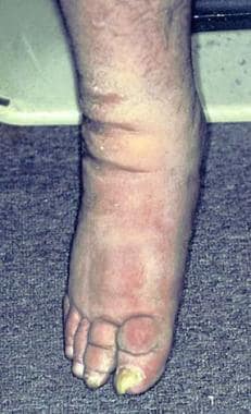 Thyroid-Associated Orbitopathy. Note the pretibial myxedema and thyroid acropachy in this image.
Classification
Numerous classification systems for thyroid-associated orbitopathy (TAO) exist. Most research studies now use either the EUGOGO (European Group of Graves' Orbitopathy) or VISA (vision, inflammation, strabismus, and appearance) classification. [34]
Types I and II
The simplest classification for thyroid-associated orbitopathy is type I and type II; these two types are not mutually exclusive. Type I is characterized by minimal inflammation and restrictive myopathy. Type II is characterized by significant orbital inflammation and restrictive myopathy.
NOSPECS
The Werner NOSPECS classification system (and its modifications) is one of the most commonly known, and it is used in many endocrine studies. NOSPECS uses a mnemonic to describe the presence or absence of signs or symptoms (NO) (upper lid retraction, no lid lag or proptosis) and grades and classifies the severity and rank order of various clinical features (SPECS: soft-tissue involvement with symptoms [excess lacrimation, sandy sensation, retrobulbar discomfort, photophobis; no diplopia], p roptosis, e xtraocular muscle involvement [often with diplopia], c orneal involvement [due mainly to lagophthalmos], and s ight loss [due to optic nerve involvement]). [35]
Unfortunately, the NOSPECS classification has some weaknesses that may limit its prognostic value. Patients may fall into more than one particular class, and they may not progress in an orderly fashion from class 1 to class 6. In addition, patients with visual loss from compressive optic neuropathy may not show marked proptosis or other signs of severe disease.
EUGOGO and VISA
To determine disease activity, the grading systems most frequently employed in clinical studies of thyroid-associated orbitopathy are the EUGOGO clinical activity score classification in Europe and the VISA classification, especially in North America. [34] Clinical activity scores greater than 3/7 on initial presentation or 4/10 on follow-up suggest active disease that might benefit from immunosuppression. VISA inflammation scores less than 4/10 are managed conservatively.
Diagnostic Considerations
Orbital and preseptal cellulitis are included in the differential diagnosis when evaluating a patient with suspected thyroid-associated orbitopathy (TAO). In orbital cellulitis, the onset of proptosis is often quicker, and the patient has other evidence of infection (eg, fever, leukocytosis). On neuroimaging, the paranasal sinuses often are opacified.
Thyroid-associated orbitopathy should not be mistaken for a dural arteriovenous malformation or a carotid cavernous fistula. In patients with carotid cavernous fistula, the patient may have a cranial bruit, and the dilated episcleral vessels extend to the limbus.
Orbital inflammatory syndrome (orbital pseudotumor) is often more painful than thyroid-associated orbitopathy, with faster progression; the tendons are involved in orbital myositis. Orbital inflammatory syndrome is associated more often with ptosis than lid retraction. Isolated enlargement of the lateral rectus muscle is more likely to represent a process such as orbital inflammatory syndrome than thyroid-associated orbitopathy.
Other causes of thickened eye muscles include immunoglobulin G 4 (IgG4)-related disease, sarcoidosis, metastases, lymphoma, amyloidosis, and acromegaly. Orbital ultrasonography can quickly confirm if the patient has thickened eye muscles or an enlarged superior ophthalmic vein.
IgG4-related orbital disease is occasionally characterized by preferential enlargement of the lateral retus, enlargement of the infraorbital nerve, increased serum IgG4, and asthma. IgG4-related disease can overlap with thyroid-associated orbitopathy.
Dorsal midbrain syndrome (Parinaud syndrome) is a condition in which patients may present with lid retraction and upgaze problems. In contrast to thyroid-associated orbitopathy, the globes in Parinaud syndrome elevate on the doll's head maneuver and the eye tends not to be injected or proptotic.
Thyroid Disease Studies
If thyroid-associated orbitopathy (TAO) is suspected, consider obtaining levels of serum thyroid-stimulating hormone (TSH) (thyrotropin), free T4 (thyroxine), and free T3 (triiodothyronine) Serum TSH is useful to establish a diagnosis of hyperthyroidism or hypothyroidism. Usually, the TSH is low in hyperthyroidism and high in hypothyroidism. The use of thyroid-stimulating immunoglobulin (TSI) is increasingly reported in the literature, but because of cost, some authors recommend initially only using the TSH level to screen for thyroid disease. One small study did not support any added benefit of TSI compared to TSH for the prediction and management of thyroid-associated orbitopathy. [36]
TSH receptor assays
The nomenclature for the various TSH receptor assays is confusing and inconsistent. Assays that measure the binding of TSH to a solubilized receptor are often referred to as TRAb (thyroid receptor antibody), TBII (TSH-binding inhibitor immunoglobulin), and LATS (long-acting thyroid stimulator) assays. Assays that measure the ability of immunoglobulin G (IgG) to bind to the TSH receptor on cells and to stimulate adenylate cyclase production have generally been referred to as the TSI assays. TSIs may show more significant association with the clinical features of thyroid-associated orbitopathy than TBII and may be regarded as functional biomarkers for thyroid-associated orbitopathy. [37]
Direct assays for TSH, free T4, and free T3 have superseded the usefulness of total T4 and T3 resin uptake testing.
Antibodies to thyroid peroxidase and thyroglobulin
Thyroid peroxidase antibodies and antibodies to thyroglobulin may be useful when trying to associate eye findings with a thyroid abnormality, such as euthyroid Graves disease.
The thyroid peroxidase test is also called the antimicrosomal antibody test and the antithyroid microsomal antibody test. The antithyroglobulin test is also known as the antithyroid antibody test.
The serum level of hyaluronan is not a sensitive indicator of its presence within the extraocular muscles.
CT Scanning and MRI
If the diagnosis of thyroid-associated orbitopathy (TAO) can be established clinically, and the patient does not have severe disease nor require surgery, computed tomography (CT) scanning and magnetic resonance imaging (MRI) are not routinely ordered. In the absence of lid retraction, orbital imaging is useful if thyroid-associated orbitopathy remains suspect. If imaging is required, axial and coronal views are useful. [38] MRI is more sensitive for showing optic nerve compression, whereas CT scanning is performed before bony decompression because it better shows bony architecture. CT scanning can be performed without iodine contrast medium for thyroid-associated orbitopathy.
Neuroimaging often reveals thick eye muscles, classically with tendon sparing. The inferior and medial rectus muscles are usually involved. Bilateral muscle enlargement is the norm; unilateral cases usually represent asymmetric involvement rather than normality of the less involved side.
Isolated rectus muscle involvement may occur in up to 6% of patients; in this subgroup of patients, the superior rectus muscle may be the most frequently involved muscle. Isolated lateral rectus muscle enlargement without other evidence of muscle enlargement is uncommon in thyroid-associated orbitopathy and suggests another disease process (eg, orbital myositis).
Neuroimaging may also show a dilated superior ophthalmic vein. In addition, apical crowding of the optic nerve is well visualized (see the image below). Occasionally, the proptosis of thyroid-associated orbitopathy results in straightening of the optic nerve.
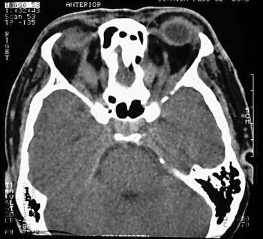 Thyroid-Associated Orbitopathy. An axial computed tomography (CT) scan in a patient with congestive thyroid-associated orbitopathy is shown. The recti muscles are thickened with apical compression. The tendons are spared.
On CT scans, orbital fat density is higher in patients with thyroid-associated orbitopathy; this is negatively correlated to fat volume but positively correlated to muscle volume and muscle density. [39]
Histologic Features
Findings on histologic examination of thyroid-associated orbitopathy (TAO) include the following:
-
Fibrosis with degenerative changes in the eye muscles
-
Lymphocytic cell infiltration
-
Enlargement of fibroblasts
-
Accumulation of mucopolysaccharides
-
Interstitial edema
-
Increased collagen production
Outpatient Management
Most patients with thyroid-associated orbitopathy (TAO) can be observed; the follow-up interval depends on the disease activity.
Monitor for visual loss from corneal exposure and optic neuropathy and for strabismus development. The author does not recommend the use of eye exercises for patients with severe restrictive strabismus as doing so may elevate intraocular pressure.
Color vision testing and perimetry are recommended at baseline, and they can be performed every 3-6 months in patients with active thyroid-associated orbitopathy.
In patients with diplopia, prisms may be beneficial for those with small-angle or relatively comitant deviations. Tape occlusion of one lens or segment of the glasses may be helpful. If this is not effective, try using an occluder or vaulted eye patch (with care not to touch the cornea or compress the orbit).
Patients with dry eye symptoms or corneal exposure should apply artificial tears during the day and lubricating ointment at night, and they should consider the use of punctal plugs.
Patient education
Inform patients that thyroid-associated orbitopathy usually has a self-limited but prolonged course over 1 or more years in most patients. Patients should also realize that no immediate cure is available. [40] Smoking cessation may reduce the risk of congestive orbitopathy.
Sleeping with the head of the bed elevated may decrease orbital congestion when the patient awakens.
Pharmacologic Treatment
For mild thyroid-associated orbitopathy (TAO), selenium and quercetin may be considered. In one study, the antioxidant selenium (200 mcg daily) was shown to help patients with mild Graves orbitopathy. [41] However, if those with thyroid-associated orbitopathy are not selenium deficient, they may not benefit from such supplementation. Quercetin, a natural plant product found in food such as capers, may inhibit proinflammatory cytokines [42] ; it has been suggested as a treatment for thyroid-associated orbitopathy.
Immunosuppressants are prescribed for patients with moderate to severe thyroid-associated orbitopathy that is in the active phase. Systemic glucocorticoids were the mainstay treatment for thyroid-associated orbitopathy, but treatment options expanded once teprotumumab became available in the United States.
The use of systemic glucocorticoids (steroids) is usually reserved for patients with moderate to severe inflammation or compressive optic neuropathy in thyroid-associated orbitopathy. The consensus statement of the European Group on Graves' Orbitopathy (EUGOGO) suggests using a total dose of 4.5 mg intravenous (IV) methylprednisolone administered once per week over 12 weeks for patients with moderate to severe thyroid-associated orbitopathy. [3] Higher doses of IV methylprednisolone can be considered for more advanced disease. [3] Liver failure does not usually occur in patients using less than 8 g of methylprednisolone.
Steroids may decrease the fibroblastic production of mucopolysaccharides. Pulse IV steroids can be considered, but they may only marginally improve long-term disease outcome. Thus, if necessary, high-dose steroids and higher IV doses are given for compressive optic neuropathy. If no response occurs after 48-72 hours, steroids probably will not be effective; at this point, the patient should have surgical decompression and maintain steroids.
The United States Food and Drug Administration approved teprotumumab (Tepezza) in January 2020 for treatment of thyroid-associated orbitopathy ("thyroid eye disease") in adults. Thyroid-associated orbitopathy is most often associated with hyperthyroidism or Graves disease; however, it arises from a separate process involving autoantibodies that activate an insulin-like growth factor (IGF) 1 receptor–mediated signaling complex on cells within the eye orbit. Teprotumumab attenuates the actions of both IGF-1 and thyroid-stimulating hormone (TSH) in fibrocytes, by blocking the induction of proinflammatory cytokines by TSH.
Approval of teprotumumab was supported by the randomized, placebo-controlled Treatment of Graves' Orbitopathy to Reduce Proptosis With Teprotumumab Infusions clinical studies (OPTIC) (phase 2, n = 76; phase 3, n = 83). Teprotumumab given intravenously (IV) every 3 weeks for three infusions was more effective than placebo in reducing proptosis and the clinical activity score in patients with thyroid-associated orbitopathy. Significantly more patients treated with teprotumumab (82.9%) had a meaningful improvement in proptosis (≥2 mm) compared with placebo (9.5%) (P ˂ 0.001) without deterioration in the fellow eye at week 24. Additional secondary endpoints were also met, including a change from baseline of at least one grade in diplopia (67.9% of patients receiving teprotumumab vs 28.6% receiving placebo; P = 0.001) at week 24. Observed adverse effects of teprotumumab included muscle spasm, hyperglycemia, and hearing impairment, most of which were transient. [43, 44]
Unfortunately, as of 2021, teprotumumab is unavailable in many parts of the world, and it remains a very expensive treatment. Alternative agents that have been described for moderate to severe thyroid-associated orbitopathy include tocilizumab, rituximab, mycophenolate, methotexate, cyclosporine, octreotide, and IV immunoglobulin (IVIg). (See the next section, Potential Medical Therapies.)
Potential Medical Therapies
If there is a good steroid response, orbital radiation may be considered. In severe cases of thyroid-associated orbitopathy (TAO), combined teprotumumab, steroids, radiation, and surgery may be required. In patients with worsening disease despite orbital decompression, intranasal steroids can be used.
Available off-label, second-line agents for thyroid-associated orbitopathy include tocilizumab, which is an inhibitor of interleukin 6 (IL-6), [45] anti-CD20 (rituximab) therapy to deplete B-cell lymphocytes, [46] and antitumor necrosis factor (anti-TNF) drugs (eg, etanercept, infliximab), but more studies are required to determine their risk-benefit ratio. Two small randomized-controlled trials of rituximab with different time windows for study enrollment showed conflicting results. [47, 48] Potential side effects of rituximab include infusion reactions and, rarely, an increased risk of infection and progressive multifocal leukoencephalopathy.
Mycophenolate, [49] methotrexate, [50] octreotide, pentoxifylline, nicotinamide, plasmapheresis, and intravenous immunoglobulin (IVIG) are not mainstream medical treatments for thyroid-associated orbitopathy. Octreotide, a potent synthetic somatostatin analogue, has a beneficial effect in this condition, especially in patients with a positive Octreoscan-111 (indium-111 [111In] pentetreotide) diagnostic study. Lanreotide is a longer-acting somatostatin analogue that is administered only once every 2 weeks; this agent may provide some benefit. Pentoxifylline and nicotinamide may be useful; both agents are believed to inhibit cytokine-induced glycosaminoglycan synthesis by the retroorbital fibroblasts.
Numerous investigational drugs are in the pipeline; they include alternative insulin-like growth factor (IGF) 1 inhibitors such as linsitinib and VRDN 001, batoclimab (neonatal crystallized fragment receptor [FcRn] monoclonal antibody), anti-CD40 (tumor necrosis factor receptor superfamily [TNFRSF] 5 antibody), thyroid-stimulating hormone (TSH) receptor antagonists (K1-70), and RVT-1401. Bimatoprost eye drops can cause prostaglandin periorbitopathy with enophthalmos; their ultimate role in the treatment of thyroid-associated orbitopathy remains to be seen.
The role of plasmapheresis and IVIg is not well delineated. One randomized trial of IVIg (1 g Ig/kg body weight × 2 consecutive days every 3 wk) versus oral prednisolone (for 20 wk, with initial dose of 100 mg/day) showed both treatments to be equally effective in patients with active thyroid-associated orbitopathy. [51] Fewer adverse effects were observed in the IVIg treatment group.
Orbital Radiation
Orbital irradiation is occasionally prescribed for moderate to severe inflammatory symptoms, diplopia, and visual loss in patients with thyroid-associated orbitopathy (TAO). The radiation (1500-2000 cGy fractionated over 10 days) is usually administered via lateral fields with posterior angulation. Radiation is believed to damage orbital fibroblasts or perhaps lymphocytes.
The administered radiation requires several weeks to take effect, and it may transiently cause increased inflammation. Thus, most patients are maintained on steroids during the first few weeks of treatment. In addition, a better response to radiation is observed in patients with active inflammation who are treated within 7 months of the onset of thyroid-associated orbitopathy. The radiation may be more effective if combined with steroid treatment.
Studies that suggest that radiotherapy is ineffective in thyroid-associated orbitopathy must be scrutinized to ensure that the radiation was administered to appropriate candidates at the appropriate time. For example, the Gorman et al study used serum thyroid-stimulating immunoglobulin (TSI) as a surrogate of active eye disease. [52] Although the blood test is an indicator of immunologic activity, it may not reflect the clinical progression of thyroid-associated orbitopathy. Furthermore, the patients in that study were enrolled at a median of 1.3 years after the onset of eye symptoms, suggesting that many of the patients in the study would not have progressive eye symptoms or signs indicative of an ongoing orbital process. [52]
Although improvement of motility disturbances can occur with radiotherapy, the radiation is limited when used in isolation to treat diplopia.
Potential adverse effects and contraindications
Cataract, radiation retinopathy, and radiation optic neuropathy are possible risks of orbital irradiation. These effects are not common if treatment is appropriately fractionated and the eyes are shielded. Marquez et al found that 12% of their study patients developed cataracts after irradiation (median follow-up, 11 y). [53]
Wakelkamp et al also believed that orbital irradiation for thyroid-associated orbitopathy is a safe treatment modality, except possibly for patients with diabetes mellitus. [54] Radiation may be a relative contraindication for patients with diabetes mellitus because of the risk of worsening retinopathy.
Although medical physics simulation studies suggest there is an up to 16% increased relative risk of brain malignancy with orbital radiation, the absolute risk of radiogenic cancer remains low. [55]
Prevention of I-131-Associated TAO
To prevent progression of thyroid-associated orbitopathy (TAO) from radioactive iodine (RAI) thyroid ablation, prophylaxis with low-dose prednisone (eg, 0.5 mg/kg/day starting with RAI treatment for 1 month, with tapering doses in the second month) can be considered if no contraindications for use of the glucocorticoids exist and this therapy is agreed to by the patient. Following administration of radioactive iodine, closely monitor the patient for the development of hypothyroidism.
Overview of Surgical Intervention
Approximately 5% of patients with thyroid-associated orbitopathy (TAO) may require surgical intervention. Alert the patient to the possibility that multiple-staged procedures may be necessary. [56, 57, 58] In elective cases, listen carefully to what the patient desires; the patient's expectations may not be realistic and these should be addressed before proceding with any surgeries.
The timing and sequence of surgery are important. Unless compressive optic neuropathy or severe corneal exposure is present, surgery is generally delayed during the active inflammatory phase of thyroid-associated orbitopathy. Rather, surgery is usually performed during the quiescent cicatricial phase of the disease.
Taking preoperative photographs is advised. With strabismus surgery, document prism measurements or fields of single binocular vision. Baseline automated perimetry is also useful. In patients with thyroid-associated orbitopathy who have proptosis and inferior scleral show, be aware that simple horizontal tightening of the lower lid will result in increased globe exposure.
The sequence of surgery is important because the outcome of each procedure may determine the necessary goals of the next. If the patient has marked proptosis, strabismus, and lid deformity, perform surgery in the following order [3] :
Orbital decompression
Strabismus surgery
Lid-lengthening surgery
Blepharoplasty
These procedures will be briefly reviewed in the following sections.
Orbital Decompression
Orbital decompression may be performed as the initial treatment of compressive optic neuropathy or used if medical treatment is ineffective. A combination of medical and surgical treatments may be required in compressive optic neuropathy. Before performing bony orbital decompression, computed tomography (CT) scans should be obtained because these studies delineate bony anatomy better than magnetic resonance imaging (MRI).
Potential complications of orbital decompression include blindness, hemorrhage, diplopia, periorbital numbness, globe malposition, sinusitis, and lid malposition.
Procedure overview
Following bony orbital decompression, open the periorbita. Little reduction in proptosis occurs until the periorbita is slit.
Due to the location of the optic foramen, optic nerve decompression requires decompression of either the medial wall or orbital roof. Medial wall removal should not extend above the frontoethmoidal suture. This averts bleeding from the ethmoidal arteries and prevents cerebrospinal fluid (CSF) leaks. Orbital roof decompression is rarely performed by ophthalmologists. Medial decompression for compressive neuropathy must be taken posteriorly all the way to the apex of the optic canal. Surgery can be approached from a transorbital or trans-sinus route. Transorbital routes include subciliary, lid crease, medial (cutaneous, transcaruncular), and coronal incisions. Trans-sinus routes include transantral approaches and endoscopy.
Most orbital decompressions involve at least two walls to provide adequate volume. A balanced decompression involves the medial and lateral orbital walls and may decrease the risk of postoperative diplopia and lid retraction.
Decompression of the medial wall and floor is also a common combination, but the latter may cause diplopia and infraorbital anesthesia. When the orbital floor is removed, preservation of a strut of bone between the ethmoid and maxillary bones may reduce strabismus from inferomedial shift in the globe position.
Lateral wall decompression does little to relieve apical compression but helps to reproduce proptosis. Valgus repositioning of the orbital wall and orbital rim-onlay, porous-polyethylene grafts are adjunctive techniques to reduce proptosis.
Four-wall decompression (with decompression of the orbital roof) requires a neurosurgical approach.
Orbital fat decompression
Orbital fat decompression without bony removal has been described for thyroid-associated orbitopathy without apical compression. Candidates for orbital fat decompression should show predominant enlargement of the orbital fat compartment—rather than the rectus muscles—on orbital imaging.
Unlike cosmetic blepharoplasty, with orbital fat decompression, fat is also removed posterior to the equator of the globe. Inferiorly, the fat is removed through a transconjunctival approach, which may be facilitated with lateral canthotomy and cantholysis. Superiorly, fat removal is through a lid crease incision, usually confined to the nasal quadrant. Transcaruncular and endoscopic approaches avoid a skin incision.
Liao and Huang confirmed that a reasonable and effective reduction in proptosis can be safely achieved by extensive orbital fat removal alone. [59] However, their study did not correlate individual case results with the extent of extraocular muscle hypertrophy compared with the degree of fat hypertrophy, and thus it greatly impacted results in individual cases. The risk of postoperative worsened strabismus was not addressed and likely still remains a (theoretical) risk. It is not advisable to rely solely on fat decompression in cases of impending or actual optic nerve compression.
Strabismus Surgery
Although successful early strabismus surgery during active thyroid-associated orbitopathy (TAO) has been described, the procedure is generally delayed until the disease is inactive and the prism measurements have been stable for at least 6 months.
Patients should realize that the goal of surgery is to minimize diplopia in primary and reading positions. Expecting binocular single vision in all positions of gaze may not be realistic. In addition, patients should be aware that multiple strabismus surgeries and prisms may be required.
Because of the restrictive myopathy of thyroid-associated orbitopathy, predominantly recessions, rather than resections, are performed. Whenever feasible, adjustable suture surgery is recommended. In patients intolerant of conscious suture adjustment, hang-back sutures can be adjusted using the corneal light reflexes. In select patients with thyroid-associated orbitopathy, strabismus surgery can be performed using topical anesthesia.
To prevent ocular ischemic syndrome, do not operate simultaneously on more than two muscles per eye.
Procedure considerations
Surgery of the inferior rectus muscle deserves special mention. Inferior rectus muscle recession may decrease upper lid retraction, but it also often results in lower lid retraction despite dissection of the lower lid retractors. Because the inferior rectus muscle has subsidiary actions (excyclotorsion and adduction), inferior rectus muscle recessions may lead to a component of intorsion and A-pattern strabismus.
If visualization during strabismus surgery is difficult, especially for the superior rectus muscle, a vertical lid split technique has been described. [56]
Some clinicians use botulinum toxin injections during the acute phase of thyroid-associated orbitopathy for lid retraction and restrictive myopathy as a temporizing measure until surgery is performed. However, optic neuropathy following a botulinum toxin injection for strabismus in a patient with thyroid-associated orbitopathy has been reported. [60]
Lid-Lengthening Surgery
If restoration of the euthyroid state does not improve lid retraction in those with thyroid-associated orbitopathy (TAO), consider lid-lengthening surgery. This procedure decreases corneal exposure and can be used to camouflage mild-to-moderate proptosis. In patients unwilling to consider lid surgery, possible alternatives to upper-lid lengthening include botulinum toxin injections to the upper lid and subconjunctival triamcinolone.
Lateral tarsorrhaphies can decrease upper and lower lid retraction, but the author does not prefer this method.
Amelioration of 2-3 mm of upper lid retraction can be done with a Müller muscle excision. Lateral levator tenotomy is often helpful to decrease the temporal flare. If further amounts of lid recession are required, levator recession can be considered.
Lower lid-lengthening usually requires a spacer material. Graft materials include human acellular dermis, tarsus and conjunctiva from the upper lid, hard palate, ear cartilage, and donor sclera.
Horizontal tightening procedures (eg, lateral tarsal strip) increase scleral show in patients with proptosis.
In the horizontally tight eyelid, lateral canthal advancement is a useful adjunct to enhance the effect of retractor recession and reduction of temporal flare.
Blepharoplasty
Blepharoplasty is the last phase of restorative surgery in thyroid-associated orbitopathy (TAO). The transconjunctival approach to lower lid blepharoplasty can be used if no excess lower lid skin is present.
Upper lid blepharoplasty is performed transcutaneously with conservative skin excision. Brow fat resection may be considered. Dacryopexy may be required if lacrimal gland prolapse occurs.
Consultations
Patients with thyroid-associated orbitopathy (TAO) benefit from consultation and follow-up care with an endocrinologist. Orbital decompression can be performed in conjunction with an otorhinolaryngologist, especially when endoscopic procedures are contemplated. Neurosurgical consultation is required when decompression of the orbital roof is performed.
Thyroid-associated orbitopathy can cause marked changes in the patient's physical appearance and psychological morbidity. [61] As such, psychiatric referral can be considered for select patients.
Pregnancy and TAO
The incidence of Graves disease in women who are pregnant has been reported to be approximately 0.2%. Information on the management of thyroid-associated orbitopathy (TAO) during pregnancy is not widely available. In 2017, the American Thyroid Association (ATA) updated its guidelines for management of thyroid disease during pregnancy. [62] Among the recommendations for management of Graves disease, the guidelines noted the following [62] :
-
Rising antibody titers following radioactive iodine treatment may contribute to worsening orbitopathy or fetal risk.
-
There is a high risk of rapid relapse of hyperthyroidism after medication withdrawal in early pregnancy in women with active orbitopathy.
If a pregnant woman with thyroid-associated orbitopathy has compressive optic neuropathy, glucocorticoids can usually be administered in consultation with the obstetrician and an endocrinologist. Ideally, surgery would be deferred until after delivery, if possible. If emergent orbital decompression is required, nonabdominal surgery may not impose the same risks to the fetus as that of abdominal surgery.
The American College of Obstetricians and Gynecologists (ACOG)/American Society of Anesthesiologists (ASA) consensus guidelines recommend against denying indicated surgery to a pregnant woman, regardless of trimester. [63] If possible, nonurgent surgery should be performed in the second trimester when preterm contractions and spontaneous abortion are least likely. [63] The American Thyroid Association (ATA) guidelines concur. [62]
Childhood TAO
Thyroid-associated orbitopathy (TAO) is less common in children and adolescents than in adults, and it tends to have a more benign disease course, with less ophthalmoplegia. In comparison with adults, children infrequently require surgical intervention.
Counsel children and their parents to avoid smoking. Secondhand smoke appears to exacerbate autoimmune thyroid disease, and passive smoking may have a deleterious effect on childhood thyroid-associated orbitopathy.
In children and adolescents, thyroid-associated orbitopathy symptoms often regress after euthyroidism is restored. In most cases, conservative management and watchful waiting is adequate. To improve quality of life, local protective agents such as lubricant eye drops and/or gels and selenium supplementation are necessary. [29]
Treatment of thyroid disease is crucial in the management of thyroid-associated orbitopathy in children and adolescents, and close collaboration between ophthalmologists and endocrinologists is required. Treatment of hyperthyroidism with antithyroid drugs can affect improvement in the ocular condition. Radioiodine is is contraindicated in children younger than 5 years because of the potentially higher risk of developing thyroid cancer; in addition, radioiodine therapy can exacerbate thyroid-associated orbitopathy. However, this complication is more common in adults; concomitant oral steroid treatment may be effective in reducing this side effect. [64]
When there is no improvement or a deterioration in ocular condition after thyroid function normalization, pharmacologic treatment may be necessary. Intravenous steroids may be considered. In adults, this route of administration has proven to have fewer side effects, but prolonged administration of steroids in children may cause side effects such as weight gain, immunosuppression, and growth failure. [29]
Because of the risk of tumor induction, retrobulbar irradiation is not recommended in children and adolescents with thyroid-associated orbitopathy. [65]
Guidelines
The 2016 American Thyroid Association (ATA) guidelines for diagnosis and management of hyperthyroidism include some recommendations for the management of thyroid-associated orbitopathy (TAO), such as the following [66] :
-
Euthyroidism should be expeditiously achieved and maintained in hyperthyroid patients with thyroid-associated orbitopathy or risk factors for the development of orbitopathy. (Strong recommendation, moderate-quality evidence)
-
Patients with thyroid-associated orbitopathy should stop smoking and may benefit from referral to a structured smoking cessation program. Second-hand smoke also increases the risk of thyroid-associated orbitopathy. (Strong recommendation, moderate-quality evidence)
-
In nonsmoking patients with Graves disease and without apparent thyroid-associated orbitopathy, radioactive iodine (RAI) therapy (without concurrent steroids), antithyroid drugs (ATDs), or thyroidectomy are equally acceptable options in regard to the risk of thyroid-associated orbitopathy. (Strong recommendation, moderate-quality evidence)
-
In smokers with Graves disease without apparent thyroid-associated orbitopathy, RAI therapy, ATDs, or thyroidectomy should be considered equally acceptable therapeutic options in regard to the risk of thyroid-associated orbitopathy. (Weak recommendation, low-quality evidence)
-
There is insufficient evidence to recommend for or against the use of prophylactic corticosteroids in smokers who receive RAI. (No recommendation, insufficient evidence)
-
In the absence of a strong contraindication to glucocorticoids, consider steroid coverage in Graves disease patients with mild active thyroid-associated orbitopathy who are treated with RAI, even in the absence of risk factors for ocular deterioration. (Weak recommendation, low-quality evidence)
-
Patients with mild thyroid-associated orbitopathy who are treated with RAI should receive steroid coverage if risk factors are present for ocular deterioration. (Strong recommendation, moderate-quality evidence)
-
In patients with active and moderate-to-severe or sight-threatening thyroid-associated orbitopathy, RAI therapy is contraindicated. Surgery or ATDs is a preferred treatment option for hyperthyroidism in these patients. (Strong recommendation, low-quality evidence)
-
In patients with inactive thyroid-associated orbitopathy, RAI therapy can be administered without steroids. However, in cases in which there is an elevated risk for reactivation (high thyroid receptor antibody [TRAb], clinical activity score [CAS] ≥1, and smokers) steroids may be considered. (Weak recommendation, low-quality evidence)
-
Thyroid-Associated Orbitopathy. This image demonstrates long-standing thyroid-associated orbitopathy with typical features of lid retraction (upper and lower) and scleral show with proptosis. This patient's chief complaint was binocular vertical diplopia. A small right hypotropia was observed on alternate cover testing.
-
Thyroid-Associated Orbitopathy. An axial computed tomography (CT) scan in a patient with congestive thyroid-associated orbitopathy is shown. The recti muscles are thickened with apical compression. The tendons are spared.
-
Thyroid-Associated Orbitopathy. Thyroid acropachy imitates the appearance of clubbing and is an uncommon finding in patients with thyroid-associated orbitopathy. This patient required bilateral orbital decompression and strabismus surgery.
-
Thyroid-Associated Orbitopathy. Note the pretibial myxedema and thyroid acropachy in this image.
Tables

What would you like to print?
- Overview
- Pathophysiology
- Etiology
- Clinical Evaluation
- Classification
- Diagnostic Considerations
- Thyroid Disease Studies
- CT Scanning and MRI
- Histologic Features
- Outpatient Management
- Pharmacologic Treatment
- Potential Medical Therapies
- Orbital Radiation
- Prevention of I-131-Associated TAO
- Overview of Surgical Intervention
- Orbital Decompression
- Strabismus Surgery
- Lid-Lengthening Surgery
- Blepharoplasty
- Consultations
- Pregnancy and TAO
- Childhood TAO
- Guidelines
- Show All
- Media Gallery
- References