Practice Essentials
Mandibulofacial dysostosis, [1] also known as Treacher Collins syndrome (TCS; entry 154500 in the Online Mendelian Inheritance in Man [OMIM] classification system), is an inherited developmental disorder with a prevalence estimated to range between 1 in 25,000 and 1 in 50,000 live births. [1, 2, 3, 4, 5] Growth of craniofacial structures derived from the first and second pharyngeal arch, groove, and pouch is diminished symmetrically and bilaterally. The condition is recognizable at birth and can also be diagnosed prenatally based on ultrasonography findings. [6] The following images are examples of the characteristic features of this condition.

Management of this condition is lengthy and requires a multidisciplinary approach focused on treatment of symptoms.
This syndrome was named after the eminent British ophthalmologist Edward Treacher Collins (1862-1932), who described the essential features of this syndrome in a paper in 1900. However, some features of this syndrome were probably first described by Thomson and Toynbee in 1846-1847 and later by Berry (1889), who is usually given credit for its discovery. [2] On the European continent, a more common name for this condition is Franceschetti-Zwahlen-Klein syndrome, based on extensive studies of mandibulofacial dysostosis published by the Swiss ophthalmologist Franceschetti and the geneticist Klein (1949).
Signs and symptoms of Treacher Collins syndrome
Epidemiology
Frequency
United States and international
Prevalence of Treacher Collins syndrome is in the range 1 per 25,000 to 1 in 50,000 live births. [2]
Race
Treacher Collins syndrome has no race predilection.
Sex
Males and females are equally affected.
Age
In the vast majority of cases, Treacher Collins syndrome is clearly diagnosed at birth. Because of typical facial dysmorphology in severe cases, it may also be diagnosed prenatally by ultrasonography. In mild cases, with minimal expression of facial features, the syndrome may be undiagnosed at birth.
-
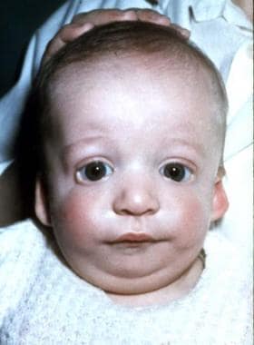
Anteroposterior view of 2-month-old boy with Treacher Collins syndrome.
-
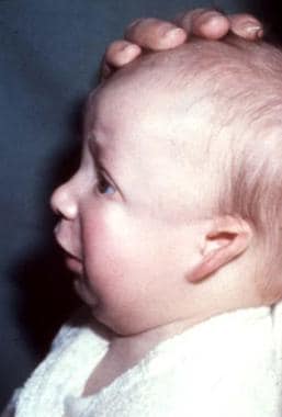
Lateral view of 2-month-old boy with Treacher Collins syndrome.
-
Anteroposterior view of 2-year-old boy with Treacher Collins syndrome.
-
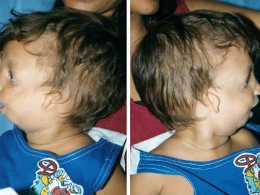
Lateral views of 2-year-old boy with Treacher Collins syndrome.
-
Anteroposterior view of 19-week-old fetus with Treacher Collins syndrome. Diagnosis was confirmed based on fetoscopy images. Tolarova M, Zwinger A. The use of fetoscopy by inborn morphological anomalies. Acta Chir Plast. 1981;23(3):139-51.
-
Lateral view of 19-week-old fetus with Treacher Collins syndrome. Diagnosis was confirmed based on fetoscopy images. Tolarova M, Zwinger A. The use of fetoscopy by inborn morphological anomalies. Acta Chir Plast. 1981;23(3):139-51.
-
Right-lateral cranial radiograph of an 18-month-old infant with Treacher Collins syndrome. Note the mandibular osteotomies and internal distraction hardware.
-
Left-lateral cranial radiograph showing mandibular osteotomy and distraction osteogeneses for micrognathia.
-
Lateral radiograph of same patient as in Media files 7 and 8. Completion of distraction; note increased mandibular length.
