Practice Essentials
Sarcoidosis is a systemic granulomatous disease that is relatively rare in children. The diagnosis is established when histopathologic evidence of noncaseating granulomata in affected organs and exclusion of other granulomatous diseases support compatible clinical and radiographic findings (see the image below). [1] Glucocorticoids are the treatment of choice for children with multisystem involvement.
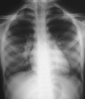 Chest radiograph showing bilateral hilar lymphadenopathy in a 10-year-old girl with sarcoidosis.
Signs and symptoms of pediatric sarcoidosis
The presentation in sarcoidosis can vary widely, depending on the extent and severity of organ involvement. In most children, the disease frequently involves the lungs, lymph nodes, eyes, skin, liver, and spleen.
Two distinct forms of childhood sarcoidosis are noted. Older children typically present with a multisystem disease similar to the adult manifestation, with frequent lymphadenopathy and pulmonary involvement, as well as generalized signs and symptoms, such as fever and malaise. In contrast, early onset childhood sarcoidosis occurs in patients who are younger than age 4 years and is characterized by the following triad:
-
Rash
-
Uveitis
-
Arthritis
See Presentation for more detail.
Diagnosis of pediatric sarcoidosis
Imaging studies
Imaging studies used in the workup of pediatric sarcoidosis include the following:
-
Chest radiography
-
High-resolution computed tomography
-
Gallium-67 scanning
Laboratory studies
No definitive laboratory test diagnostic of sarcoidosis has been identified.
Biopsy
Tissue biopsy is required for a definitive diagnosis. Biopsy specimens should be obtained from
-
Chest radiograph showing bilateral hilar lymphadenopathy in a 10-year-old girl with sarcoidosis.
-
Chest radiograph showing patchy diffuse pulmonary infiltrates involving both lung fields in a 12-year-old girl at onset of her sarcoidosis (left). A repeated study 6 months later showing almost complete resolution of the infiltrates (right).
