Practice Essentials
Panniculitis refers to a broad spectrum of diseases that involve inflammation of the subcutaneous fat layer of the skin. Weber-Christian disease is an eponym for a form of panniculitis, idiopathic nodular panniculitis, which is characterized by subcutaneous nodules, inflammatory cells in the fat lobules, and systemic symptoms. [1, 2] (See the image below.)
 Lesion of erythema nodosum: tender, erythematous, nodular lesions located over the extensor surfaces of the legs.
Signs and symptoms
Physical examination reveals erythematous, edematous, and tender subcutaneous nodules. Systemic symptoms of Weber-Christian disease include fever, malaise, nausea, vomiting, abdominal pain, weight loss, bone pain, myalgia, and arthralgia.
See Presentation for more detail.
Diagnosis
Laboratory studies
Changes in liver function test results, complete blood cell (CBC) count, and electrolyte levels reflect visceral involvement of organs. The erythrocyte sedimentation rate (ESR) and C-reactive protein (CRP) level are usually elevated.
Imaging studies
Chest radiography is used to exclude systemic inflammatory or infectious granulomatous diseases.
See Workup for more detail.
Management
No uniformly effective therapy for Weber-Christian disease is known. Clinical experience, especially in children and adolescents, has pointed to the value of corticosteroids and immunosuppressive agents.
See Treatment and Medication for more detail.
Background
The nomenclature of Weber-Christian disease and other related diseases is confusing, and some authors believe that the eponym should be abandoned and that more specific diagnoses should be made on the basis of pathogenesis, cause, or additional diagnostic testing. The history of the eponym began in 1892 when Pfeifer first described the skin condition now known as Weber-Christian disease, or idiopathic lobular panniculitis. In 1925, Weber further depicted the syndrome [3] , and Christian emphasized the significance of fever as part of the syndrome. The syndrome became known as Weber-Christian disease in 1928. [4, 5]
Increasing study and diagnostic sophistication have differentiated Weber-Christian disease from diseases such as lupus panniculitis, factitial panniculitis, panniculitis associated with pancreatic disease, histiocytic cytophagic panniculitis, and alpha1-antitrypsin deficiency panniculitis. [6] With further differentiation, additional diseases will be distinguished from Weber-Christian disease. A review of 30 Mayo Clinic patients diagnosed with Weber-Christian disease stated that: "Because separate and distinct forms of fat lesions have been described, we believe that the eponym should be abandoned and the more specific diagnoses should be made on the basis of pathogenesis or cause." [7] At this time, the eponym Weber-Christian disease still refers to cases of nodular panniculitis with systemic signs and symptoms that remain idiopathic.
Pathophysiology
Weber-Christian disease is a classic skin condition that features recurring inflammation in the subcutaneous fat layer of the skin. The involved skin areas manifest as recurrent crops of erythematous, sometimes tender, edematous, subcutaneous nodules. Lesions distribution is symmetric, and the thighs and lower legs are affected most frequently. Malaise, fever, and arthralgias often occur. Nausea, vomiting, abdominal pain, weight loss, hepatomegaly, and additional systemic features may also occur. The key pathologic finding on microscopy is a nodular inflammatory pattern of the fat lobules. Because the etiology is unknown, Weber-Christian disease is often referred to as idiopathic lobular panniculitis. [8, 9]
Etiology
Because its etiology is unknown, Weber-Christian disease is called idiopathic lobular panniculitis. Patients with Weber-Christian disease do not report a history of physical trauma.
In some patients with Weber-Christian disease, elevated levels of circulating immune complexes have been noted, suggesting an immunologically mediated reaction. [10]
Similarities between Weber-Christian disease and alpha1-antitrypsin deficiency suggest that an altered regulation of a normal inflammatory process may be involved. [5, 11] Responses to cyclosporine support a T-cell mediated inflammatory or autoinflammatory process. [12]
Infections and postviral infectious responses have been explored, and no connection has yet been established. [8, 13]
Epidemiology
United States statistics
Ambiguity surrounding Weber-Christian disease versus other closely related conditions makes it difficult to determine the frequency of the diagnosis. It is recognized as a rare condition in adults and even more rare in pediatrics. White and Winkelmann's 1998 case record review of Weber-Christian disease at the Mayo Clinic found only 30 cases in a 28 year span between 1960 and 1998. They observed that cases of primary panniculitis are rare and that physicians may see only one or a few cases in a lifetime. [7]
International statistics
The incidence and prevalence of Weber-Christian disease is unknown both in the United States and internationally. Weber-Christian disease is rare in adults and even rarer in children. For example, a chart review of children and adolescents in Brazil over a 20-year period (1983-2002) found 35 pediatric and adolescent cases of panniculitis, with only 6 cases meeting criteria for Weber-Christian disease.
Race-, sex-, and age-related demographics
No racial predilection is reported.
Weber-Christian disease occurs more often in women, who comprise approximately 75% of reported cases. The disease is rare in the pediatric population, with a slight predilection for females over males. In the Moraes and colleagues Brazilian study, 4 of the 6 cases of Weber-Christian disease were girls.
Weber-Christian disease may occur in children but is rare. It has been reported most frequently in people in the fourth to seventh decades of life, and 75% of cases occur in women after the second decade of life. [14]
Prognosis
Weber-Christian disease is a more serious form of panniculitis because of its systemic manifestations. The disease course varies, and prognosis depends on which organs are affected, the severity of organ involvement, and the response to therapy.
Weber-Christian disease may involve the lungs, [15] heart, intestines, spleen, kidney, adrenal glands, and even orbits. [16] Significant morbidity and mortality may occur in patients with inflammation involving visceral organs.
The clinical course in patients with only cutaneous manifestations may be characterized by exacerbations and remissions of the cutaneous lesions for several years before the disorder subsides.
Complications
Weber-Christian disease may involve the lungs, heart, intestines, spleen, kidney, and adrenal glands. Death may occur in patients with inflammation involving these critical visceral organs.
In patients with primarily cutaneous manifestations, the clinical course may be characterized by exacerbations and remissions of the cutaneous lesions for several years before the disorder subsides.
Patient Education
Inform patients of the risks and adverse effects of various treatment options. Select different treatment modalities on an individual basis.
-
Lesion of erythema nodosum: tender, erythematous, nodular lesions located over the extensor surfaces of the legs.
-
Standard posteroanterior chest radiograph reveals extensive bilateral hilar and mediastinal lymph node enlargement not associated with a pulmonary abnormality in a patient with sarcoidosis.
-
Young male patient with fever and cough has a focal opacity in the left lower lobe that looks like a pneumonia. This is a case of primary tuberculosis.
-
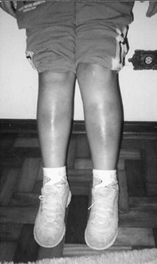
Lesion of Weber-Christian disease: tender, erythematous, nodular lesions located over the limbs with cutaneous atrophy.
-
A portion of skin is examined in multiple sections and at various magnifications. The epidermis is intact; however, it is infiltrated by small numbers of lymphocytes. A mild infiltrate of lymphocytes and histiocytes are present in the upper dermis. The most prominent change is in the subcutaneous tissue, where a prominent infiltrate of histiocytes, smaller numbers of lymphocytes, and a few plasma cells in the subcutaneous adipose tissue are noted. Occasional foam cells are also evident, and, in places, histocytes surround lipid cysts. Small clusters of necrotic cells and scattered nuclear dust are noted. Minimal extension of this infiltrate into adjacent dense collagenous tissue is observed. (Courtesy of Milton J. Finegold, MD, Professor of Pathology and Pediatrics, Baylor College of Medicine, Houston, TX).
-
Magnification of previous specimen X 100.
-
Magnification of previous specimen X 200.
-
Histopathologic features of alpha-1-antitrypsin deficiency panniculitis. (A) Scanning power shows a mostly lobular panniculitis. (B) Aggregations of neutrophils within the fat lobule are seen. (C) Neutrophils are interstitially arranged between collagen bundles of the deep reticular dermis. (A-C, hematoxylin-eosin stain; original magnifications: A, X 20; B, X 400; C, X 200).
-
Histopathologic features of late stage lesions of traumatic panniculitis. This lesion corresponds to the so-called nodular cystic fat necrosis or mobile encapsulated lipoma. A, Scanning power shows encapsulated and well-circumscribed lesion with no inflammatory infiltrate (arrow indicates area enlarged in B). B, At periphery of the lesion necrotic adipocytes appear as anucleated fat cells. (A and B, Hematoxylin-eosin stain; original magnifications: A, ×20; B, ×200.)
-
Histopathologic features of paraffinoma. A, Scanning power shows a mostly lobular panniculitis (arrow indicates area enlarged in B). B, Higher magnification demonstrates cystic spaces within the fat lobule surrounded by foamy histiocytes. (A and B, hematoxylin-eosin stain; original magnifications: A, ×20; B, ×200.)
-
Histopathologic features of subcutaneous fat necrosis of the newborn. (A) Scanning power shows a mostly lobular panniculitis (arrow indicates area enlarged in B). (B) Higher magnification demonstrated narrow needle-shaped clefts radially arranged and surrounded by histiocytes. (A and B, hematoxylin-eosin stain; original magnifications: A, X 20; B, X 200).
-
Histopathologic features of lipoatrophy secondary to subcutaneous injections of corticosteroids. (A) Low-power view showed small fat lobules (arrow indicates area enlarged in B). (B) Higher magnification demonstrates small adipocytes and prominent capillary proliferation, resembling embryonic fat. (A and B, hematoxylin-eosin stain; original magnifications: A, X 20; B, X 200).
