You are in: eMedicine Specialties >
Pediatrics: Cardiac Disease and Critical Care Medicine > Cardiology
Ventricular Septal Defect, Supracristal
Article Last Updated: Oct 10, 2008
AUTHOR AND EDITOR INFORMATION
Section 1 of 11
Author: Edward J Bayne, MD, Assistant Professor, Division of Pediatric Cardiology, Emory University School of Medicine; Consulting Staff, Sibley Heart Center Cardiology, Children's Healthcare of Atlanta
Edward J Bayne is a member of the following medical societies: Alpha Omega Alpha, American Academy of Pediatrics, American College of Cardiology, American Heart Association, and American Society of Echocardiography
Editors: Juan Carlos Alejos, MD, Associate Clinical Professor, Department of Pediatrics, Division of Cardiology, University of California at Los Angeles; Mary L Windle, PharmD, Adjunct Assistant Professor, University of Nebraska Medical Center College of Pharmacy, Pharmacy Editor, eMedicine; Hugh D Allen, MD, Professor, Department of Pediatrics, Division of Pediatric Cardiology and Department of Internal Medicine, Ohio State University College of Medicine; Gilbert Herzberg, MD, Assistant Professor, Department of Pediatrics, Section of Pediatric Cardiology, New York Medical College; Stuart Berger, MD, Professor of Pediatrics, Division of Cardiology, Medical College of Wisconsin; Chief of Pediatric Cardiology, Medical Director of Pediatric Heart Transplant Program, Medical Director of The Heart Center, Children's Hospital of Wisconsin
Author and Editor Disclosure
Synonyms and related keywords:
supracristal ventricular septal defect, doubly committed ventricular septal defect, doubly-committed VSD, subarterial ventricular septal defect, juxtaarterial ventricular septal defect, subpulmonic ventricular septal defect, conal ventricular septal defect, type I ventricular septal defect, supracristal VSD, outlet VSD, arch abnormalities, aortic insufficiency, cardiogenic shock, exercise intolerance, dyspnea, left ventricular outflow obstruction, patent ductus arteriosus, arteriovenous malformation, arteriovenous fistula
Background
Supracristal (or doubly committed) ventricular septal defect (VSD) is the least common type of VSD in the western hemisphere, accounting for approximately 5-7% of defects in the western hemisphere.1 Its location adjacent to the pulmonary and aortic valves accounts for the unique natural history associated with this defect. The spiraling course of the ventricular septum may make diagnosis of the supracristal VSD more difficult. Definition The crista supraventricularis can be considered synonymous with the infundibular (or conus) ventricular septum. It is the portion of the septum that separates the tricuspid and pulmonary valves. Defects in this part of the septum are generally referred to as supracristal defects. The term is generally reserved for defects lying immediately under the pulmonary valve, so that aortic and pulmonary valve tissue are in fibrous continuity and not separated by septal tissue. Embryology The muscular outlet septum is primarily formed from the proximal endocardial ridges (similar to endocardial cushion tissue). Semilunar valve tissue and the actual connection between the septum and the arteries is formed by the more distal endocardial ridges. Extracardiac mesenchyme, derived from neural crest tissue, condenses as prongs (which act as a welding agent) with the most superior portion of the distal cushions to form the aortopulmonary septum.2 By exposing neural crest tissue to homocysteine, supracristal VSDs have been induced in a high percentage of chick embryos. Disruption of apoptosis and myocardialization has been proposed to explain these findings.3 The frequent association between arch abnormalities and significant conal VSDs suggests a common mechanism involving a chromosome band 22q11 microdeletion. Deletions in this area have not been linked with isolated supracristal VSDs.4 Anatomy The infundibular (or conus) septum separates the tricuspid and pulmonary valves and accounts for the more superior placement of the pulmonary valve relative to the aortic valve. This portion of the septum also provides muscular fairly rigid support for the aortic valve, especially the right coronary cusp.5 Numerous synonyms indicate the confusion often associated with describing this particular type of ventricular defect. The term supracristal may be misleading because the entire conus septum (or a major portion of the septum) may be missing. However, the term is commonly used and underscores the superior location of the defect along with the close approximation of the aortic and pulmonary valve leaflets. The lack of support for the right aortic leaflet is crucial to the natural history of this type of VSD.6 The plane of the conus septum in the right ventricular outflow tract lies almost perpendicular to that of the remainder of the septum. From a surgical perspective, a defect lying in the conus septum may not be visualized from the standard right atriotomy approach, looking through the tricuspid valve.5 7 Unlike the more common perimembranous type of VSD, supracristal VSD does not lie near the tricuspid valve. Unless the supracristal defect is large, extending inferiorly to the perimembranous septum, the tricuspid valve is not involved in partial closure of the defect. Conduction system tissue lies inferior to the supracristal VSD. The conduction system may lie closer to a larger defect that crosses from the outlet septum into the perimembranous area. Natural history The natural history of supracristal VSDs depends on the location and size of the defect. Patients with small isolated supracristal VSDs may have no early symptoms or signs of congestive failure such as would be observed with a large shunt. Progressive aortic insufficiency may develop late in the first decade of life. However, larger defects of the outlet septum frequently are associated with severe forms of aortic outflow obstruction (eg, coarctation, interrupted aortic arch). In such cases, symptoms of congestive heart failure and possible circulatory collapse appear early. Patients with larger isolated supracristal VSDs may show early signs of decompensation from a large left-to-right shunt. Because these defects are not surrounded by muscular tissue, spontaneous closure is less common.8 However, the defect may decrease in size by progressive prolapse of aortic valve tissue (the right coronary cusp or, possibly, the right sinus of Valsalva).9 This valve prolapse is believed to result from negative pressure by shunt flow because of the Venturi effect. This progressive distortion of the aortic leaflet or sinus may lead to increasing aortic valve insufficiency or to formation of an aneurysm in the sinus of Valsalva.
Pathophysiology
The unique location of the supracristal ventricular septal defect with its close proximity to the aortic root accounts for the common development of aortic insufficiency with this defect. Left-to-right shunting of blood through the defect is believed to progressively pull aortic valve tissue (especially the right coronary cusp) through a Venturi effect.
Frequency
United States
VSDs account for approximately 25-30% of significant congenital heart disease. Of these, approximately 5-7% are supracristal VSDs. These rates are similar throughout the Western Hemisphere.
International
Supracristal VSDs are much more common in persons of Asian descent than in individuals of other races. Supracristal VSDs account for approximately 25% of all VSDs in patients from the Eastern Hemisphere.
Mortality/Morbidity
Morbidity/mortality is generally not the result of large left-to-right shunt. Rather, it is caused by progressive development of aortic valve insufficiency, with development of left ventricular enlargement and eventual congestive heart failure if the problem is not addressed early enough.
Race
Although the overall incidence of VSDs is no higher in Asians than in other groups, supracristal VSDs account for approximately 30% of VSDs in Asians1 and only approximately 5% of VSDs in other groups. The higher occurrence of supracristal VSDs in Asians has not been adequately explained.
Sex
No predilection based on sex is observed.
History
In patients with supracristal ventricular septal defects (VSDs), symptoms and severity are a function of size and location of the defect, the relative systemic and pulmonary vascular resistances, and presence of associated abnormalities. Symptoms may range from severe congestive failure and cardiogenic shock in patients with large conal defects and left heart obstruction to complete absence of symptoms in patients with small isolated defects. Exercise intolerance and dyspnea suggest progressive aortic insufficiency, although early detection and treatment for valve insufficiency should obviate any significant symptoms.
Physical
Because congestive heart failure is unusual in the patient with a small supracristal VSD, general examination findings should be normal, with no signs of respiratory distress or growth failure. Infants with larger defects, especially with associated left ventricular outflow obstruction (eg, doubly committed subarterial defect with interrupted aortic arch), may present within the first week of life with profound congestive heart failure and cardiogenic shock.
- The murmur from a supracristal VSD is systolic and located at the upper left sternal border (ie, second or third intercostal space). The murmur is often crescendo-decrescendo in character, unlike the holosystolic regurgitant murmur of VSDs in other locations of the septum. It may radiate laterally and posteriorly because of shunt flow directed into the pulmonary outflow tract.10
- When a patient is known to have a supracristal VSD, the examination should focus on whether aortic insufficiency is present. A high-pitched diastolic murmur beginning with the second heart sound may be heard along the left sternal border. The combined systolic and diastolic murmurs may be likened to the sound of sawing wood.
- The systolic-diastolic murmur of supracristal VSD with aortic insufficiency may be confused with a continuous murmur (eg, patent ductus arteriosus, arteriovenous malformation or fistula).
- Maneuvers to increase the diastolic murmur of aortic insufficiency include isometric handgrip, leaning forward in a sitting position, and holding the breath during expiration.10
- With increasing severity of aortic insufficiency, pulse pressure (ie, difference between systolic and diastolic blood pressures) and pulse intensity may increase.
Aortic Valve Insufficiency
Aortic Valve, Bicuspid
Aortopulmonary Septal Defect
Arteriovenous Fistulae, Pulmonary
Coronary Artery Fistula
Patent Ductus Arteriosus
Pulmonary Stenosis, Valvar
Sinus of Valsalva Aneurysm
Ventricular Septal Defect, Perimembranous
Imaging Studies
- Chest radiography
- Chest radiography may be normal in infancy if no large left-to-right shunt is present. If a large shunt is present, cardiomegaly (left heart enlargement, both left atrium and left ventricle) with increased pulmonary vascularity from increased pulmonary blood flow may be observed.
- Radiography in the older child or adult with progressive aortic insufficiency may reveal left heart enlargement (particularly left ventricular enlargement) and prominence of the ascending aorta. Shunt volume is generally smaller, thus pulmonary vascularity is generally normal unless advanced left heart failure produces pulmonary edema.
- Two-dimensional transthoracic echocardiography and color Doppler echocardiography
- Echocardiography (ECHO) provides the most efficient means to diagnose supracristal VSD accurately and the most effective means to monitor progressive aortic insufficiency.9 An accurate diagnosis can generally be made in infants and children with standard transthoracic ECHO examination findings. In the older child and adult, transthoracic ECHO findings may be inconclusive; in such cases, transesophageal ECHO may be extremely helpful.
- Two-dimensional imaging reveals the supracristal VSD in the parasternal short-axis view or the modified apical 3-chamber view (ie, left atrium, left ventricle, aortic root, and pulmonary root, equivalent to the transesophageal view with transducer at 90°). The defect can also be observed well in the subcostal parasagittal view (ie, visualizing the pulmonary and aortic outflow tracts). However, the supracristal VSD cannot be imaged from the apical 4-chamber view because of the orientation of the outlet septum. Distortion of the right aortic leaflet may be the only clue to the presence of a significant supracristal VSD because the aortic leaflet may "plug" the defect.
- Color Doppler examination, using the parasternal short-axis view, reveals left-to-right shunting with turbulent flow directed into the pulmonary outflow tract and often across the pulmonary valve. This turbulence may be confused with pulmonary stenosis; however, careful slow-motion review of color flow results (with electrocardiographic timing) may reveal early appearance of turbulent flow below the pulmonary valve.
- The best way to detect aortic insufficiency is by color Doppler in the parasternal long-axis and the apical 5-chamber views. The modified apical 3-chamber view can also be used to detect left-to-right shunting and aortic insufficiency. Numerous methods are available to provide semiquantitative information on the severity of aortic insufficiency (eg, color jet–to–outflow width ratio, pressure half time).
- The best way to identify progression of aortic insufficiency is by serial comparison of left ventricular systolic and diastolic dimensions and ventricular function (shortening fraction or ejection fraction). Progressive left atrial enlargement can be a sign of ventricular diastolic dysfunction.
- Three-dimensional echocardiography
- Three-dimensional echocardiographic imaging of VSDs closely correlates with surgical findings, although specific findings with supracristal defects have not been reported.11
- Three-dimensional echocardiography may prove useful in differentiating supracristal VSD from unruptured sinus of Valsalva aneurysm.12
- Angiography13
- The supracristal VSD is best defined in the right anterior oblique projection or the cranially tilted left anterior oblique projection. Small supracristal defects may not be identified in the standard long-axial oblique projection because of rotation of the septum. Direct continuity between the aortic and pulmonary valves is the hallmark of the supracristal type of ventricular defect. Larger defects may extend inferiorly into the perimembranous septum and sometimes through to the inlet septum.
- Distortion of the aortic valve cusp or root may be the only clue to a significant supracristal VSD, and the apparent volume of left-to-right shunt into the pulmonary outflow tract may be small.
- MRI: MRI may be used with appropriate projections and alignment to show the pulmonary outflow tract.14 Serial MRI studies can be helpful in that they do not expose the patient to ionizing radiation. Blood flow studies can be used to provide quantitative information on regurgitant volume in the assessment of aortic insufficiency.
Other Tests
- ECG findings may be normal in infancy because the defect may not be large enough to cause a significant left-to-right shunt and ventricular hypertrophy. With larger defects, the ECG may show left atrial enlargement and left and right ventricular hypertrophy.
- With progressive aortic insufficiency in the older child or adult, ECG usually reveals evidence of left heart enlargement from volume overload (ie, left atrial enlargement and left ventricular hypertrophy [tall R waves in the left precordium with or without ST-T changes]).
Procedures
- Cardiac catheterization is helpful in quantifying shunt volume and pressure resistance.13 Step-ups in oxygen saturation are often detected in the pulmonary artery rather than in the right ventricular cavity. This may be because of streaming of the shunted blood into the pulmonary outflow tract.
- If aortic valve prolapse is significant, left-to-right shunting by oximetry may be fairly unremarkable because the VSD in such cases is partially closed.
- Postcatheterization precautions include hemorrhage, vascular disruption after balloon dilation, pain, nausea and vomiting, and arterial or venous obstruction from thrombosis or spasm.
- Possible complications include rupture of blood vessel, tachyarrhythmias, bradyarrhythmias, and vascular occlusion.
Medical Care
Once the diagnosis of supracristal ventricular septal defect (VSD) has been made, carefully monitor patients for development of aortic insufficiency. This necessitates not only periodic physical examination with auscultation but also serial echocardiograms and Doppler echocardiograms because these diagnostic studies are more sensitive than auscultation in detecting valve regurgitation.
Surgical Care
Because spontaneous closure is uncommon in supracristal VSDs, and because aortic insufficiency is a common complication, surgical closure is recommended in most cases. Aortic insufficiency in supracristal VSD is usually progressive and warrants an aggressive approach with early intervention to avoid aortic valve deformity and replacement. Aortic insufficiency caused by supracristal VSD must be differentiated from that caused by an abnormal aortic valve (usually a bicuspid valve); surgical intervention is usually delayed in the latter disorder because the abnormal aortic valve typically requires replacement rather than repair in cases of aortic insufficiency. Because of the orientation of the right ventricular outflow tract, the standard surgical approach from the right atrium does not allow adequate visualization of the ventricular defect.15, 16 Incision into the main pulmonary artery, which exposes the defect through the pulmonic valve, has proved successful. Repair may be achieved with patch or suture closure, depending on the size of the defect. Aortic valvuloplasty is often, but not always, necessary, and incision through the aortic root can allow adequate visualization for valve repair (Trusler technique). The approach through the main pulmonary artery avoids the need for incision into the right ventricle. Care should be taken to avoid capturing the aortic cusp into one of the patch sutures. Intraoperative transesophageal echocardiographic monitoring can be extremely helpful in precisely defining aortic valve prolapse and the severity of valve insufficiency, which determine the necessity of valvuloplasty. Follow-up intraoperative assessment should be used to rule out residual insufficiency.17 More extensive damage to the aortic valve from long-standing prolapse and distortion may require valve replacement.
Diet
No special dietary guidelines or restrictions are necessary.
Activity
Activity level is determined by the age at which signs or symptoms develop. Infants with large left-to-right shunts, particularly with complex left heart obstruction, may have delays in development of gross motor skills that naturally may restrict activity level. Patients with small left-to-right shunts without aortic valve insufficiency or with only trivial aortic valve insufficiency generally should be allowed full activity without restriction. Older patients with more significant aortic valve insufficiency should be restricted from competitive athletics and from sustained isometric types of activities (eg, weight lifting, rope pulls, sustained heavy lifting on the job).
For patients who develop aortic valve insufficiency, surgical closure of the ventricular septal defect (VSD) and repair of valve insufficiency is the preferred treatment. If surgical repair must be postponed, afterload reducers, such as ACE inhibitors or calcium channel blockers, have proved helpful in adults and children.18
Drug Category: ACE inhibitors
These agents have proved beneficial in long-term therapy for aortic valve insufficiency. Positive effects include reduction in pulse pressure, reduction in regurgitant volume, reduction in left ventricular volume, and reduction in left ventricular mass (because of beneficial effects on ventricular remodeling).
| Drug Name | Enalapril (Vasotec) |
| Description | Reasonable first DOC in this group because of increased dosing interval (q12-24h). Competitive inhibitor of ACE. Reduces angiotensin II levels, decreasing aldosterone secretion. Available in liquid suspension. |
| Adult Dose | 2.5-5 mg PO qd or divided bid initial dose; not to exceed 40 mg/d |
| Pediatric Dose | 0.1 mg/kg/d PO qd or divided bid initial dose; may increase dose gradually to 0.5 mg/kg PO qd or divided bid |
| Contraindications | Documented hypersensitivity |
| Interactions | NSAIDs may reduce hypotensive effects of enalapril; ACE inhibitors may increase digoxin, lithium, and allopurinol levels; rifampin decreases enalapril levels; probenecid may increase enalapril levels; the hypotensive effects of ACE inhibitors may be enhanced when given concurrently with diuretics |
| Pregnancy | C - Fetal risk revealed in studies in animals but not established or not studied in humans; may use if benefits outweigh risk to fetus
|
| Precautions | Use caution in bilateral renal artery stenosis, renal impairment, valvular stenosis, or severe congestive heart failure |
| Drug Name | Lisinopril (Prinivil, Zestril) |
| Description | Reasonable first DOC in this group because of increased dosing interval (q12-24h). Prevents conversion of angiotensin I to angiotensin II, a potent vasoconstrictor, resulting in lower aldosterone secretion. |
| Adult Dose | 10 mg/d PO; increase 5-10 mg/d at 1- to 2-wk intervals; not to exceed 40 mg |
| Pediatric Dose | 0.1 mg/kg/d PO qd or divided bid initially; may increase dose gradually, not to exceed 0.5 mg/kg PO qd or divided bid |
| Contraindications | Documented hypersensitivity |
| Interactions | Lisinopril may increase digoxin, lithium, and allopurinol levels; probenecid may increase lisinopril levels; coadministration with diuretics increase hypotensive effects; hypotensive effects of lisinopril may be enhanced when given concurrently with diuretics and NSAIDs |
| Pregnancy | C - Fetal risk revealed in studies in animals but not established or not studied in humans; may use if benefits outweigh risk to fetus
|
| Precautions | Caution in renal impairment, valvular stenosis, or severe congestive heart failure |
| Drug Name | Captopril (Capoten) |
| Description | Prevents conversion of angiotensin I to angiotensin II, a potent vasoconstrictor, resulting in lower aldosterone secretion. For younger children or infants, can be formulated as a suspension if stabilized with ascorbic acid to prevent hydrolysis. Dosing interval is 6-8 h because of shorter half-life. |
| Adult Dose | 6.25-12.5 mg PO tid; may gradually titrate upward, not to exceed 150 mg tid |
| Pediatric Dose | 0.25-1 mg/kg/dose PO q8h; initiate at lower dose and gradually titrate upward if needed |
| Contraindications | Documented hypersensitivity; renal impairment |
| Interactions | NSAIDs may reduce hypotensive effects of captopril; ACE inhibitors may increase digoxin, lithium, and allopurinol levels; rifampin decreases captopril levels; probenecid may increase captopril levels; the hypotensive effects of ACE inhibitors may be enhanced when given concurrently with diuretics |
| Pregnancy | C - Fetal risk revealed in studies in animals but not established or not studied in humans; may use if benefits outweigh risk to fetus
|
| Precautions | Caution in renal impairment, valvular stenosis, or severe congestive heart failure |
Drug Category: Calcium channel blockers
Calcium channel blockers also have proved effective in reducing afterload and reducing pulse pressure and regurgitant volume. Prolonged regular use may stabilize left ventricular volume, but the effect on left ventricular muscle mass is less pronounced than that of ACE inhibitors.
| Drug Name | Nifedipine (Adalat, Procardia) |
| Description | Good first choice because of its primary action on peripheral resistance and limited effect on cardiac function and heart rate. |
| Adult Dose | 10-30 mg IR cap PO tid; not to exceed 120-180 mg/d 30-60 mg SR tab PO qd; not to exceed 90-120 mg/d |
| Pediatric Dose | 0.25-0.5 mg/kg/dose PO/SL tid/qid prn; may increase to 0.9 mg/kg PO/SL q6-8h; not to exceed 10 mg/dose or 3 mg/kg/d |
| Contraindications | Documented hypersensitivity |
| Interactions | Caution with coadministration of any agent that can lower BP, including beta-blockers and opioids; H2 blockers (eg, cimetidine) may increase toxicity |
| Pregnancy | C - Fetal risk revealed in studies in animals but not established or not studied in humans; may use if benefits outweigh risk to fetus
|
| Precautions | Use caution in congestive heart failure; may abruptly decrease blood pressure and produce postural hypotension, dizziness, or syncope; other adverse effects include peripheral edema, severe tachycardia, headache, and palpitations |
Complications
- Infective endocarditis
- Patients with supracristal ventricular septal defect (VSD) are at increased risk of infective endocarditis.
- The risk is relatively high if aortic valve insufficiency is present.
- For more information, see Antibiotic Prophylactic Regimens for Endocarditis.
- Aortic insufficiency
- The appearance of aortic insufficiency as a complication of supracristal VSD is related to age.
- Young infants and toddlers presenting with supracristal VSDs are more likely to have findings of left-to-right shunt only.
- Onset of aortic valve prolapse and progressive aortic insufficiency generally begins in children aged 6-10 years, although aortic insufficiency may develop even in infants.
Prognosis
- Prognosis should be considered good to excellent when the potential complication of aortic valve insufficiency is recognized and aggressively treated.19, 20
- Delayed recognition of or surgical treatment for progressive aortic valve insufficiency may lead to severe distortion of the aortic valve leaflet, making valve replacement more likely.21
Patient Education
- The risk for infective endocarditis is higher for supracristal VSD with aortic insufficiency than for "simple" VSD alone. Patients and families should be educated on the importance of good oral and dental hygiene. Routine prophylaxis for dental or surgical procedures is no longer recommended unless there has been a prior episode of endocarditis.22
- For excellent patient education resources, visit eMedicine's Heart Center. Also, see eMedicine's patient education article Ventricular Septal Defect.
Medical/Legal Pitfalls
- Failure to appreciate the unique natural history of supracristal ventricular septal defect (VSD) compared to other types of VSD
- VSDs less likely to close spontaneously
- VSDs more likely to be complicated by aortic insufficiency
- Delay in acting on aortic valve deformity and progressive aortic insufficiency with the goal of avoiding aortic valve replacement
- Failure to continue follow-up care after supracristal VSD repair and aortic valvuloplasty because aortic insufficiency may not be corrected completely
- Failure to diagnose supracristal VSD adequately before surgery because this type of VSD usually is not seen adequately from the standard right atrial surgical approach
Special Concerns
- Recurrence risk for offspring of mothers with supracristal VSD is estimated at 4-5%. Recurrence risk for fathers is approximately 2-3%. Detailed prenatal fetal echocardiography (ECHO) may be indicated. Supracristal VSD cannot be identified from a routine prenatal 4-chamber view.
| Media file 1:
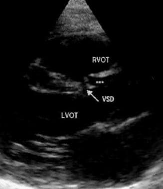
Parasternal long-axis echocardiogram view showing supracristal ventricular septal defect (arrow) with buckling and prolapse (***) of the right coronary leaflet of the aorta. |
 | View Full Size Image | |
Media type: Echo
|
| Media file 2:
Parasternal short-axis echocardiogram view with color Doppler showing proximity of ventricular septal defect jet to the aortic and pulmonic valves. The patient is an infant who has not yet developed aortic valve prolapse or aortic insufficiency. |
 | View Full Size Image | |
Media type: Echo
|
| Media file 3:
Subcostal "right ventricular inflow/outflow" view showing the close relationship between the aortic and pulmonic valves in the presence of supracristal ventricular septal defect. Turbulent shunt flow is shown directed into the main pulmonary artery. The patient is an infant who has not yet developed aortic valve prolapse and insufficiency. |
 | View Full Size Image | |
Media type: Echo
|
| Media file 4:
Transesophageal horizontal view of aortic root and right ventricle, showing sinus of Valsalva aneurysm leaking through a supracristal ventricular septal defect (VSD)(> <). Lack of supporting tissue in VSD may be one of the mechanisms involved in producing sinus of Valsalva aneurysms. |
 | View Full Size Image | |
Media type: Echo
|
- McDaniel NL, Gutgesell HP. Ventricular septal defects. In: Allen HD, Driscoll DJ, Shaddy RE, Feltes TF. Moss and Adams' Heart Disease in Infants, Children, and Adolescents. 7th ed. Philadelphia: Wolters Kluwer/ Lippincott Williams & Wilkins; 2008:667-682.
- Gittenberger-De Groot AC, Poelmann RE. Normal and abnormal cardiac development. In: Moller JH, Hoffman JI, eds. Pediatric Cardiovascular Medicine. New York, NY: Churchill Livingstone; 2000:3-20.
- Boot MJ, Steegers-Theunissen RP, Poelmann RE, van Iperen L, Gittenberger-de Groot AC. Cardiac outflow tract malformations in chick embryos exposed to homocysteine. Cardiovasc Res. Nov 1 2004;64(2):365-73. [Medline].
- Momma K, Ando M, Matsuoka R, Joo K. Interruption of the aortic arch associated with deletion of chromosome 22q11 is associated with a subarterial and doubly committed ventricular septal defect in Japanese patients. Cardiol Young. Sep 1999;9(5):463-7. [Medline].
- Anderson RH, Wilcox BR. The surgical anatomy of ventricular septal defects associated with overriding valvar orifices. J Card Surg. Mar 1993;8(2):130-42. [Medline].
- Cheung YF, Chiu CS, Yung TC. Impact of preoperative aortic cusp prolapse on long-term outcome after surgical closure of subarterial ventricular septal defect. Ann Thorac Surg. Feb 2002;73(2):622-7. [Medline].
- Ho SY, Baker EJ, Rigby ML. Color Atlas of Congenital Heart Disease: Morphologic and Clinical Correlations. St Louis, MO: Mosby-Wolfe; 1995.
- Eroglu AG, Oztunc F, Saltik L, et al. Evolution of ventricular septal defect with special reference to spontaneous closure rate, subaortic ridge and aortic valve prolapse. Pediatr Cardiol. Jan-Feb 2003;24(1):31-5. [Medline].
- Mori K, Matsuoka S, Tatara K, et al. Echocardiographic evaluation of the development of aortic valve prolapse in supracristal ventricular septal defect. Eur J Pediatr. Mar 1995;154(3):176-81. [Medline].
- Zuberbuhler JR. Ventricular septal defect. In: Clinical Diagnosis in Pediatric Cardiology. London, England: Churchill Livingstone; 1981:39-45.
- Cheng TO, Xie MX, Wang XF, Wang Y, Lu Q. Real-time 3-dimensional echocardiography in assessing atrial and ventricular septal defects: an echocardiographic-surgical correlative study. Am Heart J. Dec 2004;148(6):1091-5. [Medline].
- Masaki N, Iwatsuka R, Nagahori W, et al. Three-dimensional echocardiography could distinguish a ventricular septal defect adjacent to asymptomatic ruptured sinus of valsalva aneurysm. J Cardiol. Apr 2008;51(2):139-43. [Medline].
- Freedom RM, Mawson JB, Yoo SJ. Ventricular septal defect. In: Freedom RM, et al, eds. Congenital Heart Disease: Textbook of Angiocardiography. Futura Publishing Co; 1997:189-218.
- Wang ZJ, Reddy GP, Gotway MB, et al. Cardiovascular shunts: MR imaging evaluation. Radiographics. Oct 2003;23 Spec No:S181-94. [Medline]. [Full Text].
- Yacoub MH, Khan H, Stavri G, et al. Anatomic correction of the syndrome of prolapsing right coronary aortic cusp, dilatation of the sinus of Valsalva, and ventricular septal defect. J Thorac Cardiovasc Surg. Feb 1997;113(2):253-60; discussion 261. [Medline].
- Komai H, Naito Y, Fujiwara K, Noguchi Y, Nishimura Y, Uemura S. Surgical strategy for doubly committed subarterial ventricular septal defect with aortic cusp prolapse. Ann Thorac Surg. Oct 1997;64(4):1146-9. [Medline].
- Leung MP, Chau KT, Chiu C, Yung TC, Mok CK. Intraoperative TEE assessment of ventricular septal defect with aortic regurgitation. Ann Thorac Surg. Mar 1996;61(3):854-60. [Medline].
- Levine HJ, Gaasch WH. Vasoactive drugs in chronic regurgitant lesions of the mitral and aortic valves. J Am Coll Cardiol. Nov 1 1996;28(5):1083-91. [Medline].
- Leung MP, Beerman LB, Siewers RD, et al. Long-term follow-up after aortic valvuloplasty and defect closure in ventricular septal defect with aortic regurgitation. Am J Cardiol. Oct 1 1987;60(10):890-4. [Medline].
- Rhodes LA, Keane JF, Keane JP, et al. Long follow-up (to 43 years) of ventricular septal defect with audible aortic regurgitation. Am J Cardiol. Aug 1 1990;66(3):340-5. [Medline].
- Elgamal MA, Hakimi M, Lyons JM, Walters HL III. Risk factors for failure of aortic valvuloplasty in aortic insufficiency with ventricular septal defect. Ann Thorac Surg. Oct 1999;68(4):1350-5. [Medline].
- Wilson W, Taubert KA, Gewitz M, et al. Prevention of infective endocarditis: guidelines from the American Heart Association: a guideline from the American Heart Association Rheumatic Fever, Endocarditis, and Kawasaki Disease Committee, Council on Cardiovascular Disease in the Young, and the Council on Clinical Cardiology, Council on Cardiovascular Surgery and Anesthesia, and the Quality of Care and Outcomes Research Interdisciplinary Working Group. Circulation. Oct 9 2007;116(15):1736-54. [Medline]. [Full Text].
Ventricular Septal Defect, Supracristal excerpt Article Last Updated: Oct 10, 2008
|