You are in: eMedicine Specialties >
Pediatrics: General Medicine > Oncology
Acute Lymphoblastic Leukemia
Article Last Updated: Jul 11, 2006
AUTHOR AND EDITOR INFORMATION
Section 1 of 11
Author: Noriko Satake, MD, Clinical Fellow, Department of Pediatric Hematology-Oncology, Mattel Children's Hospital at University of California at Los Angeles
Coauthor(s):
Kathleen Sakamoto, MD, Professor, Department of Pediatrics, Mattel Children's Hospital, David Geffen School of Medicine, Division of Hematology-Oncology and Pathology and Laboratory Medicine, University of California at Los Angeles
Editors: Stephan A Grupp, MD, PhD, Director, Stem Cell Biology Program, Department of Pediatrics, Division of Oncology, Children's Hospital of Philadelphia; Associate Professor of Pediatrics, University of Pennsylvania; Mary L Windle, PharmD, Adjunct Assistant Professor, University of Nebraska Medical Center College of Pharmacy, Pharmacy Editor, eMedicine.com, Inc; Timothy P Cripe, MD, PhD, Associate Professor of Pediatric Hematology/Oncology, University of Cincinnati; Director, Translational Research Trials Office, Department of Pediatrics, Cincinnati Children's Hospital Medical Center; Samuel Gross, MD, Professor Emeritus, Department of Pediatrics, University of Florida, Clinical Professor, Department of Pediatrics, UNC, Adjunct Professor, Department of Pediatrics, Duke University; Robert J Arceci, MD, PhD, King Fahd Professor of Pediatric Oncology, Department of Oncology, Division of Pediatric Oncology, Johns Hopkins University School of Medicine
Author and Editor Disclosure
Synonyms and related keywords:
acute lymphocytic leukemia, acute lymphatic leukemia, acute lymphoid leukemia, ALL, pediatric cancer, childhood cancer, childhood malignancy, inherited genetic syndromes, lymphoblastic leukemia, leukemia, leukemic blasts, Tcell, T-cell ALL, B cell, B-lineage ALL, BCR-ABL, MLL, high-risk ALL, exposure to ionizing radiation, exposure to electromagnetic fields, allogeneic hematopoietic stem cell transplantation, HSCT, bone marrow failure, anemia, thrombocytopenia, neutropenia, petechiae, bleeding, lymphadenopathy, hepatosplenomegaly, bone pain
Background
Acute lymphoblastic leukemia (ALL) is the most common malignancy diagnosed in children, representing nearly one third of all pediatric cancers. The annual incidence of ALL is about 30 cases per million people, with a peak incidence in children aged 2-5 years. Although a few cases are associated with inherited genetic syndromes, the cause of ALL remains largely unknown.
Many environmental factors (eg, exposure to ionizing radiation and electromagnetic fields, parental use of alcohol and tobacco) have been investigated as potential risk factors, but none has been definitively shown to cause lymphoblastic leukemia. Improvements in diagnosis and treatment have produced cure rates that now exceed 80%.
Further refinements in therapy, including the use of risk-adapted treatment protocols, may improve cure rates for patients at high risk while limiting the toxicity of therapy for patients with a low risk of relapse (see Risk classification). This article summarizes advances in the diagnosis and treatment of childhood ALL.
Pathophysiology
In ALL, a lymphoid progenitor cell becomes genetically altered and subsequently undergoes dysregulated proliferation and clonal expansion. In most cases, the pathophysiology of transformed lymphoid cells reflects the altered expression of genes whose products contribute to the normal development of B cells and T cells.
Leukemic blasts have long been thought to represent the clonal expansion of hematopoietic progenitors blocked during their differentiation at discrete stages of development. Recent data challenge this theory and suggest that leukemia arises from the stem cell that acquires features of differentiated cells. Although this observation may appear to be a subtle difference, it is important because it implies the need to eradicate the leukemic stem cell, and not just the differentiated blasts, to achieve a cure. Nevertheless, leukemic blasts provide large, uniform populations of cells for molecular and functional analyses.
ALL is generally thought to arise in the bone marrow, but leukemic blasts may be systemically present at the time of presentation. They may be present in the bone marrow, thymus, liver, spleen, lymph nodes, testes, and CNS.
Frequency
United States
Each year, 2000-2500 new cases of childhood ALL are diagnosed.
International
Throughout the world, the incidence rate is thought to be similar to that in the United States.
Mortality/Morbidity
Despite overall improvements in outcome, the prognosis for patients whose leukemic blast cells carry the BCR-ABL fusion created by t(9;22) or the MLL genetic rearrangements created by translocations involving 11q23 is poor. Their estimated event-free survival (EFS) is only about 30%. Until recently, allogeneic hematopoietic stem-cell transplantation (HSCT) during the first remission was believed to be the only curative treatment option for these 2 groups of patients.
However, recent data indicate heterogeneity in each group. For example, outcomes may be good in patients whose leukemic blast cells are positive for BCR-ABL fusion and whose disease has a good initial response to prednisone. In 1 study, the estimated 4-year EFS for patients with a good response to prednisone was 55%, whereas that for patients with a poor response was 10% (Schrappe, 1998). Likewise, the estimated 4-year EFS for infants with MLL rearrangements and a good prednisone response was 41%, whereas it was only 9% in those with a poor response to prednisone.
Race
ALL occurs more frequently in Caucasians than in African Americans. The annual incidence of ALL in children and adolescents younger than 15 years in the Caucasian population is 33 per million, compared with 15 per million children and adolescents younger than 15 years in the African American population.
Sex
ALL occurs slightly more frequently in boys than in girls. This difference is most pronounced for T-cell ALL.
Age
The incidence of ALL peaks in children aged 2-5 years.
History
Children with ALL generally present with signs and symptoms that reflect bone marrow infiltration and extramedullary disease. Because leukemic blasts replace the bone marrow, patients present with signs of bone marrow failure, including anemia, thrombocytopenia, and neutropenia. Clinical manifestations include fatigue and pallor, petechiae and bleeding, and fever. In addition, leukemic spread may manifest as lymphadenopathy and hepatosplenomegaly. Other signs and symptoms of leukemia include weight loss, bone pain, and dyspnea.
Signs or symptoms of CNS involvement, even when it occurs, are rarely observed at the time of the initial diagnosis. The signs and symptoms include headache, nausea and vomiting, lethargy, irritability, nuchal rigidity, papilledema. Cranial nerve involvement, which most frequently involves the seventh, third, fourth, and sixth cranial nerves, may occur. Also, leukemia can involve as intracranial or spinal mass, which causes numerous neurologic symptoms, most of which are due to nerve compression.
Testicular involvement at diagnosis is rare. However, if present, it appears as painless testicular enlargement and is most often unilateral.
Physical
Physical findings in children with ALL reflect bone marrow infiltration and extramedullary disease. Patients present with pallor caused by anemia, petechiae, and bruising secondary to thrombocytopenia. They also have signs of infection because of neutropenia. In addition, leukemic spread may be seen as lymphadenopathy and hepatosplenomegaly.
Careful neurologic examination to look for CNS involvement is important because the treatment for leukemia with CNS involvement is different.
In male patients, testicular examination is necessary to look for testicular involvement of leukemia.
Causes
Although a small percentage of cases are associated with inherited genetic syndromes, the cause of ALL remains largely unknown.
Acute Myelocytic Leukemia
Anemia, Acute
Anemia, Fanconi
Juvenile Rheumatoid Arthritis
Leukocytosis
Mononucleosis and Epstein-Barr Virus Infection
Neuroblastoma
Non-Hodgkin Lymphoma
Osteomyelitis
Parvovirus B19 Infection
Rhabdomyosarcoma
Other Problems to be Considered
Aplastic anemia Idiopathic thrombocytopenic purpura (ITP)
Lab Studies
- Basic laboratory tests
- On initial evaluation, obtain a CBC. A hematologist or hematopathologist must evaluate the peripheral smear for the presence and morphology of lymphoblasts. An elevated leukocyte count of >10 X 109/L (>10 X 103/µL) occurs in one half of patients with ALL. The degree of leukocytic elevation (blasts) at diagnosis remains the most important predictor of the patient's prognosis. Neutropenia, anemia, and thrombocytopenia may be observed secondary to inhibition of normal hematopoiesis by leukemic infiltration. Rare cases of ALL may initially manifest with pancytopenia.
- Various metabolic abnormalities may include increased serum levels of uric acid, potassium, phosphorus, and calcium, and lactate dehydrogenase (LDH). The degree of abnormality reflects the leukemic cell burden and destruction (lysis). Although not universally performed, coagulation studies can be helpful, including tests of the prothrombin time (PT), activated partial thromboplastin time (aPTT), fibrinogen level, and D-dimer level to assess for disseminated intravascular coagulation; these studies are particularly important in a child who is acutely toxic.
- Immunophenotyping
- Complete morphologic, immunologic, and genetic examination of the bone marrow is necessary to establish the diagnosis of ALL.
- An important advancement in the classification of ALL was the observation that malignant lymphoblasts share many of the features of normal lymphoid progenitors. ALL cells rearrange their immunoglobulin and T-cell receptor (TCR) genes and express antigen receptor molecules in ways that correspond to such processes in normal developing B and T lymphocytes. However, lymphoblasts can also have aberrant gene expression with resultant phenotypes that differ from those of normal lymphocyte progenitors. Nevertheless, ALL can be classified broadly as B- or T-lineage ALL.
- The diagnosis of B-cell leukemia, which accounts for only about 3% of ALLs, depends on the detection of surface immunoglobulin on leukemic blasts. Lymphoblasts with this phenotype have a distinctive morphology, with deeply basophilic cytoplasm containing prominent vacuoles. This morphologic pattern is designated L3 in the French-American-British (FAB) system (see Histologic Findings below). Prominent clinical features include extramedullary lymphomatous masses in the abdomen or head and neck and frequent involve the CNS.
- Approximately 80% of childhood ALLs involve lymphoblasts with phenotypes that correspond to those of B-cell progenitors. These cases can be identified by their cell-surface expression of 2 or more B-lineage–associated antigens, ie, CD19, CD20, CD24, CD22, CD21, or CD79. Only CD79 is specific for B-lineage ALL. In addition, about one fourth of B-cell precursor cases express cytoplasmic immunoglobulin µ heavy-chain proteins and are designated pre–B-cell ALL. Cases related to B-cell precursors can be subclassified as early pre–B-cell, pre–B-cell, or transitional pre–B-cell cases. Although mature B-cell ALL should be differentiated from B-precursor cases, distinguishing the subtypes of B-precursor ALL is probably not clinically relevant.
- T-cell ALL is identified by the expression of T-cell–associated surface antigens, of which cytoplasmic CD3 is specific. T-cell ALL cases can be classified by early, mid, or late thymocytes. Clinical features most closely associated with T-cell ALL are high blood leukocyte counts and CNS involvement. About one half of patients have a mediastinal mass at the time of diagnosis. The prognosis of patients with T-cell ALL has historically been worse than that of patients with B-lineage ALL. However, the outlook for patients with T-cell leukemia appears to improve with intensive chemotherapy.
- Cytogenetic and molecular diagnosis
- In more than 90% of ALLs, specific genetic alterations can be found in the leukemic blasts. These alterations include changes in chromosome number (ploidy) and structure; about half of all childhood ALLs involve recurrent translocations. Standard cytogenetic analysis is an essential tool in the workup of all patients with leukemia because the karyotype of the leukemic cells has important diagnostic, therapeutic, and prognostic implications. In addition, molecular techniques, including fluorescence in situ hybridization (FISH), reverse transcriptase-polymerase chain reaction (RT-PCR), and Southern blot analysis help improve diagnostic accuracy. Molecular analysis can be used to identify translocations not detected on routine karyotype analysis and to distinguish lesions that appear cytogenetically identical but molecularly different
- Clinically important genetic alterations in B-precursor ALL include chromosomal translocations (BCR-ABL, E2A-PBX1, TEL-AML1 gene fusions), a variety of MLL gene rearrangements, and hyperdiploidy. Hyperdiploidy, defined as a DNA index (DI) of >1.16, occurs in about 20% of B-precursor cases and is a favorable prognostic factor. The good outcome of patients with hyperdiploid blasts is probably due to a combination of factors, including increased accumulation of methotrexate (MTX) polyglutamates by leukemic blast cells, sensitivity to antimetabolites, and a propensity for apoptosis. Heterogeneity in the hyperdiploid group is demonstrated by the fact that the outcomes are better in patients with hyperdiploidy and trisomies of chromosomes 4 and 10 than in patients with hyperdiploidy but without both trisomies.
- Molecular techniques show that TEL-AML1 fusion, created by t(12;21), is the most common genetic abnormality observed in childhood ALL thus far. This fusion occurs in about 20% of patients and mainly in children aged 3-5 years. This fusion occurs only in B-precursor ALLs, and 50% of these cases express myeloid-associated antigens (CD13, CD33, or both). Many studies have shown that TEL-AML1 fusion is associated with an excellent prognosis, but investigators from 2 studies of relapses question this finding. They reported an incidence of TEL-AML1 of about 20%. Three studies showed a <10% incidence of TEL-AML1 in relapses; this finding is consistent with a favorable prognosis in TEL-AML1–positive ALL.
- Therefore, TEL-AML1 fusion appears to be useful in identifying a large subset of patients with B-precursor ALL who have a favorable prognosis. Additional studies are needed to determine whether these patients can be treated successfully with relatively nonintensive, antimetabolite-based therapy.
- Assessment of minimal residual disease
- Although still experimental, molecular analysis promises to play a role in the diagnosis and treatment of ALL and in monitoring patients' responses to therapy. Studies of minimal residual disease (MRD) may be based on the detection of chimeric transcripts generated by fusion genes, the detection of clonal TCR or immunoglobulin heavy-chain (IgH) gene rearrangements, or the identification of a phenotype specific to the leukemic blasts.
- Recent studies have demonstrated that both the presence and level of MRD are correlated with the outcome. A prerequisite to MRD detection in protocol treatment is the ability to apply detection methods in all patients. The author's recent study indicated that immunologic and molecular techniques are equally reliable in detecting clinically significant levels of MRD and that they achieve concordant results. They can be applied in tandem for universal monitoring of MRD in childhood ALL.
- Risk classification
- With the recognition of distinct prognostic subgroups, contemporary protocols stratify children with B-precursor ALL according to their risk of relapse; low-, standard-, and high-risk groups are generally recognized. Risk classification is partly based on clinical features, the most important of which are age and leukocyte count at the time of diagnosis. Participants at a workshop sponsored by the National Cancer Institute defined the standard-risk group as children aged 1-10 years with an initial leukocyte count of <50 X 109/L; all other patients were considered to have high-risk ALL. When these criteria are used, estimated 4-year EFSs 80% for the standard-risk group and 65% for the high-risk group. However, the EFS for hyperdiploid patients in both risk groups is approximately 89%. This finding suggests that genetic factors may be more accurate than age and leukocyte count as a predictor of outcome.
- Further evidence that genetic features of leukemic blasts may be the best factors for risk classification comes from patients with TEL-AM1 expression, who generally have excellent outcomes regardless of their age or leukocyte count. Likewise, infants were previously considered to be a high-risk group. However, now, only infants with MLL rearrangements are assigned to this classification. Outcomes of the 20% of infants without this genetic feature may be similar to those of children older than 1 year. Therefore, a risk-classification scheme based on a combination of clinical features, genetic features, and responses to therapy is used.
- According to this scheme, patients with B-lineage ALL and hyperdiploidy or the TEL-AM1 fusion make up the low-risk group, whereas infants with MLL gene rearrangements and patients with BCR-ABL expression make up the high-risk group. All other patients with B-lineage leukemia and all patients with T-cell leukemia are placed into the standard-risk group. However, even specific genetic features are not perfect predictors of outcome. Therefore, additional clinical and biologic information, including rate of cytoreduction, helps refine this classification system and improves the ability to direct treatment.
Imaging Studies
- Chest radiography: Evaluate for a mediastinal mass. In general, no other imaging studies are required. However, if the physical examination reveals enlarged testes, perform ultrasonography to evaluate for testicular infiltration.
- Testicular ultrasonography: Perform testicular ultrasonography if the testes are enlarged on physical examination.
- Renal ultrasonography: Some clinicians prefer to evaluate for leukemic kidney involvement to assess the risk of tumor lysis syndrome.
- Echocardiography and ECG: Obtain an echocardiogram and an ECG before anthracyclines are administered.
Procedures
- Bone marrow aspirate and biopsy: The results confirm the diagnosis of ALL. In addition, special stains (immunohistochemistry), immunophenotyping, cytogenetic analysis, and molecular analysis help in classifying each case.
- Lumbar puncture with cytospin morphologic analysis: These tests are performed before systemic chemotherapy is administered to assess for CNS involvement and to administer intrathecal chemotherapy.
Histologic Findings
Although it is not correlated with the immunophenotypic and cytogenetic classification information, the FAB system is generally well accepted and used. According to this system, ALL is classified into 3 groups based on morphology.
- L1: Cells are usually small, with scant cytoplasm and inconspicuous nucleoli. L1 accounts for 85% of all cases of childhood ALL.
- L2: Cells are larger, than in L1. The cells demonstrate considerable heterogeneity in size, with prominent nucleoli, and abundant cytoplasm. L2 accounts for 14% of all childhood ALLs.
- L3: Cells are large and notable for their deep cytoplasmic basophilia. They frequently have prominent cytoplasmic vacuolation and are morphologically identical to Burkitt lymphoma cells. L3 accounts for 1% of childhood ALLs.
Medical Care
Because leukemia is a systemic disease, therapy is primarily based on chemotherapy. Different forms of ALL require different approaches for optimal results. For example, B-cell ALL does not respond well to the chemotherapy traditionally used for childhood ALL. However, outstanding results, with EFS estimates of nearly 90%, have been obtained with treatments designed for Burkitt lymphoma, which emphasize cyclophosphamide and the rapid rotation of antimetabolites in high dosages. Therefore, B-cell ALL was the first form of ALL to be recognized as a distinct clinical entity on the basis of immunophenotypic and cytogenetic features, and it was the first to be treated by using separate protocols designed specifically for the unique features of this leukemia.
- Tumor lysis syndrome
- Before and during the initial induction phase of chemotherapy, patients may develop tumor lysis syndrome, which refers to the metabolic derangements caused by the systemic and rapid release of intracellular contents as chemotherapy destroys leukemic blasts. Because some cells can die before therapy, such derangements can occur even before therapy begins.
- Primary features of tumor lysis syndrome include hyperuricemia (due to metabolism of purines), hyperphosphatemia, hypocalcemia, and hyperkalemia. The hyperuricemia can lead to crystal formation with tubular obstruction and, possibly, acute renal failure requiring dialysis. Therefore, electrolyte and uric acid levels should be monitored closely throughout initial therapy.
- To prevent complications of tumor lysis syndrome, all patients should initially receive intravenous (IV) fluids at twice the maintenance rates, usually without potassium. Sodium bicarbonate is added to the IV fluid to achieve moderate alkalinization of the urine to pH 7.5-8 to enhance the excretion of phosphate and uric acid. Avoid a urine pH higher than this to prevent crystallization of hypoxanthine or calcium phosphate. Administer allopurinol or rasburicase to prevent or correct hyperuricemia.
- Phases of therapy
- The treatment of childhood ALL, with the exception of B-cell ALL, has 5 components: induction, consolidation, interim maintenance, delayed intensification, and maintenance. The goal of induction is to achieve remission or <5% blasts in the bone marrow. Induction therapy generally consists of 3-4 drugs, which may include a glucocorticoid, a vincristine, an asparaginase, and possibly an anthracycline. This type of therapy induces complete remission in more than 98% of patients.
- Consolidation (ie, intensification) therapy is given soon after remission is achieved to further reduce the leukemic cell burden before the emergence of drug resistance and relapse in sanctuary sites (eg, testes, CNS). In this phase of therapy, the drugs given at doses higher than those used during induction, or the patient is given different drugs (eg, high-dose MTX and 6-mercaptopurine (6-MP), epipodophyllotoxins with cytarabine, or multiagent combination therapy). Consolidation therapy, first used successfully to treat patients with high-risk disease, also appears to improve the long-term survival of patients with standard-risk disease. The addition of intensive reinduction therapy (administered soon after remission is achieved) is similarly beneficial for patients in both risk groups.
- In interim maintenance, oral medications are administered to maintain remission and allow the bone marrow to recover. This occurs for 4 weeks and is followed by delayed intensification, which is aimed at treating any remaining resistant leukemia cells.
- The last phase of treatment is maintenance. This consists of LPs with intrathecal MTX every 3 months, monthly vincristine, daily 6-MP, and weekly MTX.
- Duration of therapy:
- Whereas B-cell ALL is treated with a 2- to 8-month course of intensive therapy, achieving acceptable cure rates for patients with B-precursor and T-cell ALL requires approximately 2-2.5 years of continuation therapy. Attempts to reduce this time result in high relapse rates after therapy is stopped.
- Most contemporary protocols include a continuation phase based on weekly parenterally administered MTX given with daily, orally administered 6-MP interrupted by monthly pulses of vincristine and a glucocorticoid. Although these pulses improve outcomes, they are associated with avascular necrosis of the bone. Patients with high-risk ALL also may benefit from intensified continuation therapy that includes the rotational use of drug pairs.
- Improvements in relapse-free survival gained by intensification with anthracyclines or epipodophyllotoxins must be weighed against the late sequelae of these agents, which include cardiotoxicity and treatment-related acute myeloid leukemia.
- CNS disease:
- Treatment of subclinical CNS leukemia is an essential component of ALL therapy.
- Although cranial irradiation effectively prevents overt CNS relapse, concern about subsequent neurotoxicity and brain tumors has led many investigators to replace irradiation with intensive intrathecal and systemic chemotherapy for most patients. This strategy has produced excellent results, with CNS relapse rates of <2% in some studies.
- Whether cranial irradiation is necessary for patients with very high-risk ALL (patients with BCR-ABL or MLL gene rearrangements) is unclear.
- High-risk patients:
- Optimal treatment for patients with very high-risk ALL has not been found.
- Many institutions treat these patients with allogeneic stem-cell transplantation (SCT) soon after first remission is achieved. For patients without a matched family donor, transplantation of marrow from an unrelated donor is a reasonable treatment option. Results of SCT, often reported from single institutions, have been inconsistent and sometimes disappointing. Large, multi-institutional, controlled trials are clearly needed to determine the effectiveness of this therapy for patients without a matched donor.
- Treatment of relapse: In general, relapsed ALL cells acquire resistance to exposed chemotherapy drugs. Therefore, treatment of relapse is intensive and often including SCT and new drugs. However, the outcome of relapse is poor.
- Molecular targeted therapy
- The principle that a drug targeted at the underlying molecular defect which is unique to certain leukemias can have potent and specific antileukemic activity while producing minimal toxicity to normal cells.
- This approach has been evolving in the treatment of leukemia.
- The best example is imatinib mesylate, a selective BCR-ABL tyrosine kinase inhibitor. Imatinib mesylate has demonstrated significant anti-leukemic activity and is now a standard frontline treatment for Ph-positive chronic myeloid leukemia (CML). Imatinib mesylate is also effective in Ph-positive ALL, and combination regimens with imatinib mesylate and conventional chemotherapy or SCT have been evaluated in trials.
- Impact of genetic studies and future challenges:
- More than 80% of children with ALL now can be cured. However, the cause of treatment failure in the remaining 20% of patients is largely unknown.
- Because of the diverse nature of the disease, use of risk-directed therapy for all patients on the basis of molecular and pharmacogenetic characterization of the leukemic cells at the time of diagnosis is favored.
- Microarray, improved multiparameter flow-cytometric analysis, quantitative RT-PCR, genomics, proteomics and sophisticated bioinformatics, and computing technologies hold real promise for providing important clues to the mechanisms behind leukemogenesis and response and resistance to current therapies.
- Future goals include the use of these technologies to identify additional biologic subsets of ALL that require specifically targeted therapies.
Surgical Care
Surgical care is generally not required in the treatment of ALL, except for the placement of a central venous catheter. Such catheters are used for administering chemotherapy, blood products, and antibiotics, and for drawing blood samples.
Consultations
A number of consultations should be obtained depending on the clinical circumstances of patients with newly diagnosed ALL.
- Pediatric oncologist: Refer all patients to a subspecialist to direct their care.
- Pediatric surgeon: Patients require placement of a central venous catheter.
- Psychosocial team: Involve psychologists and social workers in the care of patients with ALL to aid them and their families in navigating all of the difficult issues surrounding their care.
- Radiation oncologist: Depending on their risk group, some patients require craniospinal radiation as part of the treatment plan.
- Other subspecialists: Consultations with other specialists (eg, infectious disease specialist, nephrologist) may be appropriate depending on the clinical circumstances.
Diet
Because of the use of MTX, avoid folate supplementation.
Drugs commonly used during remission induction therapy include dexamethasone or prednisone, vincristine, asparaginase, and daunorubicin. Consolidation therapy often includes MTX and 6-MP. Drugs used for intensification or continuation include cytarabine, cyclophosphamide, etoposide, dexamethasone, asparaginase, doxorubicin, MTX, 6-MP, and vincristine. Intrathecal chemotherapy includes MTX, hydrocortisone, and cytarabine.
Drug Category: Antineoplastics agents
Cancer chemotherapy is based on an understanding of tumor cell growth and how drugs affect this growth. After cells divide, they enter a period of growth (ie, phase G1), followed by DNA synthesis (ie, phase S). The next phase is a premitotic phase (ie, G2), then finally a mitotic cell division (ie, phase M).
Cell-division rates vary for different tumors. Most common cancers grow slowly compared with normal tissues, and the rate may be decreased in large tumors. This difference allows normal cells to recover from chemotherapy more quickly than malignant ones and is the rationale behind current cyclic dosage schedules.
Antineoplastic agents interfere with cell reproduction. Some agents are specific to phases of the cell cycle, whereas others (eg, alkylating agents, anthracyclines, cisplatin) are not. Cellular apoptosis (ie, programmed cell death) is another potential mechanism of many antineoplastic agents.
| Drug Name | Prednisone (Deltasone) |
| Description | Corticosteroid. Important chemotherapeutic agent in treatment of ALL. Used in induction and reinduction therapy. Also given as intermittent pulses during continuation therapy. |
| Adult Dose | 20-25 mg PO tid |
| Pediatric Dose | 40 mg/m2/d PO divided tid |
| Contraindications | Documented hypersensitivity; serious infections (excluding meningitis and septic shock) and fungal infections; varicella infections |
| Interactions | May potentiate thrombogenic effects of asparaginase; barbiturates, phenytoin; rifampin may decrease effectiveness |
| Pregnancy | B - Usually safe but benefits must outweigh the risks.
|
| Precautions | Gradual tapering of dose required after prolonged treatment (ie, > 2 wk); toxicity includes fluid retention, hypertension, increased appetite, transient diabetes, acne, striae, personality changes, peptic ulcer, immunosuppression, osteoporosis, growth retardation; caution in diabetes, fungal infections, and osteonecrosis |
| Drug Name | Dexamethasone (Decadron, Dexone) |
| Description | Corticosteroid. Important chemotherapeutic agent in treatment of ALL. Used in induction and reinduction therapy. Also given as intermittent pulses during continuation therapy. |
| Adult Dose | 6-8 mg/m2/d PO divided tid |
| Pediatric Dose | Administer as in adults |
| Contraindications | Documented hypersensitivity; serious infections (excluding meningitis and septic shock) and fungal infections; varicella infections |
| Interactions | May potentiate thrombogenic effects of asparaginase; barbiturates, phenytoin; rifampin may decrease effectiveness |
| Pregnancy | C - Safety for use during pregnancy has not been established.
|
| Precautions | Gradually taper after prolonged use; adverse effects include gastritis, hypertension, hyperglycemia, salt and water retention, personality changes, growth retardation, osteoporosis; caution in diabetes and osteonecrosis |
| Drug Name | Vincristine (Oncovin, Vincasar) |
| Description | Chemotherapeutic agent derived from periwinkle plant. Inhibits microtubule formation in mitotic spindle, causing metaphase arrest. |
| Adult Dose | Induction therapy: 2 mg IV qwk Continuation therapy: 2 mg IV every mo |
| Pediatric Dose | 1.5 mg/m2 IV; not to exceed 2 mg/dose |
| Contraindications | Documented hypersensitivity; demyelinating form of Charcot-Marie-Tooth syndrome; intrathecal administration |
| Interactions | Acute pulmonary reaction may occur with concurrent mitomycin-C; asparaginase, cytochrome P450 (CYP) 3A4 inhibitors (eg, itraconazole, quinupristin/dalfopristin, sertraline, ritonavir), granulocyte-macrophage colony-stimulating factor (GM-CSF, eg, sargramostim, filgrastim), or nifedipine increase toxicity; CYP3A4 inducers (eg, carbamazepine, phenytoin, phenobarbital, rifampin) may decrease effects; zidovudine increases risk of bone marrow suppression |
| Pregnancy | D - Unsafe in pregnancy
|
| Precautions | Peripheral neuropathy manifested by constipation, ileus, ptosis, vocal cord paralysis, jaw pain, abdominal pain, loss of deep tendon reflexes; reduce dosage with severe peripheral neuropathy; bone marrow depression; local ulceration with extravasation, syndrome of inappropriate antidiuretic hormone secretion (SIADH) |
| Drug Name | Asparaginase (Elspar, Kidrolase) |
| Description | Extracts of Escherichia coli or Erwinia L-asparaginase impair asparagine synthesis. Lethal to cells that cannot synthesize essential amino acid asparagine. |
| Adult Dose | Induction therapy: 6000-25,000 U/m2 IM 3 times/wk Continuation therapy: Administer qwk |
| Pediatric Dose | Administer as in adults |
| Contraindications | Documented hypersensitivity; history of pancreatitis |
| Interactions | Possible inhibition of MTX effect; possible increased toxicity with vincristine or prednisone |
| Pregnancy | C - Safety for use during pregnancy has not been established.
|
| Precautions | Hypersensitivity reactions with local rash, hives, anaphylaxis; bone marrow depression, hyperglycemia, hepatotoxicity, and bleeding may occur |
| Drug Name | Daunorubicin (Cerubidine) |
| Description | Anthracycline that intercalates with DNA and interferes with DNA synthesis. |
| Adult Dose | 25 mg/m2 IV qwk during induction therapy |
| Pediatric Dose | Administer as in adults |
| Contraindications | Documented hypersensitivity; congestive heart failure, arrhythmias, or cardiopathy |
| Interactions | Coadministration of trastuzumab increases cardiotoxic effects |
| Pregnancy | D - Unsafe in pregnancy
|
| Precautions | Myelosuppression and thrombocytopenia; may cause cardiac arrhythmias immediately after administration and cardiomyopathy after long-term use; nausea, vomiting, stomatitis, and alopecia; extravasation may occur, resulting in severe tissue necrosis; caution in impaired hepatic, renal, or biliary function |
| Drug Name | MTX (Folex PFS) |
| Description | Folate analog that competitively inhibits dihydrofolate reductase, inhibiting DNA, RNA, and protein synthesis. |
| Adult Dose | 20-8000 mg/m2 PO/IV/IM qwk to every mo, depending on protocol |
| Pediatric Dose | Administer as in adults |
| Contraindications | Documented hypersensitivity; alcoholism, hepatic insufficiency, documented immunodeficiency syndromes, preexisting blood dyscrasias (eg, bone marrow hypoplasia, leukopenia, thrombocytopenia, significant anemia) |
| Interactions | Concurrent PO aminoglycosides may decrease absorption and blood levels; charcoal lowers levels; coadministration with etretinate may increase hepatotoxicity; folic acid or its derivatives contained in some vitamins may decrease response; coadministration with nonsteroidal anti-inflammatory drugs (NSAIDs) may be fatal; indomethacin and phenylbutazone can increase plasma levels; may decrease phenytoin serum levels; probenecid, salicylates, procarbazine, and sulfonamides, including trimethoprim-sulfamethoxazole (TMP-SMZ), may increase effects and toxicity; may increase plasma levels of thiopurines |
| Pregnancy | X - Contraindicated in pregnancy
|
| Precautions | Hematologic, renal, GI, pulmonary, and neurologic systems; discontinue if blood counts substantially decrease; aspirin, NSAIDs, or low-dose steroids may be administered concomitantly; increased toxicity with NSAIDs, including salicylates, not tested |
| Drug Name | 6-MP (Purinethol) |
| Description | Synthetic purine analog that kills cells by incorporating into DNA as false base. |
| Adult Dose | 50-75 mg/m2/dose PO qd |
| Pediatric Dose | Administer as in adults |
| Contraindications | Documented hypersensitivity |
| Interactions | Increased toxicity with allopurinol; increased hepatic toxicity when combined with doxorubicin |
| Pregnancy | D - Unsafe in pregnancy
|
| Precautions | Renal or hepatic impairment; high risk of pancreatitis; monitor for myelosuppression |
| Drug Name | Cytarabine (Cytosar-U) |
| Description | Synthetic analog of nucleoside deoxycytidine. Undergoes phosphorylation to arabinofuranosyl-cytarabine-triphosphate (ara-CTP), competitive inhibitor of DNA polymerase. |
| Adult Dose | Induction therapy: 300-3000 mg/m2 IV qid Continuation therapy: <qmo |
| Pediatric Dose | Administer as in adults |
| Contraindications | Documented hypersensitivity; cerebellar toxicity |
| Interactions | Decreased effects of gentamicin and flucytosine; increased toxicity with other alkylating agents and radiation |
| Pregnancy | D - Unsafe in pregnancy
|
| Precautions | Severe leukopenia and thrombocytopenia; immunosuppression, nausea, vomiting, anorexia, stomatitis, GI ulceration, fever, alopecia, and rash; cerebellar toxicity and ataxia may develop |
| Drug Name | Etoposide (Toposar, VePesid) |
| Description | Inhibits topoisomerase II and breaks DNA strands, causing cell proliferation to arrest in late S or early G2 portion of cell cycle. |
| Adult Dose | 300 mg/m2 IV, frequency depends on protocol; often not used |
| Pediatric Dose | Administer as in adults |
| Contraindications | Documented hypersensitivity; IT administration may cause death |
| Interactions | May prolong effects of warfarin and increase clearance of MTX; with cyclosporine, has additive effects on cytotoxicity of tumor cells |
| Pregnancy | D - Unsafe in pregnancy
|
| Precautions | Myelosuppression; secondary acute myeloid leukemia |
| Drug Name | Cyclophosphamide (Cytoxan) |
| Description | Chemically related to nitrogen mustards. As alkylating agent, mechanism of action of active metabolites may involve cross-linking of DNA, which may interfere with growth of normal and neoplastic cells. |
| Adult Dose | Induction therapy: 300-1000 mg/m2 IV once Continuation therapy: <qmo |
| Pediatric Dose | Administer as in adults |
| Contraindications | Documented hypersensitivity; severely depressed bone marrow function |
| Interactions | Possibly increased risk of bleeding or infection and enhanced myelosuppressive effects with coadministration of allopurinol; may potentiate doxorubicin-induced cardiotoxicity; may reduce digoxin serum levels and antimicrobial effects of quinolones; chloramphenicol may increase half-life while decreasing metabolite concentrations; may increase effect of anticoagulants; coadministration with high doses of phenobarbital may increase rate of metabolism and leukopenic activity of cyclophosphamide; thiazide diuretics may prolong cyclophosphamide-induced leukopenia and neuromuscular blockade by inhibiting cholinesterase activity. |
| Pregnancy | D - Unsafe in pregnancy
|
| Precautions | Alopecia, nausea, vomiting, stomatitis, diarrhea, myelosuppression, immunosuppression, hemorrhagic cystitis, SIADH; may cause sterility in male patients |
| Drug Name | Nelarabine (Arranon) |
| Description | Prodrug of deoxyguanosine analog 9-beta-D-arabinofuranosylguanine (ara-G). Converted to active 5'-triphosphate, arabinofuranosyl-guanine-5'-triphosphate (ara-GTP), T-cell–selective nucleoside analog. Leukemic blast cells accumulate ara-GTP. This allows for incorporation into DNA, leading to inhibition of DNA synthesis and cell death. Approved by US Food and Drug Administration [FDA] as orphan drug to treat T-cell lymphoblastic lymphoma (type of non-Hodgkin lymphoma [NHL]) that does not respond or that relapsing with at least 2 chemotherapy regimens. |
| Adult Dose | 1500 mg/m2 IV (infuse over 2 h) on days 1, 3, and 5; repeat q21d |
| Pediatric Dose | 650 mg/m2 IV (infuse over 1 h) qd for 5 consecutive days; repeat q21d |
| Contraindications | Documented hypersensitivity |
| Interactions | None reported |
| Pregnancy | D - Fetal risk shown; may use if benefits outweigh risk to fetus.
|
| Precautions | Common adverse effects include hematologic toxicity (eg, leukopenia, thrombocytopenia, anemia, neutropenia), hypokalemia, hypoalbuminemia, hyperbilirubinemia, fatigue, nausea, vomiting, and diarrhea; severe neurologic events reported and include extreme somnolence, convulsions, demyelination, ascending peripheral neuropathies similar to Guillain-Barré syndrome, and peripheral neuropathy ranging from numbness and paresthesia to motor weakness and paralysis; do not dilute before administration; preventive measures for hyperuricemia of tumor lysis syndrome (eg, hydration, urine alkalinization, allopurinol prophylaxis) must be taken |
| Drug Name | Clofarabine (Clolar) |
| Description | Purine nucleoside antimetabolite that inhibits DNA synthesis. Pools of cellular deoxynucleotide triphosphate decreased by inhibiting ribonucleotide reductase and terminating DNA chain elongation and repair. Also disrupts mitochondrial membrane integrity. Indicated for relapsed or refractory ALL in pediatric patients. |
| Adult Dose | >21 years: Not established |
| Pediatric Dose | <1 year: Not established 1-21 years: 52 mg/m2 IV infused over 2 h qd for 5 consecutive days; repeat cycle after recovery or return to baseline organ function (about q2-6wk) |
| Contraindications | None known |
| Interactions | Avoid coadministration with drugs toxic to kidneys or liver (eg, aminoglycosides, amphotericin B, loop diuretics, inhaled anesthetics, high doses of acetaminophen) |
| Pregnancy | D - Unsafe in pregnancy
|
| Precautions | Because of rapid reduction in leukemia cells after treatment, may cause tumor lysis syndrome and cytokine release (eg, tachypnea, tachycardia, hypotension, pulmonary edema) that may develop into systemic inflammatory response syndrome or capillary leak syndrome and organ dysfunction; may cause bone marrow depression and risk of severe opportunistic infections; may cause vomiting, diarrhea, and subsequent dehydration |
Drug Category: Prophylactic antimicrobials
These drugs are given to prevent infection in patients receiving chemotherapy.
| Drug Name | SMZ and TMP (Cotrim, Septra, Bactrim) |
| Description | Inhibits bacterial growth by inhibiting synthesis of dihydrofolic acid. All immunocompromised patients should be treated with cotrimoxazole to prevent Pneumocystis carinii pneumonia (PCP). |
| Adult Dose | 2 tabs PO bid 3 d/wk; alternatively 1 double-strength tab bid 3 d/wk |
| Pediatric Dose | 5-10 mg/kg/d (based on TMP component) PO divided q12h 3 times/wk |
| Contraindications | Documented hypersensitivity; megaloblastic anemia due to folate deficiency |
| Interactions | May increase PT when used with warfarin (perform coagulation tests and adjust dose accordingly); most other interactions minor in severity when dosed 3 times/wk |
| Pregnancy | C - Safety for use during pregnancy has not been established.
|
| Precautions | Discontinue at first appearance of rash or sign of adverse reaction; caution in folate deficiency; hemolysis may occur in individuals with glucose-6-phosphate dehydrogenase (G-6-PD) deficiency; patients with AIDS may not tolerate or respond to TMP-SMZ |
| Drug Name | Nystatin (Nilstat) |
| Description | Used to prevent fungal infections in mucositis. Fungicidal and fungistatic antibiotic from Streptomyces noursei; effective against various yeasts and yeastlike fungi. Changes permeability of fungal cell membrane after binding to cell membrane sterols, causing cellular contents to leak. Treatment should continue until 48 h after symptoms disappear. Not substantially absorbed from GI tract. |
| Adult Dose | 10 mL PO swish and swallow qid |
| Pediatric Dose | 5 mL PO swish and swallow qid |
| Contraindications | Documented hypersensitivity |
| Interactions | None reported |
| Pregnancy | C - Safety for use during pregnancy has not been established.
|
| Precautions | Not for treatment of systemic fungal infections |
| Drug Name | Clotrimazole troches (Mycelex) |
| Description | May be used instead of nystatin to prevent fungal infections. Broad-spectrum antifungal agent that inhibits yeast growth by altering cell membrane permeability, causing death of fungal cells. |
| Adult Dose | 1 troche dissolved PO qid |
| Pediatric Dose | Administer as in adults |
| Contraindications | Documented hypersensitivity |
| Interactions | None reported |
| Pregnancy | B - Usually safe but benefits must outweigh the risks.
|
| Precautions | Not for treatment of systemic fungal infections; avoid contact with eyes; if irritation or sensitivity develops, discontinue and start appropriate therapy |
| Drug Name | Itraconazole (Sporanox) |
| Description | Used to prevent fungal infections in high-risk patients. Fungistatic activity. Synthetic triazole antifungal agent that slows fungal cell growth by inhibiting CYP-dependent synthesis of ergosterol, vital component of fungal cell membranes. Bioavailability greater for PO solution than for cap. |
| Adult Dose | 200-400 mg/d PO |
| Pediatric Dose | 10 mg/kg/d PO |
| Contraindications | Documented hypersensitivity; coadministration with cisapride may cause adverse cardiovascular effects (possibly death) |
| Interactions | Inhibits CYP3A4; antacids may reduce absorption; edema may occur with coadministration of calcium channel blockers (eg, amlodipine, nifedipine); hypoglycemia may occur with sulfonylureas; may increase tacrolimus and cyclosporine plasma concentrations when high doses are used; rhabdomyolysis may occur with coadministration of 3-hydroxy-3-methylgluatryl coenzyme A reductase (HMG-CoA) reductase inhibitors (lovastatin or simvastatin); coadministration with cisapride can cause cardiac rhythm abnormalities and death; may increase digoxin levels; coadministration may increase plasma levels of CYP3A4 substrates (eg, midazolam, triazolam, cyclosporine); phenytoin and rifampin may reduce levels (may alter phenytoin metabolism) |
| Pregnancy | C - Safety for use during pregnancy has not been established.
|
| Precautions | Caution in hepatic insufficiencies |
Further Inpatient Care
- Frequent hospitalizations may be required to deal with complications of therapy, including the need for blood transfusions or antibiotics.
- Immediately admit any patient who is neutropenic and who develops chills or fever to administer IV broad-spectrum antibiotics.
Further Outpatient Care
- Frequent clinic visits are required to administer outpatient chemotherapy, to monitor blood counts, and to evaluate new symptoms.
In/Out Patient Meds
- Pneumocystis prophylaxis: All patients should be on TMP-SMZ to prevent PCP.
- Fungal prophylaxis: Patients should be given oral nystatin or clotrimazole (Mycelex) troches to prevent candidiasis. Patients with a high risk of relapse should also be taking daily itraconazole.
- Mouth care: Patients should swish and spit with an antimicrobial, such as chlorhexidine (Peridex) or antibacterial enzymatic mouthwash (Biotene), 4 times a day.
Transfer
- Initially transfer patients to a facility in which they can be in the care of a pediatric oncologist, preferably a center that participates in multi-institutional clinical trials.
Deterrence/Prevention
- Because the cause of ALL is unknown, no method of prevention is known.
Complications
- Complications of leukemia and its therapy include the following:
- Tumor lysis syndrome
- Renal failure
- Sepsis
- Bleeding
- Thrombosis
- Typhlitis
- Neuropathy
- Encephalopathy
- Seizures
- Secondary malignancy
- Short stature (if craniospinal radiation)
- Growth hormone deficiency
- Cognitive defects
Prognosis
- Overall, the cure rate for childhood ALL is over 80%.
- However, the prognosis depends on the clinical and laboratory features described above.
- In general, the prognosis is best for children aged 1-10 years.
- Adolescents have intermediate outcomes.
- Infants younger than 1 year have a poor outcome, with cure rates of about 30%.
Patient Education
- Ensure that the patient's parents and guardians have a reasonable understanding of the expected adverse effects of each medication.
- In addition, parents and guardians must be able to recognize signs and symptoms that require medical attention, such as signs and symptoms of anemia, thrombocytopenia, and especially infection.
- Parents must know how to quickly access medical help from the oncology team.
- For excellent patient education resources, visit eMedicine's Cancer and Tumors Center. Also, see eMedicine's patient education article Leukemia.
Medical/Legal Pitfalls
- Failure to recognize signs and symptoms of ALL can lead to delays in treatment.
- ALL is a life-threatening disease, and delays in diagnosis can lead to death.
| Media file 1:
Bone marrow aspirate from a child with B-precursor acute lymphoblastic leukemia. The marrow is replaced primarily with small, immature lymphoblasts that show open chromatin, scant cytoplasm, and a high nuclear-cytoplasmic ratio. |
 | View Full Size Image | |
Media type: Photo
|
| Media file 2:
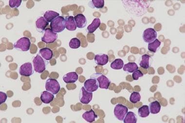
Bone marrow aspirate from a child with T-cell acute lymphoblastic leukemia. The marrow is replaced with lymphoblasts of various sizes. No myeloid or erythroid precursors are seen. Megakaryocytes are absent. |
 | View Full Size Image | |
Media type: Photo
|
| Media file 3:
Bone marrow aspirate from a child with B-cell acute lymphoblastic leukemia. The lymphoblasts are large and have basophilic cytoplasm with prominent vacuoles. |
 | View Full Size Image | |
Media type: Photo
|
- Cave H, van der Werff ten Bosch J, Suciu S, et al. Clinical significance of minimal residual disease in childhood acute lymphoblastic leukemia. European Organization for Research and Treatment of Cancer--Childhood Leukemia Cooperative Group. N Engl J Med. Aug 27 1998;339(9):591-8. [Medline].
- Cheok MH, Evans WE. Acute lymphoblastic leukaemia: a model for the pharmacogenomics of cancer therapy. Nat Rev Cancer. Feb 2006;6(2):117-29. [Medline].
- Coustan-Smith E, Behm FG, Sanchez J, et al. Immunological detection of minimal residual disease in children with acute lymphoblastic leukaemia. Lancet. Feb 21 1998;351(9102):550-4. [Medline].
- Dordelmann M, Reiter A, Borkhardt A, et al. Prednisone response is the strongest predictor of treatment outcome in infant acute lymphoblastic leukemia. Blood. Aug 15 1999;94(4):1209-17. [Medline].
- Gaynon PS. Childhood acute lymphoblastic leukaemia and relapse. Br J Haematol. Dec 2005;131(5):579-87. [Medline].
- Giordano P, Santoro N, Del Vecchio GC, et al. T-immunophenotype is associated with an increased prevalence of thrombosis in children with acute lymphoblastic leukemia. A retrospective study. Haematologica. Sep 2003;88(9):1079-80. [Medline].
- Greaves MF. Aetiology of acute leukaemia. Lancet. Feb 1 1997;349(9048):344-9. [Medline].
- Jones LK, Saha V. Philadelphia positive acute lymphoblastic leukaemia of childhood. Br J Haematol. Aug 2005;130(4):489-500. [Medline].
- Lee S, Kim YJ, Min CK, et al. The effect of first-line imatinib interim therapy on the outcome of allogeneic stem cell transplantation in adults with newly diagnosed Philadelphia chromosome-positive acute lymphoblastic leukemia. Blood. May 1 2005;105(9):3449-57. [Medline].
- Linet MS, Hatch EE, Kleinerman RA, et al. Residential exposure to magnetic fields and acute lymphoblastic leukemia in children. N Engl J Med. Jul 3 1997;337(1):1-7. [Medline].
- Margolin JF, Steuber CP, Poplack DG. Acute lymphoblastic leukemia. In: Principles and Practice of Pediatric Oncology. 15th ed. 2006:538-90.
- Pui CH, Evans WE. Treatment of acute lymphoblastic leukemia. N Engl J Med. Jan 12 2006;354(2):166-78. [Medline].
- Pui CH, Relling MV, Downing JR. Acute lymphoblastic leukemia. N Engl J Med. Apr 8 2004;350(15):1535-48. [Medline].
- Rivera GK, Pinkel D, Simone JV, et al. Treatment of acute lymphoblastic leukemia. 30 years'' experience at St. Jude Children''s Research Hospital. N Engl J Med. Oct 28 1993;329(18):1289-95. [Medline].
- Rubnitz JE, Behm FG, Downing JR. 11q23 rearrangements in acute leukemia. Leukemia. Jan 1996;10(1):74-82. [Medline].
- Rubnitz JE, Pui CH. Molecular diagnostics in the treatment of leukemia. Curr Opin Hematol. Jul 1999;6(4):229-35. [Medline].
- Rubnitz JE, Pui CH, Downing JR. The role of TEL fusion genes in pediatric leukemias. Leukemia. Jan 1999;13(1):6-13. [Medline].
- Schrappe M, Arico M, Harbott J, et al. Philadelphia chromosome-positive (Ph+) childhood acute lymphoblastic leukemia: good initial steroid response allows early prediction of a favorable treatment outcome. ALYSIS. Oct 15 1998;92(8):2730-41. [Medline].
- Smith M, Arthur D, Camitta B, et al. Uniform approach to risk classification and treatment assignment for children with acute lymphoblastic leukemia. J Clin Oncol. Jan 1996;14(1):18-24. [Medline].
- Thomas DA, Faderl S, Cortes J, et al. Treatment of Philadelphia chromosome-positive acute lymphocytic leukemia with hyper-CVAD and imatinib mesylate. Blood. Jun 15 2004;103(12):4396-407. [Medline].
- Tsutsumi S, Taketani T, Nishimura K, et al. Two distinct gene expression signatures in pediatric acute lymphoblastic leukemia with MLL rearrangements. Cancer Res. Aug 15 2003;63(16):4882-7. [Medline].
Acute Lymphoblastic Leukemia excerpt Article Last Updated: Jul 11, 2006
|