Background
Café au lait spots, or café au lait (CAL) macules (CALMs), are hyperpigmented lesions that may vary in color from light brown to dark brown (see the images below) [1] ; this is reflected in the name of the condition, which means "coffee with milk." The borders may be smooth or irregular.
Café au lait skin lesions widely vary widely in size and number and are usually the earliest manifestations of neurofibromatosis. [2] The macules may be observed in infancy, though they are typically very light in infants and can be difficult to appreciate. The skin lesions develop in early infancy, and they may enlarge in size and become obvious after age 2 years.
CALMs are observed in 95% of patients with neurofibromatosis type 1 (NF1), which is the neurocutaneous syndrome with which they are most often associated. [3] These spots may also be observed in patients without NF1. Other conditions in which they may be observed include McCune-Albright syndrome, tuberous sclerosis, Fanconi anemia, and Coffin-Siris syndrome, characterized by developmental delay, speech impairment, distinctive facial features, hypertrichosis, hypoplasia of the distal phalanx of the fifth digit, and agenesis of the corpus callosum. [4] CALMs may be a marker for RASopathies, disorders related to RAS mutations. [5, 6]
Pathophysiology
Café au lait spots are caused by an increase in melanin content, often with the presence of giant melanosomes. A significant increase in melanocyte density is noted in the CALMs of patients with NF1 compared with patients who have isolated CALMs without NF1 involvement. Also, an increase in stem cell factor cytokines is more frequently observed in NF1 CALMs than in non-NF1 CALMs.
Etiology
CALMs associated with NF1 result from an autosomal dominant disorder with high penetrance and variability in the expression of clinical features.
The NF1 gene is localized to the pericentromeric region of the long arm of chromosome 17. The gene encodes for neurofibromin, which is a GTP-ase activating protein that downregulates the cellular proto-oncogene p21-ras.
About 50% of individuals with NF1 have a spontaneous mutation. This high incidence is thought to result from the large size of the gene, which increases the likelihood of spontaneous mutations.
Occasionally, patients who have larger gene deletions have a higher incidence of intellectual disability and earlier appearance of cutaneous neurofibromas.
Epidemiology
United States and international statistics
In the newborn period, solitary café au lait spots may occur in 0.3% of Whites, 3% of Hispanics, and in 18% of Blacks. [7] In childhood, solitary CALMs occur in 13% of Whites and 27% of Blacks. Two or more CALMs were not observed in any of 4000 White newborns but were found in 8% of Black newborns. Café au lait spots that confirm the diagnosis of NF1 occur at an estimated frequency of 1 in 3500 persons. [8]
Solitary café au lait spots have been reported to occur in 0.5% of Arab newborns and in 0.4% of Chinese newborns. [7]
Age-, sex-, and race-related demographics
Typically, café au lait spots are present at birth, though they may be difficult to appreciate. A Wood lamp may improve the ability to visualize these faint spots. By the time the child is aged 2-3 years, CALMs are clearly visible. The size and number of CALMs increase with patient age in patients with NF1.
No sexual predilection is recognized.
Café au lait spots are more frequently observed in Black children.
Prognosis
CALMs have not been demonstrated to undergo malignant change. They are benign and produce no mortality or morbidity, though the syndromes associated with them may have significant manifestations.
-
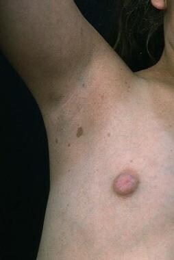
Axillary freckling showing café au lait spots.
-
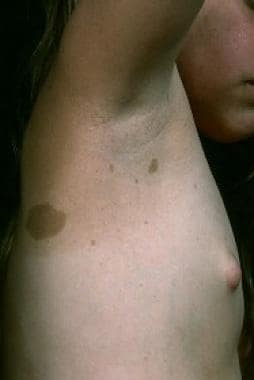
Multiple irregular sized and shaped café au lait lesions.
-
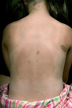
Café au lait lesions.
-
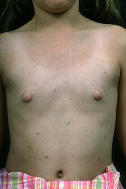
Café au lait lesions.
